Loading
PDBj
メニューPDBj@FacebookPDBj@TwitterPDBj@YouTubewwPDB FoundationwwPDB
RCSB PDBPDBeBMRBAdv. SearchSearch help
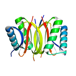
Search by PDB author
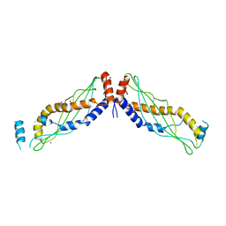
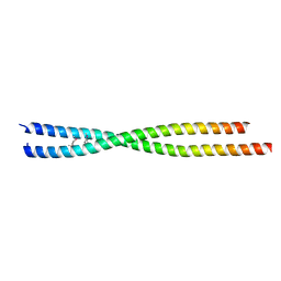
2V66
DownloadVisualize
BU of 2v66 by Molmil
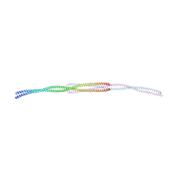
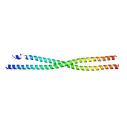
Crystal Structure of the coiled-coil domain of Ndel1 (a.a. 58 to 169) C
分子名称: MERCURY (II) ION, NUCLEAR DISTRIBUTION PROTEIN NUDE-LIKE 1
著者Tarricone, C, Perrina, F, Musacchio, A.
登録日2007-07-13
公開日2007-11-27
最終更新日2024-05-08
実験手法X-RAY DIFFRACTION (2.1 Å)
主引用文献The Structure of the Coiled-Coil Domain of Ndel1 and the Basis of its Interaction with Lis1, the Causal Protein of Miller-Dieker Lissencephaly.
Structure, 15, 2007
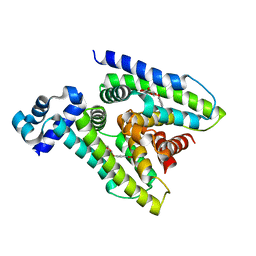
4DS1
DownloadVisualize
BU of 4ds1 by Molmil
The Structure of a Yeast Dyn2-Nup159 Complex and the Molecular Basis for the Dynein Light Chain - Nuclear Pore Interaction
分子名称: Dynein light chain 1, cytoplasmic, Nucleoporin NUP159
著者Slep, K.C, Romes, E.M.
登録日2012-02-17
公開日2012-03-21
最終更新日2023-09-13
実験手法X-RAY DIFFRACTION (1.851 Å)
主引用文献Structure of a yeast Dyn2-Nup159 complex and molecular basis for dynein light chain-nuclear pore interaction.
J.Biol.Chem., 287, 2012
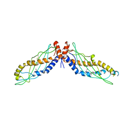
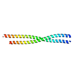
2V71
DownloadVisualize
BU of 2v71 by Molmil
Coiled-coil region of NudEL
分子名称: NUCLEAR DISTRIBUTION PROTEIN NUDE-LIKE 1
著者Derewenda, U, Cooper, D.R, Kim, M.H, Derewenda, Z.S.
登録日2007-07-25
公開日2007-11-27
最終更新日2017-06-28
実験手法X-RAY DIFFRACTION (2.24 Å)
主引用文献The Structure of the Coiled-Coil Domain of Ndel1 and the Basis of its Interaction with Lis1, the Causal Protein of Miller-Dieker Lissencephaly.
Structure, 15, 2007
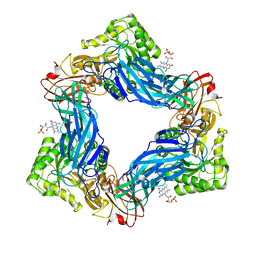
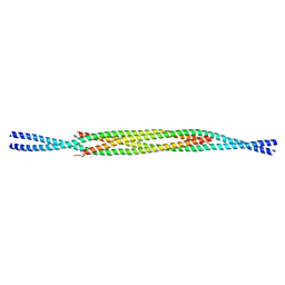
6WY1
DownloadVisualize
BU of 6wy1 by Molmil
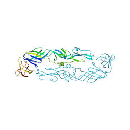
Crystal structure of an engineered thermostable dengue virus 2 envelope protein dimer
分子名称: 2-acetamido-2-deoxy-beta-D-glucopyranose, Dengue 2 soluble recombinant envelope
著者Kudlacek, S.T, Lakshmanane, P, Kuhlman, B.
登録日2020-05-12
公開日2021-11-10
最終更新日2023-10-18
実験手法X-RAY DIFFRACTION (3.42 Å)
主引用文献Designed, highly expressing, thermostable dengue virus 2 envelope protein dimers elicit quaternary epitope antibodies.
Sci Adv, 7, 2021
3P8H
DownloadVisualize
BU of 3p8h by Molmil
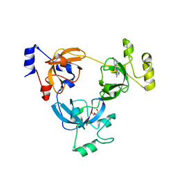
Crystal structure of L3MBTL1 (MBT repeat) in complex with a nicotinamide antagonist
分子名称: 3-bromo-5-[(4-pyrrolidin-1-ylpiperidin-1-yl)carbonyl]pyridine, GLYCEROL, Lethal(3)malignant brain tumor-like protein, ...
著者Lam, R, Herold, J.M, Ouyang, H, Tempel, W, Gao, C, Ravichandran, M, Senisterra, G, Bountra, C, Weigelt, J, Arrowsmith, C.H, Edwards, A.M, Vedadi, M, Kireev, D, Frye, S.V, Brown, P.J, Structural Genomics Consortium (SGC)
登録日2010-10-13
公開日2010-11-03
最終更新日2023-09-06
実験手法X-RAY DIFFRACTION (2.55 Å)
主引用文献Small-molecule ligands of methyl-lysine binding proteins.
J.Med.Chem., 54, 2011
7TJL
DownloadVisualize
BU of 7tjl by Molmil
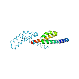
Crystal structure of de novo designed protein, SEWN0.1
分子名称: De novo designed protein, SEWN0.1
著者Cummins, M.C, Jacobs, T.M, Kuhlman, B.
登録日2022-01-16
公開日2022-06-29
最終更新日2024-04-03
実験手法X-RAY DIFFRACTION (1.87 Å)
主引用文献AlphaFold accurately predicts distinct conformations based on the oligomeric state of a de novo designed protein
Protein Sci., 31, 2022
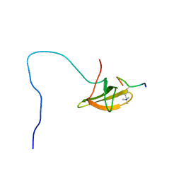
2M0O
DownloadVisualize
BU of 2m0o by Molmil
The solution structure of human PHF1 in complex with H3K36me3
分子名称: H3K36me3 peptide, PHD finger protein 1
著者Song, J, Patel, D.J.
登録日2012-10-30
公開日2013-01-16
最終更新日2013-02-27
実験手法SOLUTION NMR
主引用文献An H3K36 Methylation-Engaging Tudor Motif of Polycomb-like Proteins Mediates PRC2 Complex Targeting.
Mol.Cell, 49, 2013
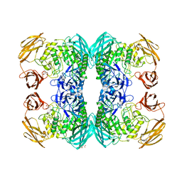
6MVH
DownloadVisualize
BU of 6mvh by Molmil
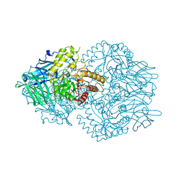
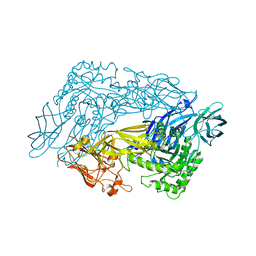
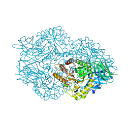
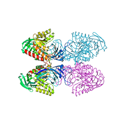
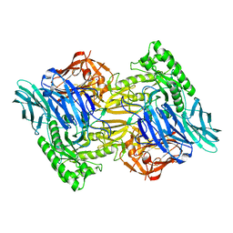
Crystal structure of FMN-binding beta-glucuronidase from Roseburia hominis
分子名称: Beta-galactosidase, CALCIUM ION, FLAVIN MONONUCLEOTIDE
著者Pellock, S.J, Redinbo, M.R.
登録日2018-10-25
公開日2019-01-30
最終更新日2024-03-13
実験手法X-RAY DIFFRACTION (2.4 Å)
主引用文献Discovery and Characterization of FMN-Binding beta-Glucuronidases in the Human Gut Microbiome.
J. Mol. Biol., 431, 2019
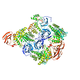
6MVF
DownloadVisualize
BU of 6mvf by Molmil
Crystal structure of FMN-binding beta-glucuronidase from Facaelibacterium prausnitzii L2-6
分子名称: Beta-galactosidase/beta-glucuronidase, FLAVIN MONONUCLEOTIDE
著者Pellock, S.J, Redinbo, M.R.
登録日2018-10-25
公開日2019-01-30
最終更新日2023-10-11
実験手法X-RAY DIFFRACTION (2.55 Å)
主引用文献Discovery and Characterization of FMN-Binding beta-Glucuronidases in the Human Gut Microbiome.
J. Mol. Biol., 431, 2019
6MVG
DownloadVisualize
BU of 6mvg by Molmil
Crystal structure of FMN-binding beta-glucuronidase from Ruminococcus gnavus
分子名称: CALCIUM ION, FLAVIN MONONUCLEOTIDE, beta-glucuronidase
著者Pellock, S.J, Redinbo, M.R.
登録日2018-10-25
公開日2019-01-30
最終更新日2024-03-13
実験手法X-RAY DIFFRACTION (2.8 Å)
主引用文献Discovery and Characterization of FMN-Binding beta-Glucuronidases in the Human Gut Microbiome.
J. Mol. Biol., 431, 2019
4KGH
DownloadVisualize
BU of 4kgh by Molmil
Crystal Structure of human splunc1 lacking the secretion signal sequence
分子名称: AMMONIUM ION, BPI fold-containing family A member 1, GLYCEROL, ...
著者Betts, L, Walton, W.G.
登録日2013-04-29
公開日2013-09-25
最終更新日2017-11-15
実験手法X-RAY DIFFRACTION (2.806 Å)
主引用文献Molecular basis for pH-dependent mucosal dehydration in cystic fibrosis airways.
Proc.Natl.Acad.Sci.USA, 110, 2013
4KGO
DownloadVisualize
BU of 4kgo by Molmil
Crystal Structure of double Leucine to Methionine mutant human splunc1 lacking the secretion signal sequence
分子名称: BPI fold-containing family A member 1
著者Betts, L, Walton, W.G.
登録日2013-04-29
公開日2013-09-25
最終更新日2017-11-15
実験手法X-RAY DIFFRACTION (3.194 Å)
主引用文献Molecular basis for pH-dependent mucosal dehydration in cystic fibrosis airways.
Proc.Natl.Acad.Sci.USA, 110, 2013
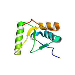
2HTF
DownloadVisualize
BU of 2htf by Molmil
The solution structure of the BRCT domain from human polymerase reveals homology with the TdT BRCT domain
分子名称: DNA polymerase mu
著者DeRose, E.F, Clarkson, M.W, Gilmore, S.A, Ramsden, D.A, Mueller, G.A, London, R.E, Lee, A.L.
登録日2006-07-25
公開日2007-02-27
最終更新日2024-05-29
実験手法SOLUTION NMR
主引用文献Solution structure of polymerase mu's BRCT Domain reveals an element essential for its role in nonhomologous end joining.
Biochemistry, 46, 2007
6DXU
DownloadVisualize
BU of 6dxu by Molmil
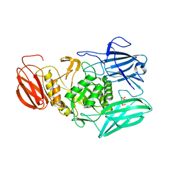
Crystal Structure of Parabacteroides merdae Beta-Glucuronidase (GUS)
分子名称: BICINE, Glycosyl hydrolase family 2, TIM barrel domain protein, ...
著者Little, M.S, Redinbo, M.R.
登録日2018-06-30
公開日2019-02-27
最終更新日2024-03-13
実験手法X-RAY DIFFRACTION (1.9 Å)
主引用文献Active site flexibility revealed in crystal structures of Parabacteroides merdae beta-glucuronidase from the human gut microbiome.
Protein Sci., 27, 2018
6ED2
DownloadVisualize
BU of 6ed2 by Molmil
Faecalibacterium prausnitzii beta-glucuronidase
分子名称: FORMIC ACID, GLYCEROL, Glycosyl hydrolase family 2, ...
著者Pellock, S.J, Biernat, K.A, Redinbo, M.R.
登録日2018-08-08
公開日2019-02-13
最終更新日2024-03-13
実験手法X-RAY DIFFRACTION (2.3 Å)
主引用文献Structure, function, and inhibition of drug reactivating human gut microbial beta-glucuronidases.
Sci Rep, 9, 2019
6ED1
DownloadVisualize
BU of 6ed1 by Molmil
Bacteroides dorei Beta-glucuronidase
分子名称: Glycosyl hydrolase family 2, sugar binding domain protein, SODIUM ION
著者Biernat, K.A, Redinbo, M.R.
登録日2018-08-08
公開日2019-02-13
最終更新日2023-10-11
実験手法X-RAY DIFFRACTION (2.9 Å)
主引用文献Structure, function, and inhibition of drug reactivating human gut microbial beta-glucuronidases.
Sci Rep, 9, 2019
6EC6
DownloadVisualize
BU of 6ec6 by Molmil
Ruminococcus gnavus Beta-glucuronidase
分子名称: Beta-glucuronidase, CHLORIDE ION, GLYCEROL
著者Biernat, K.A, Redinbo, M.R.
登録日2018-08-07
公開日2019-02-13
最終更新日2023-10-11
実験手法X-RAY DIFFRACTION (2.85 Å)
主引用文献Structure, function, and inhibition of drug reactivating human gut microbial beta-glucuronidases.
Sci Rep, 9, 2019
6D6W
DownloadVisualize
BU of 6d6w by Molmil
Bacteroides uniformis beta-glucuronidase 1 bound to glucuronate
分子名称: Beta-galactosidase/beta-glucuronidase, CHLORIDE ION, GLYCEROL, ...
著者Walton, W.G, Pellock, S.J, Redinbo, M.R.
登録日2018-04-23
公開日2018-10-17
最終更新日2024-03-13
実験手法X-RAY DIFFRACTION (1.8 Å)
主引用文献Three structurally and functionally distinct beta-glucuronidases from the human gut microbeBacteroides uniformis.
J. Biol. Chem., 293, 2018
6D8G
DownloadVisualize
BU of 6d8g by Molmil
D341A D367A calcium binding mutant of Bacteroides uniformis beta-glucuronidase 2
分子名称: Glycosyl hydrolases family 2, sugar binding domain protein, SODIUM ION
著者Walton, W.G, Pellock, S.J, Redinbo, M.R.
登録日2018-04-26
公開日2018-10-17
最終更新日2023-10-04
実験手法X-RAY DIFFRACTION (3 Å)
主引用文献Three structurally and functionally distinct beta-glucuronidases from the human gut microbeBacteroides uniformis.
J. Biol. Chem., 293, 2018
6AP0
DownloadVisualize
BU of 6ap0 by Molmil
Crystal structure of human FLASH N-terminal domain C54S/C83A (Crystal form 2)
分子名称: CASP8-associated protein 2
著者Aik, W.S, Tong, L.
登録日2017-08-16
公開日2017-11-15
最終更新日2023-10-04
実験手法X-RAY DIFFRACTION (2.581 Å)
主引用文献The N-terminal domains of FLASH and Lsm11 form a 2:1 heterotrimer for histone pre-mRNA 3'-end processing.
PLoS ONE, 12, 2017
6AZ6
DownloadVisualize
BU of 6az6 by Molmil
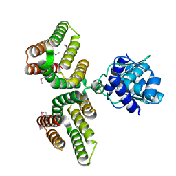
Streptococcus agalactiae GntR
分子名称: GntR family transcriptional regulator
著者Pellock, S.J, Redinbo, M.R.
登録日2017-09-10
公開日2017-12-20
最終更新日2019-12-04
実験手法X-RAY DIFFRACTION (1.909 Å)
主引用文献Structural basis for the regulation of beta-glucuronidase expression by human gut Enterobacteriaceae.
Proc. Natl. Acad. Sci. U.S.A., 115, 2018
6AZH
DownloadVisualize
BU of 6azh by Molmil
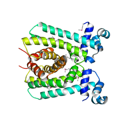
Clostridium perfringens putative fatty acid metabolism regulator
分子名称: TetR family transcriptional regulator
著者William, W.G, Redinbo, M.R.
登録日2017-09-11
公開日2017-12-20
最終更新日2018-01-17
実験手法X-RAY DIFFRACTION (1.75 Å)
主引用文献Structural basis for the regulation of beta-glucuronidase expression by human gut Enterobacteriaceae.
Proc. Natl. Acad. Sci. U.S.A., 115, 2018
6ANO
DownloadVisualize
BU of 6ano by Molmil
Crystal structure of human FLASH N-terminal domain
分子名称: CASP8-associated protein 2
著者Aik, W.S, Tong, L.
登録日2017-08-14
公開日2017-11-15
実験手法X-RAY DIFFRACTION (2.61 Å)
主引用文献The N-terminal domains of FLASH and Lsm11 form a 2:1 heterotrimer for histone pre-mRNA 3'-end processing.
PLoS ONE, 12, 2017
6AYI
DownloadVisualize
BU of 6ayi by Molmil
Escherichia coli GusR
分子名称: 4-nitrophenyl beta-D-glucopyranosiduronic acid, HTH-type transcriptional regulator UidR
著者Little, M.S, Pellock, S.J.
登録日2017-09-08
公開日2017-12-20
最終更新日2024-03-13
実験手法X-RAY DIFFRACTION (2.09 Å)
主引用文献Structural basis for the regulation of beta-glucuronidase expression by human gut Enterobacteriaceae.
Proc. Natl. Acad. Sci. U.S.A., 115, 2018
6AOZ
DownloadVisualize
BU of 6aoz by Molmil
Crystal structure of human FLASH N-terminal domain C54S/C83A (Crystal form 1)
分子名称: 1,2-ETHANEDIOL, CASP8-associated protein 2
著者Aik, W.S, Tong, L.
登録日2017-08-16
公開日2017-11-15
最終更新日2023-10-04
実験手法X-RAY DIFFRACTION (2.1 Å)
主引用文献The N-terminal domains of FLASH and Lsm11 form a 2:1 heterotrimer for histone pre-mRNA 3'-end processing.
PLoS ONE, 12, 2017

222415

件を2024-07-10に公開中

PDB statisticsPDBj update infoContact PDBjnumon