8TDW
 
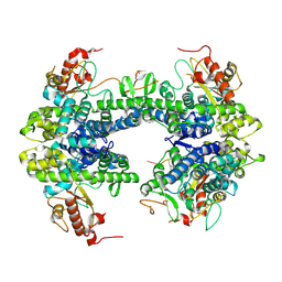 | | ssRNA bound SAMHD1 T open | | 分子名称: | Deoxynucleoside triphosphate triphosphohydrolase SAMHD1, FE (III) ION, RNA (5'-R(P*CP*CP*GP*AP*CP*C)-3'), ... | | 著者 | Sung, M, Huynh, K, Han, S. | | 登録日 | 2023-07-05 | | 公開日 | 2023-11-22 | | 最終更新日 | 2023-12-20 | | 実験手法 | ELECTRON MICROSCOPY (3.04 Å) | | 主引用文献 | Guanine-containing ssDNA and RNA induce dimeric and tetrameric structural forms of SAMHD1.
Nucleic Acids Res., 51, 2023
|
|
8TDV
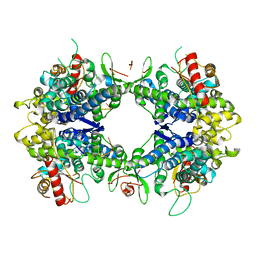 | | ssRNA bound SAMHD1 T closed | | 分子名称: | Deoxynucleoside triphosphate triphosphohydrolase SAMHD1, FE (III) ION, RNA (5'-R(P*CP*CP*GP*AP*CP*CP*C)-3'), ... | | 著者 | Sung, M, Huynh, K, Han, S. | | 登録日 | 2023-07-05 | | 公開日 | 2023-12-20 | | 実験手法 | ELECTRON MICROSCOPY (3.44 Å) | | 主引用文献 | Guanine-containing ssDNA and RNA induce dimeric and tetrameric structural forms of SAMHD1.
Nucleic Acids Res., 51, 2023
|
|
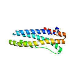
5V5F
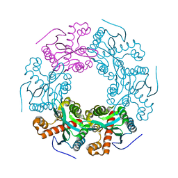 | | Crystal structure of RICE1 (PNT2) | | 分子名称: | At3g11770 | | 著者 | Li, P. | | 登録日 | 2017-03-14 | | 公開日 | 2017-09-13 | | 最終更新日 | 2024-03-06 | | 実験手法 | X-RAY DIFFRACTION (2.945 Å) | | 主引用文献 | RISC-interacting clearing 3'- 5' exoribonucleases (RICEs) degrade uridylated cleavage fragments to maintain functional RISC in Arabidopsis thaliana.
Elife, 6, 2017
|
|
3U9J
 | |
3U9M
 | |
4HZ8
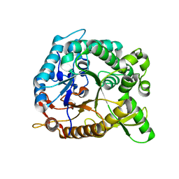 | |
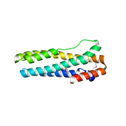
4HZ7
 | | Crystal structure of BglB with glucose | | 分子名称: | beta-D-glucopyranose, beta-glucosidase | | 著者 | Hwang, K.Y, Nam, K.H. | | 登録日 | 2012-11-14 | | 公開日 | 2012-12-19 | | 最終更新日 | 2023-11-08 | | 実験手法 | X-RAY DIFFRACTION (2 Å) | | 主引用文献 | Structural insights into the substrate recognition properties of beta-glucosidase.
Biochem.Biophys.Res.Commun., 391, 2010
|
|
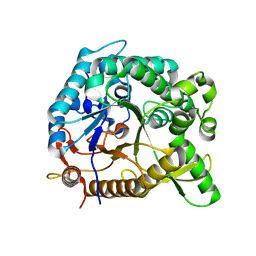
4HZ6
 | | crystal structure of BglB | | 分子名称: | Beta-glucosidase, GLYCEROL | | 著者 | Hwang, K.Y, Nam, K.H. | | 登録日 | 2012-11-14 | | 公開日 | 2012-12-19 | | 最終更新日 | 2023-11-08 | | 実験手法 | X-RAY DIFFRACTION (1.4 Å) | | 主引用文献 | Structural insights into the substrate recognition properties of beta-glucosidase.
Biochem.Biophys.Res.Commun., 391, 2010
|
|


