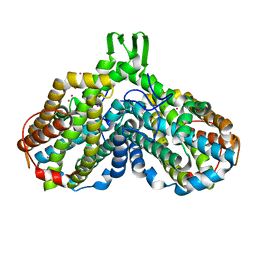
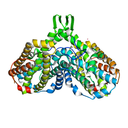
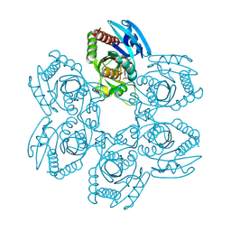
3ZZW
 
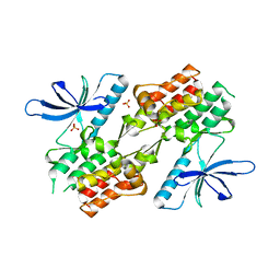 | | Crystal structure of the kinase domain of ROR2 | | 分子名称: | CHLORIDE ION, SULFATE ION, TYROSINE-PROTEIN KINASE TRANSMEMBRANE RECEPTOR ROR2 | | 著者 | Tresaugues, L, Moche, M, Arrowsmith, C.H, Berglund, H, Bountra, C, Edwards, A.M, Ekblad, T, Graslund, S, Karlberg, T, Nyman, T, Schuler, H, Thorsell, A.G, Weigelt, J, Nordlund, P, Structural Genomics Consortium (SGC) | | 登録日 | 2011-09-05 | | 公開日 | 2011-09-14 | | 最終更新日 | 2023-12-20 | | 実験手法 | X-RAY DIFFRACTION (2.9 Å) | | 主引用文献 | Crystal Structure of the Kinase Domain of Ror2
To be Published
|
|
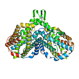
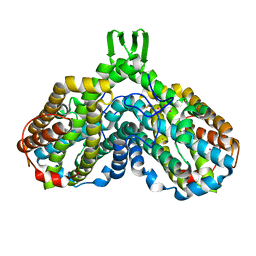
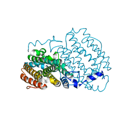
4A9C
 | | Crystal structure of human SHIP2 in complex with biphenyl 2,3',4,5',6- pentakisphosphate | | 分子名称: | BIPHENYL 2,3',4,5',6-PENTAKISPHOSPHATE, PHOSPHATIDYLINOSITOL-3,4,5-TRISPHOSPHATE 5-PHOSPHATASE 2 | | 著者 | Tresaugues, L, Arrowsmith, C.H, Berglund, H, Bountra, C, Edwards, A.M, Ekblad, T, Graslund, S, Karlberg, T, Mills, S.J, Moche, M, Nyman, T, Persson, C, Potter, B.V.L, Schuler, H, Thorsell, A.G, Weigelt, J, Nordlund, P. | | 登録日 | 2011-11-25 | | 公開日 | 2012-05-30 | | 最終更新日 | 2023-12-20 | | 実験手法 | X-RAY DIFFRACTION (2.1 Å) | | 主引用文献 | A Synthetic Polyphosphoinositide Headgroup Surrogate in Complex with Ship2 Provides a Rationale for Drug Discovery.
Acs Chem.Biol., 7, 2012
|
|
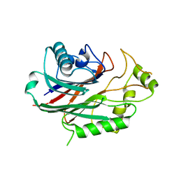
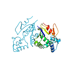
4AQL
 | | HUMAN GUANINE DEAMINASE IN COMPLEX WITH VALACYCLOVIR | | 分子名称: | 2-[(2-amino-6-oxo-1,6-dihydro-9H-purin-9-yl)methoxy]ethyl L-valinate, GUANINE DEAMINASE, ZINC ION | | 著者 | Welin, M, Egeblad, L, Arrowsmith, C.H, Berglund, H, Bountra, C, Collins, R, Edwards, A.M, Flodin, S, Graslund, S, Hammarstrom, M, Johansson, I, Karlberg, T, Kotenyova, T, Moche, M, Nyman, T, Persson, C, Schuler, H, Thorsell, A.G, Tresaugues, L, Weigelt, J, Nordlund, P. | | 登録日 | 2012-04-18 | | 公開日 | 2012-05-02 | | 最終更新日 | 2023-12-20 | | 実験手法 | X-RAY DIFFRACTION (1.99 Å) | | 主引用文献 | Pan-Pathway Based Interaction Profiling of Fda-Approved Nucleoside and Nucleobase Analogs with Enzymes of the Human Nucleotide Metabolism.
Plos One, 7, 2012
|
|
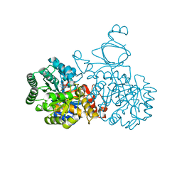
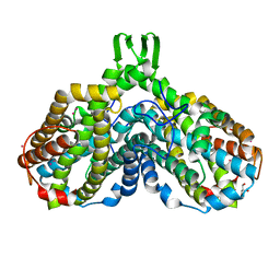
4A1N
 | | Human Mitochondrial endo-exonuclease | | 分子名称: | CHLORIDE ION, MAGNESIUM ION, NUCLEASE EXOG, ... | | 著者 | Welin, M, Moche, M, Arrowsmith, C.H, Berglund, H, Bountra, C, Collins, R, Edwards, A.M, Flodin, S, Graslund, S, Hammarstrom, M, Johansson, I, Karlberg, T, Kotenyova, T, Nyman, T, Persson, C, Schuler, H, Thorsell, A.G, Tresaugues, L, Weigelt, J, Nordlund, P. | | 登録日 | 2011-09-16 | | 公開日 | 2012-02-29 | | 最終更新日 | 2023-12-20 | | 実験手法 | X-RAY DIFFRACTION (2.8 Å) | | 主引用文献 | Human Mitochondrial Endo-Exonuclease
To be Published
|
|
1JPR
 | |
1JQC
 | |
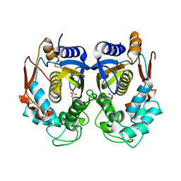
1PFR
 | | RIBONUCLEOSIDE-DIPHOSPHATE REDUCTASE 1 BETA CHAIN | | 分子名称: | FE (III) ION, MERCURY (II) ION, PROTEIN R2 OF RIBONUCLEOTIDE REDUCTASE | | 著者 | Logan, D.T, Su, X.D, Aberg, A, Regnstrom, K, Hajdu, J, Eklund, H, Nordlund, P. | | 登録日 | 1996-12-03 | | 公開日 | 1997-03-12 | | 最終更新日 | 2024-05-22 | | 実験手法 | X-RAY DIFFRACTION (2.2 Å) | | 主引用文献 | Crystal structure of reduced protein R2 of ribonucleotide reductase: the structural basis for oxygen activation at a dinuclear iron site.
Structure, 4, 1996
|
|
1PHR
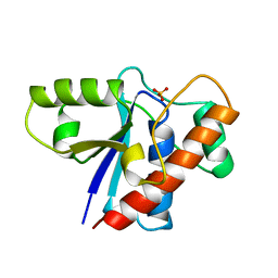 | | THE CRYSTAL STRUCTURE OF A LOW MOLECULAR PHOSPHOTYROSINE PROTEIN PHOSPHATASE | | 分子名称: | LOW MOLECULAR WEIGHT PHOSPHOTYROSINE PROTEIN PHOSPHATASE, SULFATE ION | | 著者 | Su, X.-D, Taddei, N, Stefani, M, Ramponi, G, Nordlund, P. | | 登録日 | 1994-07-05 | | 公開日 | 1995-07-31 | | 最終更新日 | 2024-02-14 | | 実験手法 | X-RAY DIFFRACTION (2.1 Å) | | 主引用文献 | The crystal structure of a low-molecular-weight phosphotyrosine protein phosphatase.
Nature, 370, 1994
|
|
1RNR
 | |
1RSV
 | | azide complex of the diferrous E238A mutant R2 subunit of ribonucleotide reductase | | 分子名称: | AZIDE ION, FE (III) ION, MERCURY (II) ION, ... | | 著者 | Assarsson, M, Andersson, M.E, Hogbom, M, Persson, B.O, Sahlin, M, Barra, A.L, Sjoberg, B.M, Nordlund, P, Graslund, A. | | 登録日 | 2003-12-10 | | 公開日 | 2003-12-23 | | 最終更新日 | 2024-05-29 | | 実験手法 | X-RAY DIFFRACTION (2.2 Å) | | 主引用文献 | Restoring proper radical generation by azide binding to the iron site of the E238A mutant R2 protein of ribonucleotide reductase from Escherichia coli.
J.Biol.Chem., 276, 2001
|
|
1RSR
 | | azide complex of the diferrous F208A mutant R2 subunit of ribonucleotide reductase | | 分子名称: | AZIDE ION, FE (II) ION, MERCURY (II) ION, ... | | 著者 | Andersson, M.E, Hogbom, M, Rinaldo-Matthis, A, Andersson, K.K, Sjoberg, B.M, Nordlund, P. | | 登録日 | 2003-12-10 | | 公開日 | 2003-12-23 | | 最終更新日 | 2024-05-29 | | 実験手法 | X-RAY DIFFRACTION (2 Å) | | 主引用文献 | The Crystal Structure of an Azide Complex of the Diferrous R2 Subunit of Ribonucleotide Reductase Displays a Novel Carboxylate Shift with Important Mechanistic Implications for Diiron-Catalyzed Oxygen Activation
J.Am.Chem.Soc., 121, 1999
|
|
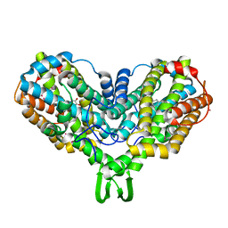
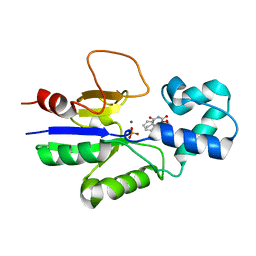
1MH9
 | | Crystal Structure Analysis of deoxyribonucleotidase | | 分子名称: | MAGNESIUM ION, PHOSPHATE ION, deoxyribonucleotidase | | 著者 | Rinaldo-Matthis, A, Rampazzo, C, Reichard, P, Bianchi, V, Nordlund, P. | | 登録日 | 2002-08-19 | | 公開日 | 2002-10-30 | | 最終更新日 | 2024-03-13 | | 実験手法 | X-RAY DIFFRACTION (1.8 Å) | | 主引用文献 | Crystal structure of a human mitochondrial deoxyribonucleotidase.
Nat.Struct.Biol., 9, 2002
|
|
1MXR
 | | High resolution structure of Ribonucleotide reductase R2 from E. coli in its oxidised (Met) form | | 分子名称: | FE (III) ION, GLYCEROL, MERCURY (II) ION, ... | | 著者 | Andersson, M.A, Hogbom, M, Nordlund, P. | | 登録日 | 2002-10-03 | | 公開日 | 2003-03-25 | | 最終更新日 | 2023-10-25 | | 実験手法 | X-RAY DIFFRACTION (1.42 Å) | | 主引用文献 | Displacement of the tyrosyl radical cofactor in ribonucleotide reductase obtained by single-crystal high-field EPR and 1.4-A x-ray data.
Proc.Natl.Acad.Sci.Usa, 100, 2003
|
|
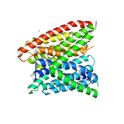
1MTY
 | | METHANE MONOOXYGENASE HYDROXYLASE FROM METHYLOCOCCUS CAPSULATUS (BATH) | | 分子名称: | FE (III) ION, METHANE MONOOXYGENASE HYDROXYLASE | | 著者 | Rosenzweig, A.C, Nordlund, P, Lippard, S.J, Frederick, C.A. | | 登録日 | 1996-07-10 | | 公開日 | 1997-04-21 | | 最終更新日 | 2024-05-22 | | 実験手法 | X-RAY DIFFRACTION (1.7 Å) | | 主引用文献 | Crystal structures of the methane monooxygenase hydroxylase from Methylococcus capsulatus (Bath): implications for substrate gating and component interactions.
Proteins, 29, 1997
|
|
1Q91
 | | Crystal structure of human mitochondrial deoxyribonucleotidase in complex with the inhibitor DPB-T | | 分子名称: | (S)-1-[2'-DEOXY-3',5'-O-(1-PHOSPHONO)BENZYLIDENE-B-D-THREO-PENTOFURANOSYL]THYMINE, 5(3)-deoxyribonucleotidase, MAGNESIUM ION, ... | | 著者 | Rinaldo-Matthis, A, Rampazzo, C, Balzarini, J, Reichard, P, Bianchi, V, Nordlund, P. | | 登録日 | 2003-08-22 | | 公開日 | 2004-04-20 | | 最終更新日 | 2023-10-25 | | 実験手法 | X-RAY DIFFRACTION (1.6 Å) | | 主引用文献 | Crystal structures of the mitochondrial deoxyribonucleotidase in complex with two specific inhibitors
Mol.Pharmacol., 65, 2004
|
|
1Q92
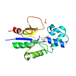 | | Crystal structure of human mitochondrial deoxyribonucleotidase in complex with the inhibitor PMcP-U | | 分子名称: | 5(3)-deoxyribonucleotidase, GLYCEROL, MAGNESIUM ION, ... | | 著者 | Rinaldo-Matthis, A, Rampazzo, C, Balzarini, J, Reichard, P, Bianchi, V, Nordlund, P. | | 登録日 | 2003-08-22 | | 公開日 | 2004-04-20 | | 最終更新日 | 2023-10-25 | | 実験手法 | X-RAY DIFFRACTION (1.4 Å) | | 主引用文献 | Crystal structures of the mitochondrial deoxyribonucleotidase in complex with two specific inhibitors
Mol.Pharmacol., 65, 2004
|
|
5ZNC
 | |
5ZNI
 | |
1SYY
 | | Crystal structure of the R2 subunit of ribonucleotide reductase from Chlamydia trachomatis | | 分子名称: | FE (III) ION, LEAD (II) ION, Ribonucleoside-diphosphate reductase beta chain | | 著者 | Hogbom, M, Stenmark, P, Voevodskaya, N, McClarty, G, Graslund, A, Nordlund, P. | | 登録日 | 2004-04-02 | | 公開日 | 2004-07-13 | | 最終更新日 | 2024-02-14 | | 実験手法 | X-RAY DIFFRACTION (1.7 Å) | | 主引用文献 | The radical site in chlamydial ribonucleotide reductase defines a new R2 subclass.
Science, 305, 2004
|
|
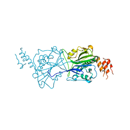
4JA3
 | | Partially occluded inward open conformation of the xylose transporter XylE from E. coli | | 分子名称: | CADMIUM ION, D-xylose-proton symporter, LUTETIUM (III) ION | | 著者 | Quistgaard, E.M, Low, C, Moberg, P, Tresaugues, L, Nordlund, P. | | 登録日 | 2013-02-18 | | 公開日 | 2013-05-01 | | 最終更新日 | 2024-02-28 | | 実験手法 | X-RAY DIFFRACTION (3.8 Å) | | 主引用文献 | Structural basis for substrate transport in the GLUT-homology family of monosaccharide transporters.
Nat.Struct.Mol.Biol., 20, 2013
|
|
4JA4
 | | Inward open conformation of the xylose transporter XylE from E. coli | | 分子名称: | CADMIUM ION, D-xylose-proton symporter | | 著者 | Quistgaard, E.M, Low, C, Moberg, P, Tresaugues, L, Nordlund, P. | | 登録日 | 2013-02-18 | | 公開日 | 2013-05-01 | | 最終更新日 | 2024-02-28 | | 実験手法 | X-RAY DIFFRACTION (4.2 Å) | | 主引用文献 | Structural basis for substrate transport in the GLUT-homology family of monosaccharide transporters.
Nat.Struct.Mol.Biol., 20, 2013
|
|
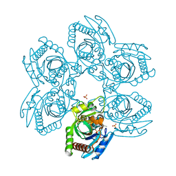
4JZL
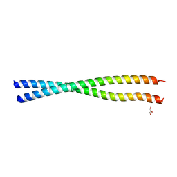 | | Crystal structure of BAP31 vDED at alkaline pH | | 分子名称: | B-cell receptor-associated protein 31, GLYCEROL | | 著者 | Quistgaard, E.M, Low, C, Moberg, P, Guettou, F, Maddi, K, Nordlund, P. | | 登録日 | 2013-04-03 | | 公開日 | 2013-09-25 | | 最終更新日 | 2023-09-20 | | 実験手法 | X-RAY DIFFRACTION (2.2 Å) | | 主引用文献 | Structural and Biophysical Characterization of the Cytoplasmic Domains of Human BAP29 and BAP31.
Plos One, 8, 2013
|
|
4JZP
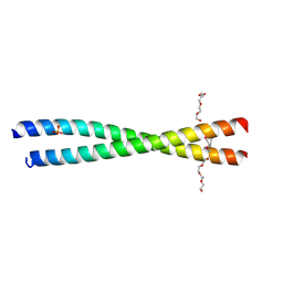 | | Crystal structure of BAP31 vDED at acidic pH | | 分子名称: | B-cell receptor-associated protein 31, SULFATE ION, TETRAETHYLENE GLYCOL | | 著者 | Quistgaard, E.M, Low, C, Moberg, P, Guettou, F, Maddi, K, Nordlund, P. | | 登録日 | 2013-04-03 | | 公開日 | 2013-09-25 | | 実験手法 | X-RAY DIFFRACTION (2.1 Å) | | 主引用文献 | Structural and Biophysical Characterization of the Cytoplasmic Domains of Human BAP29 and BAP31.
Plos One, 8, 2013
|
|
4LEP
 | | Structural insights into substrate recognition in proton dependent oligopeptide transporters | | 分子名称: | N-[(1R)-1-phosphonoethyl]-L-alaninamide, Proton:oligopeptide symporter POT family, ZINC ION | | 著者 | Guettou, F, Quistgaard, E.M, Tresaugues, L, Moberg, P, Jegerschold, C, Zhu, L, Jong, A.J, Nordlund, P, Low, C. | | 登録日 | 2013-06-26 | | 公開日 | 2013-07-10 | | 最終更新日 | 2024-02-28 | | 実験手法 | X-RAY DIFFRACTION (3.2 Å) | | 主引用文献 | Structural insights into substrate recognition in proton-dependent oligopeptide transporters.
Embo Rep., 14, 2013
|
|
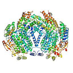
5X5A
 | | Human thymidylate synthase bound with phosphate ion | | 分子名称: | PHOSPHATE ION, Thymidylate synthase | | 著者 | Chen, D, Jansson, A, Larsson, A, Nordlund, P. | | 登録日 | 2017-02-15 | | 公開日 | 2017-06-28 | | 最終更新日 | 2023-11-22 | | 実験手法 | X-RAY DIFFRACTION (2.39 Å) | | 主引用文献 | Structural analyses of human thymidylate synthase reveal a site that may control conformational switching between active and inactive states
J. Biol. Chem., 292, 2017
|
|


