Loading
PDBj
メニューPDBj@FacebookPDBj@X(formerly Twitter)PDBj@BlueSkyPDBj@YouTubewwPDB FoundationwwPDBDonate
RCSB PDBPDBeBMRBAdv. SearchSearch help
Search by PDB author
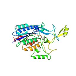
3TXA
DownloadVisualize
BU of 3txa by Molmil
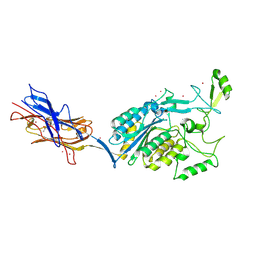
Structural Analysis of Adhesive Tip pilin, GBS104 from Group B Streptococcus agalactiae
分子名称: CADMIUM ION, Cell wall surface anchor family protein, LITHIUM ION, ...
著者Krishnan, V, Narayana, S.V.L.
登録日2011-09-23
公開日2013-03-27
最終更新日2024-10-30
実験手法X-RAY DIFFRACTION (2.619 Å)
主引用文献Structure of Streptococcus agalactiae tip pilin GBS104: a model for GBS pili assembly and host interactions.
Acta Crystallogr.,Sect.D, 69, 2013
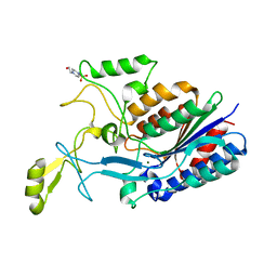
3TW0
DownloadVisualize
BU of 3tw0 by Molmil
Structural Analysis of Adhesive Tip pilin, GBS104 from Group B Streptococcus agalactiae
分子名称: ACETATE ION, Cell wall surface anchor family protein, MAGNESIUM ION
著者Krishnan, V, Narayana, S.V.L.
登録日2011-09-21
公開日2013-03-27
最終更新日2024-10-30
実験手法X-RAY DIFFRACTION (2 Å)
主引用文献Structure of Streptococcus agalactiae tip pilin GBS104: a model for GBS pili assembly and host interactions.
Acta Crystallogr.,Sect.D, 69, 2013
3TVY
DownloadVisualize
BU of 3tvy by Molmil
Structural Analysis of Adhesive Tip pilin, GBS104 from Group B Streptococcus agalactiae
分子名称: 4-(2-HYDROXYETHYL)-1-PIPERAZINE ETHANESULFONIC ACID, Cell wall surface anchor family protein, MAGNESIUM ION
著者Krishnan, V, Narayana, S.V.L.
登録日2011-09-21
公開日2013-03-27
最終更新日2024-02-28
実験手法X-RAY DIFFRACTION (2 Å)
主引用文献Structure of Streptococcus agalactiae tip pilin GBS104: a model for GBS pili assembly and host interactions.
Acta Crystallogr.,Sect.D, 69, 2013
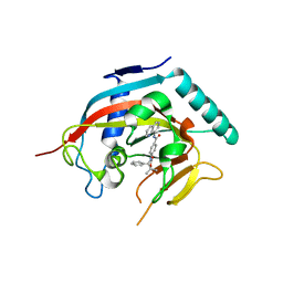
4K4F
DownloadVisualize
BU of 4k4f by Molmil
Co-crystal structure of TNKS1 with compound 18 [4-[(4S)-5,5-dimethyl-2-oxo-4-phenyl-1,3-oxazolidin-3-yl]-N-(quinolin-8-yl)benzamide]
分子名称: 4-[(4S)-5,5-dimethyl-2-oxo-4-phenyl-1,3-oxazolidin-3-yl]-N-(quinolin-8-yl)benzamide, Tankyrase-1, ZINC ION
著者Huang, X, Bregman, H, Wilson, C, DiMauro, E, Gunaydin, H.
登録日2013-04-12
公開日2013-06-05
最終更新日2024-02-28
実験手法X-RAY DIFFRACTION (2.9 Å)
主引用文献Discovery of novel, induced-pocket binding oxazolidinones as potent, selective, and orally bioavailable tankyrase inhibitors.
J.Med.Chem., 56, 2013
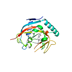
4K4E
DownloadVisualize
BU of 4k4e by Molmil
Co-crystal structure of tnks1 with compound 52 [N~2-(5-chloro-2-methoxyphenyl)-N-[trans-4-(2-oxo-2,3-dihydro-1H-benzimidazol-1-yl)cyclohexyl]glycinamide]
分子名称: N~2~-(5-chloro-2-methoxyphenyl)-N-[trans-4-(2-oxo-2,3-dihydro-1H-benzimidazol-1-yl)cyclohexyl]glycinamide, Tankyrase-1, ZINC ION
著者Huang, X.
登録日2013-04-12
公開日2013-06-05
最終更新日2024-02-28
実験手法X-RAY DIFFRACTION (2.3 Å)
主引用文献Discovery of novel, induced-pocket binding oxazolidinones as potent, selective, and orally bioavailable tankyrase inhibitors.
J.Med.Chem., 56, 2013
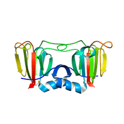
3ENT
DownloadVisualize
BU of 3ent by Molmil
Crystal structure of Nitrollin, a betagamma-crystallin from Nitrosospira multiformis-in alternate space group (P65)
分子名称: Putative uncharacterized protein
著者Aravind, P, Sankaranarayanan, R.
登録日2008-09-26
公開日2009-03-31
最終更新日2024-10-16
実験手法X-RAY DIFFRACTION (2.14 Å)
主引用文献Three-dimensional domain swapping in nitrollin, a single-domain betagamma-crystallin from Nitrosospira multiformis, controls protein conformation and stability but not dimerization
J.Mol.Biol., 385, 2009
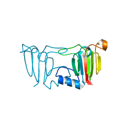
3ENU
DownloadVisualize
BU of 3enu by Molmil
Crystal structure of Nitrollin, a betagamma-crystallin from Nitrosospira multiformis
分子名称: Putative uncharacterized protein
著者Aravind, P, Sankaranarayanan, R.
登録日2008-09-26
公開日2009-03-31
最終更新日2024-11-20
実験手法X-RAY DIFFRACTION (1.86 Å)
主引用文献Three-dimensional domain swapping in nitrollin, a single-domain betagamma-crystallin from Nitrosospira multiformis, controls protein conformation and stability but not dimerization
J.Mol.Biol., 385, 2009
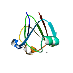
3I9H
DownloadVisualize
BU of 3i9h by Molmil
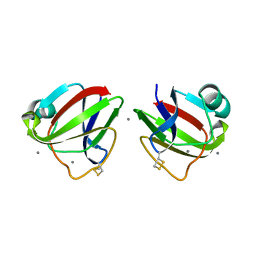
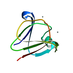
Crystal structure of a betagamma-crystallin domain from Clostridium beijerinckii
分子名称: Beta and gamma crystallin, CALCIUM ION
著者Aravind, P, Sankaranarayanan, R.
登録日2009-07-11
公開日2009-12-01
最終更新日2023-11-01
実験手法X-RAY DIFFRACTION (2 Å)
主引用文献betagamma-Crystallin superfamily contains a universal motif for binding calcium.
Biochemistry, 2009
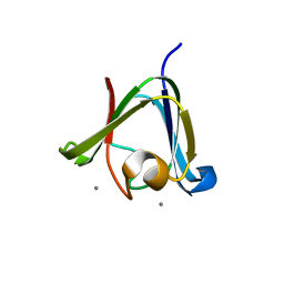
3HZ2
DownloadVisualize
BU of 3hz2 by Molmil
Crystal structure of a betagamma-crystallin from an Archaea
分子名称: Beta/gama crystallin family protein, CALCIUM ION
著者Aravind, P, Sankaranarayanan, R.
登録日2009-06-23
公開日2009-12-01
最終更新日2023-11-01
実験手法X-RAY DIFFRACTION (1.86 Å)
主引用文献The betagamma-crystallin superfamily contains a universal motif for binding calcium
Biochemistry, 48, 2009
3HZB
DownloadVisualize
BU of 3hzb by Molmil
Crystal structure of a betagamma-crystallin domain from Flavobacterium johnsoniae
分子名称: CALCIUM ION, Carbohydrate binding protein
著者Aravind, P, Sankaranarayanan, R.
登録日2009-06-23
公開日2009-12-01
最終更新日2024-10-30
実験手法X-RAY DIFFRACTION (1.74 Å)
主引用文献The betagamma-crystallin superfamily contains a universal motif for binding calcium
Biochemistry, 48, 2009
3IAJ
DownloadVisualize
BU of 3iaj by Molmil
Crystal structure of a betagamma-crystallin domain from Clostridium beijerinckii-in alternate space group I422
分子名称: Beta and gamma crystallin, CALCIUM ION
著者Aravind, P, Sankaranarayanan, R.
登録日2009-07-14
公開日2009-12-01
最終更新日2023-11-01
実験手法X-RAY DIFFRACTION (2.1 Å)
主引用文献betagamma-Crystallin superfamily contains a universal motif for binding calcium.
Biochemistry, 2009
4JL9
DownloadVisualize
BU of 4jl9 by Molmil
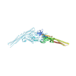
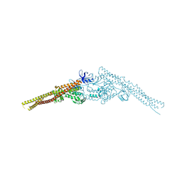
Crystal structure of mouse TBK1 bound to BX795
分子名称: N-(3-{[5-iodo-4-({3-[(thiophen-2-ylcarbonyl)amino]propyl}amino)pyrimidin-2-yl]amino}phenyl)pyrrolidine-1-carboxamide, Serine/threonine-protein kinase TBK1
著者Li, P, Shu, C.
登録日2013-03-12
公開日2013-06-19
最終更新日2024-02-28
実験手法X-RAY DIFFRACTION (3.0999 Å)
主引用文献Structural Insights into the Functions of TBK1 in Innate Antimicrobial Immunity.
Structure, 21, 2013
4JLC
DownloadVisualize
BU of 4jlc by Molmil
Crystal structure of mouse TBK1 bound to SU6668
分子名称: 3-{2,4-dimethyl-5-[(Z)-(2-oxo-1,2-dihydro-3H-indol-3-ylidene)methyl]-1H-pyrrol-3-yl}propanoic acid, Serine/threonine-protein kinase TBK1
著者Li, P.
登録日2013-03-12
公開日2013-06-19
最終更新日2023-09-20
実験手法X-RAY DIFFRACTION (3 Å)
主引用文献Structural Insights into the Functions of TBK1 in Innate Antimicrobial Immunity.
Structure, 21, 2013
9G0L
DownloadVisualize
BU of 9g0l by Molmil
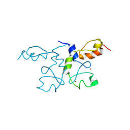
Crystal structure of the RING-ZnF1 fragment of SIAH1
分子名称: E3 ubiquitin-protein ligase SIAH1, ZINC ION
著者Coste, F, Suskiewicz, M.J.
登録日2024-07-08
公開日2025-02-19
最終更新日2025-06-18
実験手法X-RAY DIFFRACTION (1.9 Å)
主引用文献RING dimerisation drives higher-order organisation of SINA/SIAH E3 ubiquitin ligases.
Febs J., 292, 2025
<12


238582

件を2025-07-09に公開中

PDB statisticsPDBj update infoContact PDBjnumon