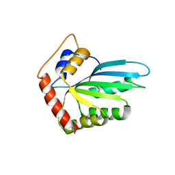
8S0S
 
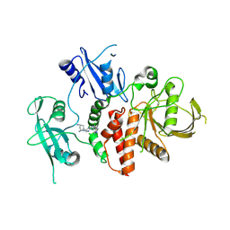 | | A fragment-based inhibitor of SHP2 | | 分子名称: | (1R,5S)-8-[7-(4-chloranyl-2-methyl-indazol-5-yl)-5H-pyrrolo[2,3-b]pyrazin-3-yl]-8-azabicyclo[3.2.1]octan-3-amine, Tyrosine-protein phosphatase non-receptor type 11 | | 著者 | Cleasby, A, Price, A. | | 登録日 | 2024-02-14 | | 公開日 | 2024-03-20 | | 最終更新日 | 2024-04-10 | | 実験手法 | X-RAY DIFFRACTION (1.94 Å) | | 主引用文献 | Fragment-Based Discovery of Allosteric Inhibitors of SH2 Domain-Containing Protein Tyrosine Phosphatase-2 (SHP2).
J.Med.Chem., 67, 2024
|
|
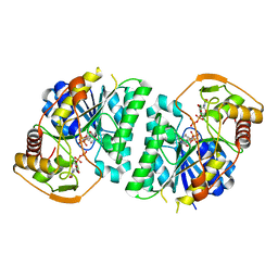
6L8S
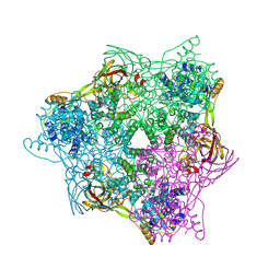 | | High resolution crystal structure of crustacean hemocyanin. | | 分子名称: | 1,2-ETHANEDIOL, 2-acetamido-2-deoxy-beta-D-glucopyranose, CHLORIDE ION, ... | | 著者 | Masuda, T, Mikami, B, Baba, S. | | 登録日 | 2019-11-07 | | 公開日 | 2020-05-27 | | 最終更新日 | 2023-11-22 | | 実験手法 | X-RAY DIFFRACTION (1.58 Å) | | 主引用文献 | The high-resolution crystal structure of lobster hemocyanin shows its enzymatic capability as a phenoloxidase.
Arch.Biochem.Biophys., 688, 2020
|
|
6KOS
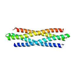 | |
5ZC6
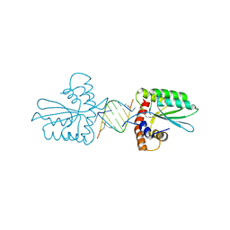
 | | Solution structure of H-RasT35S mutant protein in complex with KBFM123 | | 分子名称: | 3-oxidanyl-~{N}-[[(2~{R})-oxolan-2-yl]methyl]naphthalene-2-carboxamide, GTPase HRas, MAGNESIUM ION, ... | | 著者 | Matsumoto, S, Hayashi, Y, Hiraga, T, Matsuo, K, Kataoka, T. | | 登録日 | 2018-02-15 | | 公開日 | 2018-09-12 | | 最終更新日 | 2024-05-01 | | 実験手法 | SOLUTION NMR | | 主引用文献 | Molecular Basis for Allosteric Inhibition of GTP-Bound H-Ras Protein by a Small-Molecule Compound Carrying a Naphthalene Ring
Biochemistry, 57, 2018
|
|
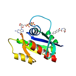
5Z98
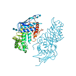
 | | Crystal Structure of the Primate APOBEC3H Dimer mediated by RNA Duplex | | 分子名称: | Apolipoprotein B mRNA editing enzyme catalytic polypeptide-like protein 3H, RNA (5'-R(*AP*UP*AP*CP*CP*CP*GP*GP*CP*A)-3'), RNA (5'-R(P*CP*UP*GP*CP*CP*GP*GP*GP*UP*A)-3'), ... | | 著者 | Matsuoka, T, Nagae, T, Ode, H, Watanabe, N, Iwatani, Y. | | 登録日 | 2018-02-02 | | 公開日 | 2018-08-15 | | 最終更新日 | 2023-11-22 | | 実験手法 | X-RAY DIFFRACTION (2.2 Å) | | 主引用文献 | Structural basis of chimpanzee APOBEC3H dimerization stabilized by double-stranded RNA.
Nucleic Acids Res., 46, 2018
|
|
5BRO
 | | Crystal structure of modified HexB (modB) | | 分子名称: | 2-acetamido-2-deoxy-beta-D-glucopyranose-(1-4)-2-acetamido-2-deoxy-beta-D-glucopyranose, Beta-hexosaminidase subunit beta, FORMIC ACID, ... | | 著者 | Kitakaze, K, Maita, N, Itoh, K. | | 登録日 | 2015-06-01 | | 公開日 | 2016-05-04 | | 最終更新日 | 2020-07-29 | | 実験手法 | X-RAY DIFFRACTION (2.4 Å) | | 主引用文献 | Protease-resistant modified human beta-hexosaminidase B ameliorates symptoms in GM2 gangliosidosis model.
J.Clin.Invest., 126, 2016
|
|
8WOW
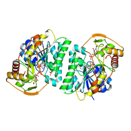 | | Crystal structure of Arabidopsis thaliana UDP-glucose 4-epimerase 2 (AtUGE2) complexed with UDP, I160L/G233A mutant | | 分子名称: | NICOTINAMIDE-ADENINE-DINUCLEOTIDE, SULFATE ION, UDP-glucose 4-epimerase 2, ... | | 著者 | Matsumoto, M, Umezawa, A, Kotake, T, Fushinobu, S. | | 登録日 | 2023-10-08 | | 公開日 | 2024-05-15 | | 最終更新日 | 2024-07-10 | | 実験手法 | X-RAY DIFFRACTION (2.6 Å) | | 主引用文献 | Cytosolic UDP-L-arabinose synthesis by bifunctional UDP-glucose 4-epimerases in Arabidopsis.
Plant J., 119, 2024
|
|
8WOV
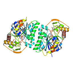 | | Crystal structure of Arabidopsis thaliana UDP-glucose 4-epimerase 2 (AtUGE2) complexed with UDP, G233A mutant | | 分子名称: | NICOTINAMIDE-ADENINE-DINUCLEOTIDE, UDP-glucose 4-epimerase 2, URIDINE-5'-DIPHOSPHATE | | 著者 | Matsumoto, M, Umezawa, A, Kotake, T, Fushinobu, S. | | 登録日 | 2023-10-07 | | 公開日 | 2024-05-15 | | 最終更新日 | 2024-07-10 | | 実験手法 | X-RAY DIFFRACTION (2.25 Å) | | 主引用文献 | Cytosolic UDP-L-arabinose synthesis by bifunctional UDP-glucose 4-epimerases in Arabidopsis.
Plant J., 119, 2024
|
|
8WOP
 | | Crystal structure of Arabidopsis thaliana UDP-glucose 4-epimerase 2 (AtUGE2) complexed with UDP, wild-type | | 分子名称: | NICOTINAMIDE-ADENINE-DINUCLEOTIDE, UDP-glucose 4-epimerase 2, URIDINE-5'-DIPHOSPHATE | | 著者 | Matsumoto, M, Umezawa, A, Kotake, T, Fushinobu, S. | | 登録日 | 2023-10-07 | | 公開日 | 2024-05-08 | | 最終更新日 | 2024-07-10 | | 実験手法 | X-RAY DIFFRACTION (2.35 Å) | | 主引用文献 | Cytosolic UDP-L-arabinose synthesis by bifunctional UDP-glucose 4-epimerases in Arabidopsis.
Plant J., 119, 2024
|
|
6A6M
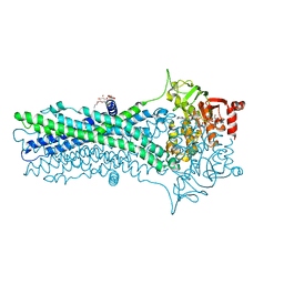 | | Crystal structure of an outward-open nucleotide-bound state of the eukaryotic ABC multidrug transporter CmABCB1 | | 分子名称: | ATP-binding cassette, sub-family B, member 1, ... | | 著者 | Kato, H, Nakatsu, T, Kodan, A. | | 登録日 | 2018-06-28 | | 公開日 | 2019-02-20 | | 最終更新日 | 2024-03-27 | | 実験手法 | X-RAY DIFFRACTION (1.9 Å) | | 主引用文献 | Inward- and outward-facing X-ray crystal structures of homodimeric P-glycoprotein CmABCB1.
Nat Commun, 10, 2019
|
|
6A6N
 | | Crystal structure of an inward-open apo state of the eukaryotic ABC multidrug transporter CmABCB1 | | 分子名称: | 2-AMINO-2-HYDROXYMETHYL-PROPANE-1,3-DIOL, ATP-binding cassette, sub-family B, ... | | 著者 | Kato, H, Nakatsu, T, Kodan, A. | | 登録日 | 2018-06-28 | | 公開日 | 2019-01-16 | | 最終更新日 | 2023-11-22 | | 実験手法 | X-RAY DIFFRACTION (3.02 Å) | | 主引用文献 | Inward- and outward-facing X-ray crystal structures of homodimeric P-glycoprotein CmABCB1.
Nat Commun, 10, 2019
|
|
3WUS
 | | Crystal Structure of the Vif-Binding Domain of Human APOBEC3F | | 分子名称: | DNA dC->dU-editing enzyme APOBEC-3F, ZINC ION | | 著者 | Nakashima, M, Kawamura, T, Ode, H, Watanabe, N, Iwatani, Y. | | 登録日 | 2014-05-02 | | 公開日 | 2015-06-24 | | 最終更新日 | 2023-11-08 | | 実験手法 | X-RAY DIFFRACTION (2.54 Å) | | 主引用文献 | Structural Insights into HIV-1 Vif-APOBEC3F Interaction.
J.Virol., 90, 2015
|
|


