4J9D
 
 | |
4J9B
 | |
4JJB
 | |
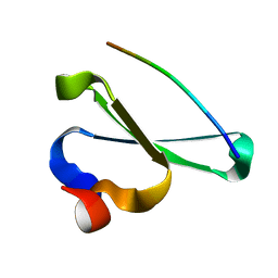
4C9I
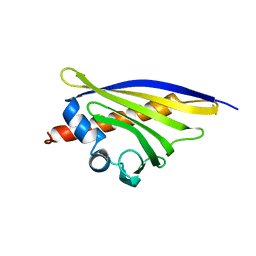 | | Crystal Structure of the Strawberry Pathogenesis-Related 10 (PR-10) Fra a 1E protein (Form B) | | 分子名称: | (2R,3S)-2-(3,4-dihydroxyphenyl)-3,4-dihydro-2H-chromene-3,5,7-triol, GLYCEROL, MAJOR STRAWBERRY ALLERGEN FRA A 1-E | | 著者 | Casanal, A, Zander, U, Valpuesta, V, Marquez, J.A. | | 登録日 | 2013-10-02 | | 公開日 | 2013-10-16 | | 最終更新日 | 2023-12-20 | | 実験手法 | X-RAY DIFFRACTION (3.1 Å) | | 主引用文献 | The Strawberry Pathogenesis-Related 10 (Pr-10) Fra a Proteins Control Flavonoid Biosynthesis by Binding to Metabolic Intermediates.
J.Biol.Chem., 288, 2013
|
|
4C94
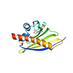 | | Crystal Structure of the Strawberry Pathogenesis-Related 10 (PR-10) Fra a 3 protein in complex with Catechin | | 分子名称: | (2R,3S)-2-(3,4-dihydroxyphenyl)-3,4-dihydro-2H-chromene-3,5,7-triol, FRA A 3 ALLERGEN | | 著者 | Casanal, A, Zander, U, Valpuesta, V, Marquez, J.A. | | 登録日 | 2013-10-02 | | 公開日 | 2013-10-16 | | 最終更新日 | 2023-12-20 | | 実験手法 | X-RAY DIFFRACTION (3 Å) | | 主引用文献 | The Strawberry Pathogenesis-Related 10 (Pr-10) Fra a Proteins Control Flavonoid Biosynthesis by Binding to Metabolic Intermediates.
J.Biol.Chem., 288, 2013
|
|
4C9C
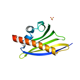 | | Crystal Structure of the Strawberry Pathogenesis-Related 10 (PR-10) Fra a 1E protein (Form A) | | 分子名称: | GLYCEROL, MAJOR STRAWBERRY ALLERGEN FRA A 1-E, SULFATE ION | | 著者 | Casanal, A, Zander, U, Valpuesta, V, Marquez, J.A. | | 登録日 | 2013-10-02 | | 公開日 | 2013-10-16 | | 最終更新日 | 2023-12-20 | | 実験手法 | X-RAY DIFFRACTION (2.2 Å) | | 主引用文献 | The Strawberry Pathogenesis-Related 10 (Pr-10) Fra a Proteins Control Flavonoid Biosynthesis by Binding to Metabolic Intermediates.
J.Biol.Chem., 288, 2013
|
|
3FJ5
 | | Crystal structure of the c-src-SH3 domain | | 分子名称: | ACETATE ION, GLYCEROL, Proto-oncogene tyrosine-protein kinase Src, ... | | 著者 | Camara-Artigas, A. | | 登録日 | 2008-12-14 | | 公開日 | 2009-03-03 | | 最終更新日 | 2023-11-01 | | 実験手法 | X-RAY DIFFRACTION (1.65 Å) | | 主引用文献 | Intertwined dimeric structure for the SH3 domain of the c-Src tyrosine kinase induced by polyethylene glycol binding
Febs Lett., 583, 2009
|
|
2KQ0
 | | Human NEDD4 3rd WW Domain Complex with Ebola Zaire Virus Matrix Protein VP40 Derived Peptide ILPTAPPEYMEA | | 分子名称: | 12-mer from Matrix protein VP40, E3 ubiquitin-protein ligase NEDD4 | | 著者 | Iglesias-Bexiga, M, Macias, M, Bonet, R, Blanco, F.J, Cobos, E.S, Luque, I. | | 登録日 | 2009-10-23 | | 公開日 | 2010-11-03 | | 最終更新日 | 2024-05-01 | | 実験手法 | SOLUTION NMR | | 主引用文献 | Human NEDD4 3rd WW Domain Complex with Ebola Zaire Virus Matrix Protein VP40 Derived Peptide
To be Published
|
|
6GIS
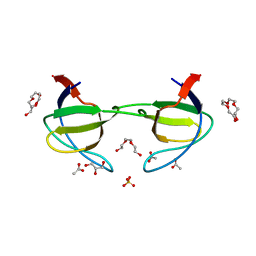
 | | Structural basis of human clamp sliding on DNA | | 分子名称: | DNA (5'-D(P*AP*TP*AP*CP*GP*AP*TP*GP*GP*G)-3'), DNA (5'-D(P*CP*CP*CP*AP*TP*CP*GP*TP*AP*T)-3'), Proliferating cell nuclear antigen | | 著者 | De March, M, Merino, N, Barrera-Vilarmau, S, Crehuet, R, Onesti, S, Blanco, F.J, De Biasio, A. | | 登録日 | 2018-05-15 | | 公開日 | 2018-05-30 | | 最終更新日 | 2024-10-23 | | 実験手法 | X-RAY DIFFRACTION (2.82 Å) | | 主引用文献 | Structural basis of human PCNA sliding on DNA.
Nat Commun, 8, 2017
|
|
4ZTD
 | | Crystal Structure of Human PCNA in complex with a TRAIP peptide | | 分子名称: | ALA-GLY-ALA-GLY-ALA, ALA-PHE-GLN-ALA-LYS-LEU-ASP-THR-PHE-LEU-TRP-SER, Proliferating cell nuclear antigen | | 著者 | Montoya, G, Mortuza, G.B, Blanco, F.J, Ibanez de Opakua, A. | | 登録日 | 2015-05-14 | | 公開日 | 2015-12-16 | | 最終更新日 | 2024-01-10 | | 実験手法 | X-RAY DIFFRACTION (2.199 Å) | | 主引用文献 | TRAIP is a PCNA-binding ubiquitin ligase that protects genome stability after replication stress.
J.Cell Biol., 212, 2016
|
|


