3T7S
 
 | |
4IAH
 | |
4IAM
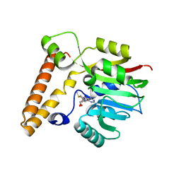
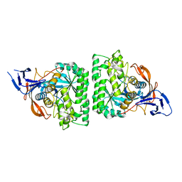
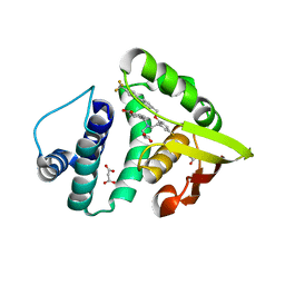
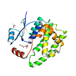
 | | Crystal Structure of the C139A mutant of nostoc H-NOX domain | | 分子名称: | Alr2278 protein, MALONATE ION, PROTOPORPHYRIN IX CONTAINING FE | | 著者 | Kumar, V, van den Akker, F. | | 登録日 | 2012-12-06 | | 公開日 | 2013-11-20 | | 最終更新日 | 2023-09-20 | | 実験手法 | X-RAY DIFFRACTION (1.99 Å) | | 主引用文献 | Insights into BAY 60-2770 Activation and S-Nitrosylation-Dependent Desensitization of Soluble Guanylyl Cyclase via Crystal Structures of Homologous Nostoc H-NOX Domain Complexes.
Biochemistry, 52, 2013
|
|
4KIR
 | |
4KQN
 | |
4E50
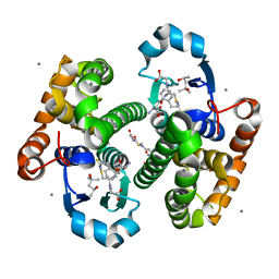
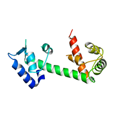 | | Calmodulin and Ng peptide complex | | 分子名称: | Calmodulin, Linker, IQ motif of Neurogranin | | 著者 | Kumar, V, Sivaraman, J. | | 登録日 | 2012-03-13 | | 公開日 | 2013-03-20 | | 最終更新日 | 2024-03-20 | | 実験手法 | X-RAY DIFFRACTION (2.7 Å) | | 主引用文献 | Structural basis for the interaction of unstructured neuron specific substrates neuromodulin and neurogranin with calmodulin
Sci Rep, 3, 2013
|
|
4E53
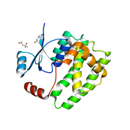
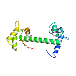 | | Calmodulin and Nm peptide complex | | 分子名称: | Calmodulin, Linker, IQ motif of Neuromodulin | | 著者 | Kumar, V, Sivaraman, J. | | 登録日 | 2012-03-13 | | 公開日 | 2013-03-20 | | 最終更新日 | 2024-03-20 | | 実験手法 | X-RAY DIFFRACTION (2.69 Å) | | 主引用文献 | Structural basis for the interaction of unstructured neuron specific substrates neuromodulin and neurogranin with calmodulin
Sci Rep, 3, 2013
|
|
4HEX
 | |
4IAE
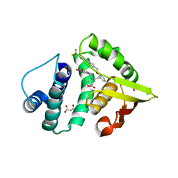
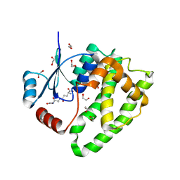
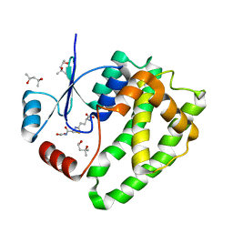
 | | Crystal structure of BAY 60-2770 bound to nostoc H-NOX domain | | 分子名称: | 4-({(4-carboxybutyl)[2-(5-fluoro-2-{[4'-(trifluoromethyl)biphenyl-4-yl]methoxy}phenyl)ethyl]amino}methyl)benzoic acid, Alr2278 protein, MALONATE ION | | 著者 | Kumar, V, van den Akker, F, Martin, F. | | 登録日 | 2012-12-06 | | 公開日 | 2013-11-20 | | 最終更新日 | 2023-09-20 | | 実験手法 | X-RAY DIFFRACTION (2.05 Å) | | 主引用文献 | Insights into BAY 60-2770 Activation and S-Nitrosylation-Dependent Desensitization of Soluble Guanylyl Cyclase via Crystal Structures of Homologous Nostoc H-NOX Domain Complexes.
Biochemistry, 52, 2013
|
|
5JCU
 | |
5JCW
 | | Crystal Structure of hGSTP1-1 with Glutathione Adduct of Phenethyl Isothiocyanate | | 分子名称: | (4S)-2-METHYL-2,4-PENTANEDIOL, ACETATE ION, CALCIUM ION, ... | | 著者 | Kumari, V, Ji, X. | | 登録日 | 2016-04-15 | | 公開日 | 2016-10-12 | | 最終更新日 | 2023-09-27 | | 実験手法 | X-RAY DIFFRACTION (1.945 Å) | | 主引用文献 | Irreversible Inhibition of Glutathione S-Transferase by Phenethyl Isothiocyanate (PEITC), a Dietary Cancer Chemopreventive Phytochemical.
Plos One, 11, 2016
|
|
6ATR
 | |
6ATP
 | |
6ATQ
 | |
6ATO
 | |
6AP9
 | |
6VJE
 | | Crystal structure of Pseudomonas aeruginosa penicillin-binding protein 3 (PBP3) complexed with ceftobiprole | | 分子名称: | (2R)-2-[(1R)-1-{[(2Z)-2-(5-amino-1,2,4-thiadiazol-3-yl)-2-(hydroxyimino)acetyl]amino}-2-oxoethyl]-5-({2-oxo-1-[(3R)-pyr rolidin-3-yl]-2,5-dihydro-1H-pyrrol-3-yl}methyl)-3,6-dihydro-2H-1,3-thiazine-4-carboxylic acid, CHLORIDE ION, Peptidoglycan D,D-transpeptidase FtsI | | 著者 | van den Akker, F, Kumar, V. | | 登録日 | 2020-01-15 | | 公開日 | 2020-03-11 | | 最終更新日 | 2023-10-11 | | 実験手法 | X-RAY DIFFRACTION (1.76 Å) | | 主引用文献 | Structural Insights into Ceftobiprole Inhibition of Pseudomonas aeruginosa Penicillin-Binding Protein 3.
Antimicrob.Agents Chemother., 64, 2020
|
|
4RMP
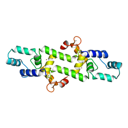
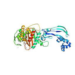
 | | Crystal structure of allophycocyanin from marine cyanobacterium Phormidium sp. A09DM | | 分子名称: | Allophycocyanin, PHYCOCYANOBILIN | | 著者 | Kumar, V, Gupta, G.D, Sonani, R.R, Madamwar, D. | | 登録日 | 2014-10-22 | | 公開日 | 2015-05-13 | | 最終更新日 | 2023-09-20 | | 実験手法 | X-RAY DIFFRACTION (2.506 Å) | | 主引用文献 | Crystal Structure of Allophycocyanin from Marine Cyanobacterium Phormidium sp. A09DM.
Plos One, 10, 2015
|
|
1KSI
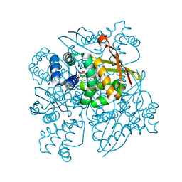
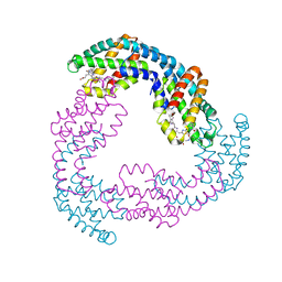
 | | CRYSTAL STRUCTURE OF A EUKARYOTIC (PEA SEEDLING) COPPER-CONTAINING AMINE OXIDASE AT 2.2A RESOLUTION | | 分子名称: | 2-acetamido-2-deoxy-beta-D-glucopyranose, 2-acetamido-2-deoxy-beta-D-glucopyranose-(1-4)-2-acetamido-2-deoxy-beta-D-glucopyranose, COPPER (II) ION, ... | | 著者 | Wilce, M.C.J, Kumar, V, Freeman, H.C, Guss, J.M. | | 登録日 | 1996-07-20 | | 公開日 | 1997-12-24 | | 最終更新日 | 2020-07-29 | | 実験手法 | X-RAY DIFFRACTION (2.2 Å) | | 主引用文献 | Crystal structure of a eukaryotic (pea seedling) copper-containing amine oxidase at 2.2 A resolution.
Structure, 4, 1996
|
|
4JQH
 | |
7LY1
 | |
8SDR
 | |
8SDN
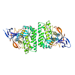
 | | Crystal structure of PDC-3 Y221H beta-lactamase | | 分子名称: | Beta-lactamase, ISOPROPYL ALCOHOL | | 著者 | Kumar, V, van den Akker, F. | | 登録日 | 2023-04-07 | | 公開日 | 2023-08-16 | | 最終更新日 | 2023-11-22 | | 実験手法 | X-RAY DIFFRACTION (2.1 Å) | | 主引用文献 | Natural protein engineering in the Omega-loop: the role of Y221 in ceftazidime and ceftolozane resistance in Pseudomonas -derived cephalosporinase.
Antimicrob.Agents Chemother., 67, 2023
|
|
8SDV
 | | Crystal structure of PDC-3 Y221H beta-lactamase in complex with the boronic acid inhibitor S02030 | | 分子名称: | 1-{(2R)-2-(dihydroxyboranyl)-2-[(thiophen-2-ylacetyl)amino]ethyl}-1H-1,2,3-triazole-4-carboxylic acid, Beta-lactamase, DIMETHYL SULFOXIDE, ... | | 著者 | Kumar, V, van den Akker, F. | | 登録日 | 2023-04-07 | | 公開日 | 2023-08-16 | | 最終更新日 | 2023-11-22 | | 実験手法 | X-RAY DIFFRACTION (1.42 Å) | | 主引用文献 | Natural protein engineering in the Omega-loop: the role of Y221 in ceftazidime and ceftolozane resistance in Pseudomonas -derived cephalosporinase.
Antimicrob.Agents Chemother., 67, 2023
|
|
8SDT
 | |


