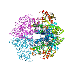
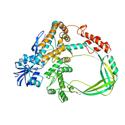
4RXR
 
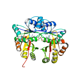
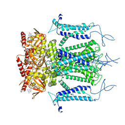
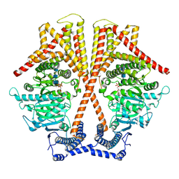
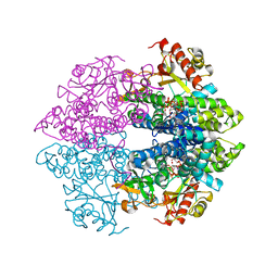 | | The structure of GTP-dCTP-bound SAMHD1 | | 分子名称: | 2'-DEOXYCYTIDINE-5'-TRIPHOSPHATE, Deoxynucleoside triphosphate triphosphohydrolase SAMHD1, GUANOSINE-5'-TRIPHOSPHATE, ... | | 著者 | Zhu, C.F, Wei, W, Peng, X, Dong, Y.H, Gong, Y, Yu, X.F. | | 登録日 | 2014-12-11 | | 公開日 | 2015-03-11 | | 最終更新日 | 2023-09-20 | | 実験手法 | X-RAY DIFFRACTION (2.117 Å) | | 主引用文献 | The mechanism of substrate-controlled allosteric regulation of SAMHD1 activated by GTP.
Acta Crystallogr.,Sect.D, 71, 2015
|
|
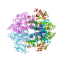
4RXS
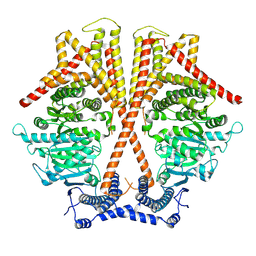
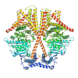
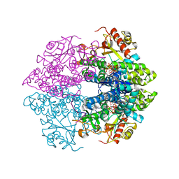 | | The structure of GTP-dTTP-bound SAMHD1 | | 分子名称: | Deoxynucleoside triphosphate triphosphohydrolase SAMHD1, GUANOSINE-5'-TRIPHOSPHATE, MAGNESIUM ION, ... | | 著者 | Zhu, C.F, Wei, W, Peng, X, Dong, Y.H, Gong, Y, Yu, X.F. | | 登録日 | 2014-12-11 | | 公開日 | 2015-03-11 | | 最終更新日 | 2023-09-20 | | 実験手法 | X-RAY DIFFRACTION (2.2 Å) | | 主引用文献 | The mechanism of substrate-controlled allosteric regulation of SAMHD1 activated by GTP.
Acta Crystallogr.,Sect.D, 71, 2015
|
|
4RXO
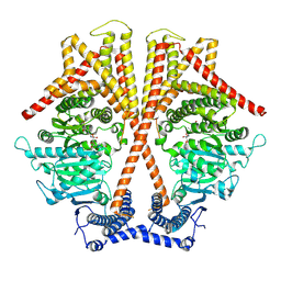
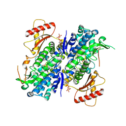 | | The structure of GTP-bound SAMHD1 | | 分子名称: | Deoxynucleoside triphosphate triphosphohydrolase SAMHD1, GUANOSINE-5'-TRIPHOSPHATE, PHOSPHATE ION, ... | | 著者 | Zhu, C.F, Wei, W, Peng, X, Dong, Y.H, Gong, Y, Yu, X.F. | | 登録日 | 2014-12-11 | | 公開日 | 2015-03-11 | | 最終更新日 | 2023-09-20 | | 実験手法 | X-RAY DIFFRACTION (2.6 Å) | | 主引用文献 | The mechanism of substrate-controlled allosteric regulation of SAMHD1 activated by GTP.
Acta Crystallogr.,Sect.D, 71, 2015
|
|
4RXQ
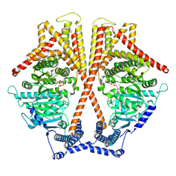
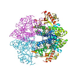 | | The structure of GTP-dUTP-bound SAMHD1 | | 分子名称: | DEOXYURIDINE-5'-TRIPHOSPHATE, Deoxynucleoside triphosphate triphosphohydrolase SAMHD1, GUANOSINE-5'-TRIPHOSPHATE, ... | | 著者 | Zhu, C.F, Wei, W, Peng, X, Dong, Y.H, Gong, Y, Yu, X.F. | | 登録日 | 2014-12-11 | | 公開日 | 2015-03-11 | | 最終更新日 | 2023-09-20 | | 実験手法 | X-RAY DIFFRACTION (2.1 Å) | | 主引用文献 | The mechanism of substrate-controlled allosteric regulation of SAMHD1 activated by GTP.
Acta Crystallogr.,Sect.D, 71, 2015
|
|
4RXP
 | | The structure of GTP-dATP-bound SAMHD1 | | 分子名称: | 2'-DEOXYADENOSINE 5'-TRIPHOSPHATE, Deoxynucleoside triphosphate triphosphohydrolase SAMHD1, GUANOSINE-5'-TRIPHOSPHATE, ... | | 著者 | Zhu, C.F, Wei, W, Peng, X, Dong, Y.H, Gong, Y, Yu, X.F. | | 登録日 | 2014-12-11 | | 公開日 | 2015-03-11 | | 最終更新日 | 2023-09-20 | | 実験手法 | X-RAY DIFFRACTION (2.1 Å) | | 主引用文献 | The mechanism of substrate-controlled allosteric regulation of SAMHD1 activated by GTP.
Acta Crystallogr.,Sect.D, 71, 2015
|
|
3LWI
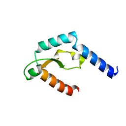
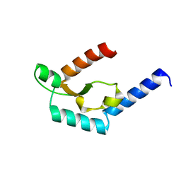
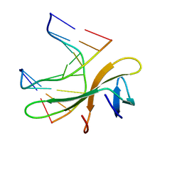 | | Crystal structure of Cren7-dsDNA complex | | 分子名称: | Chromatin protein Cren7, DNA (5'-D(*GP*CP*GP*AP*TP*CP*GP*C)-3') | | 著者 | Zhang, Z.F, Gong, Y, Guo, L, Jiang, T, Huang, L. | | 登録日 | 2010-02-23 | | 公開日 | 2010-05-26 | | 最終更新日 | 2023-11-01 | | 実験手法 | X-RAY DIFFRACTION (2.3 Å) | | 主引用文献 | Structural insights into the interaction of the crenarchaeal chromatin protein Cren7 with DNA
Mol.Microbiol., 76, 2010
|
|
3LWH
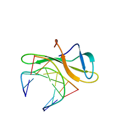 | | Crystal structure of Cren7-dsDNA complex | | 分子名称: | Chromatin protein Cren7, DNA (5'-D(*GP*TP*AP*AP*TP*TP*AP*C)-3') | | 著者 | Zhang, Z.F, Gong, Y, Guo, L, Jiang, T, Huang, L. | | 登録日 | 2010-02-23 | | 公開日 | 2010-05-26 | | 最終更新日 | 2023-11-01 | | 実験手法 | X-RAY DIFFRACTION (1.9 Å) | | 主引用文献 | Structural insights into the interaction of the crenarchaeal chromatin protein Cren7 with DNA
Mol.Microbiol., 76, 2010
|
|
4MZ7
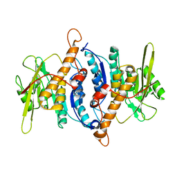
 | | Structural insight into dGTP-dependent activation of tetrameric SAMHD1 deoxynucleoside triphosphate triphosphohydrolase | | 分子名称: | 2'-DEOXYADENOSINE 5'-TRIPHOSPHATE, 2'-DEOXYGUANOSINE-5'-TRIPHOSPHATE, Deoxynucleoside triphosphate triphosphohydrolase SAMHD1, ... | | 著者 | Zhu, C, Gao, W, Zhao, K, Qin, X, Zhang, Y, Peng, X, Zhang, L, Dong, Y, Zhang, W, Li, P, Wei, W, Gong, Y, Yu, X.F. | | 登録日 | 2013-09-29 | | 公開日 | 2013-11-20 | | 最終更新日 | 2024-10-30 | | 実験手法 | X-RAY DIFFRACTION (1.8 Å) | | 主引用文献 | Structural insight into dGTP-dependent activation of tetrameric SAMHD1 deoxynucleoside triphosphate triphosphohydrolase
Nat Commun, 4, 2013
|
|
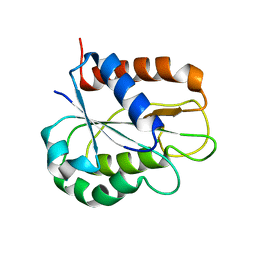
5HW4
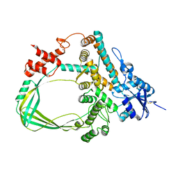
 | | Crystal structure of Escherichia coli 16S rRNA methyltransferase RsmI in complex with AdoMet | | 分子名称: | Ribosomal RNA small subunit methyltransferase I, S-ADENOSYLMETHIONINE | | 著者 | Zhao, M, Zhang, H, Dong, Y, Gong, Y. | | 登録日 | 2016-01-28 | | 公開日 | 2016-10-19 | | 最終更新日 | 2023-11-08 | | 実験手法 | X-RAY DIFFRACTION (2.211 Å) | | 主引用文献 | Structural Insights into the Methylation of C1402 in 16S rRNA by Methyltransferase RsmI
Plos One, 11, 2016
|
|
2F7K
 | |
8WKH
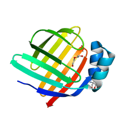 | | Crystal structure of group 13 allergen from Blomia tropicalis | | 分子名称: | 2-AMINO-2-HYDROXYMETHYL-PROPANE-1,3-DIOL, DI(HYDROXYETHYL)ETHER, Fatty acid-binding protein | | 著者 | Zhu, K.L, Gong, Y, Cui, Y.B. | | 登録日 | 2023-09-27 | | 公開日 | 2023-11-22 | | 最終更新日 | 2023-11-29 | | 実験手法 | X-RAY DIFFRACTION (2 Å) | | 主引用文献 | Immunobiological properties and structure analysis of group 13 allergen from Blomia tropicalis and its IgE-mediated cross-reactivity.
Int.J.Biol.Macromol., 254, 2023
|
|
5H0M
 | | Crystal structure of deep-sea thermophilic bacteriophage GVE2 HNH endonuclease with zinc ion | | 分子名称: | HNH endonuclease, ZINC ION | | 著者 | Zhang, L.K, Xu, D.D, Huang, Y.C, Gong, Y. | | 登録日 | 2016-10-05 | | 公開日 | 2017-03-08 | | 最終更新日 | 2024-03-20 | | 実験手法 | X-RAY DIFFRACTION (1.52 Å) | | 主引用文献 | Structural and functional characterization of deep-sea thermophilic bacteriophage GVE2 HNH endonuclease
Sci Rep, 7, 2017
|
|
5H3O
 | | Structure of a eukaryotic cyclic nucleotide-gated channel | | 分子名称: | CYCLIC GUANOSINE MONOPHOSPHATE, Cyclic nucleotide-gated cation channel, SODIUM ION | | 著者 | Li, M, Zhou, X, Wang, S, Michailidis, I, Gong, Y, Su, D, Li, H, Li, X, Yang, J. | | 登録日 | 2016-10-26 | | 公開日 | 2017-01-25 | | 最終更新日 | 2024-05-29 | | 実験手法 | ELECTRON MICROSCOPY (3.5 Å) | | 主引用文献 | Structure of a eukaryotic cyclic-nucleotide-gated channel.
Nature, 542, 2017
|
|
7WJM
 | |
7WJO
 | | CryoEM structure of chitin synthase 1 from Phytophthora sojae complexed with nikkomycin Z | | 分子名称: | (2S)-{[(2S,3S,4S)-2-amino-4-hydroxy-4-(5-hydroxypyridin-2-yl)-3-methylbutanoyl]amino}[(2R,3S,4R,5R)-5-(2,4-dioxo-3,4-dihydropyrimidin-1(2H)-yl)-3,4-dihydroxyoxolan-2-yl]acetic acid (non-preferred name), Chitin synthase | | 著者 | Chen, W, Cao, P, Gong, Y, Yang, Q. | | 登録日 | 2022-01-07 | | 公開日 | 2022-09-28 | | 最終更新日 | 2024-06-26 | | 実験手法 | ELECTRON MICROSCOPY (3.2 Å) | | 主引用文献 | Structural basis for directional chitin biosynthesis.
Nature, 610, 2022
|
|
7WJN
 | | CryoEM structure of chitin synthase 1 mutant E495A from Phytophthora sojae complexed with UDP-GlcNAc | | 分子名称: | Chitin synthase, MANGANESE (II) ION, URIDINE-DIPHOSPHATE-N-ACETYLGLUCOSAMINE | | 著者 | Chen, W, Cao, P, Gong, Y, Yang, Q. | | 登録日 | 2022-01-07 | | 公開日 | 2022-09-28 | | 最終更新日 | 2024-06-26 | | 実験手法 | ELECTRON MICROSCOPY (3.3 Å) | | 主引用文献 | Structural basis for directional chitin biosynthesis.
Nature, 610, 2022
|
|
5H0O
 | | Crystal structure of deep-sea thermophilic bacteriophage GVE2 HNH endonuclease with manganese ion | | 分子名称: | HNH endonuclease, MANGANESE (II) ION | | 著者 | Zhang, L.K, Xu, D.D, Huang, Y.C, Gong, Y. | | 登録日 | 2016-10-06 | | 公開日 | 2017-03-08 | | 最終更新日 | 2023-11-08 | | 実験手法 | X-RAY DIFFRACTION (1.53 Å) | | 主引用文献 | Structural and functional characterization of deep-sea thermophilic bacteriophage GVE2 HNH endonuclease.
Sci Rep, 7, 2017
|
|
7X05
 | | CryoEM structure of chitin synthase 1 from Phytophthora sojae complexed with the nascent chitooligosaccharide | | 分子名称: | 2-acetamido-2-deoxy-beta-D-glucopyranose-(1-4)-2-acetamido-2-deoxy-beta-D-glucopyranose-(1-4)-2-acetamido-2-deoxy-beta-D-glucopyranose, Chitin synthase, MANGANESE (II) ION, ... | | 著者 | Chen, W, Cao, P, Gong, Y, Yang, Q. | | 登録日 | 2022-02-21 | | 公開日 | 2022-09-28 | | 最終更新日 | 2024-06-26 | | 実験手法 | ELECTRON MICROSCOPY (3.9 Å) | | 主引用文献 | Structural basis for directional chitin biosynthesis.
Nature, 610, 2022
|
|
7X06
 | | CryoEM structure of chitin synthase 1 from Phytophthora sojae complexed with UDP | | 分子名称: | Chitin synthase, MAGNESIUM ION, URIDINE-5'-DIPHOSPHATE | | 著者 | Chen, W, Cao, P, Gong, Y, Yang, Q. | | 登録日 | 2022-02-21 | | 公開日 | 2022-09-28 | | 最終更新日 | 2024-06-26 | | 実験手法 | ELECTRON MICROSCOPY (3.1 Å) | | 主引用文献 | Structural basis for directional chitin biosynthesis.
Nature, 610, 2022
|
|
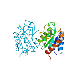
6K8N
 | | Crystal structure of the Sulfolobus solfataricus topoisomerase III | | 分子名称: | ZINC ION, topoisomerase III | | 著者 | Wang, H.Q, Zhang, J.H, Zheng, X, Zheng, Z.F, Dong, Y.H, Huang, L, Gong, Y. | | 登録日 | 2019-06-13 | | 公開日 | 2020-06-24 | | 最終更新日 | 2023-11-22 | | 実験手法 | X-RAY DIFFRACTION (2.1 Å) | | 主引用文献 | Crystal structures of the Sulfolobus solfataricus topoisomerase III reveal that its C-terminal novel zinc finger part is a unique decatenation domain
To Be Published
|
|
6K8O
 | | Crystal structure of the Sulfolobus solfataricus topoisomerase III in complex with DNA | | 分子名称: | DNA (5'-D(*GP*CP*AP*AP*GP*GP*TP*C)-3'), ZINC ION, topoisomerase III | | 著者 | Wang, H.Q, Zhang, J.H, Zheng, X, Zheng, Z.F, Dong, Y.H, Huang, L, Gong, Y. | | 登録日 | 2019-06-13 | | 公開日 | 2020-06-24 | | 最終更新日 | 2024-10-16 | | 実験手法 | X-RAY DIFFRACTION (2.5 Å) | | 主引用文献 | Crystal structures of the Sulfolobus solfataricus topoisomerase III reveal that its C-terminal novel zinc finger part is a unique decatenation domain
To Be Published
|
|
8HGR
 | | The apo-flavodoxin monomer from Synechococcus elongatus PCC 7942 | | 分子名称: | CHLORIDE ION, Flavodoxin, MAGNESIUM ION | | 著者 | Liu, S.W, Chen, Y.Y, Gong, Y, Cao, P. | | 登録日 | 2022-11-15 | | 公開日 | 2022-12-14 | | 最終更新日 | 2023-11-29 | | 実験手法 | X-RAY DIFFRACTION (1.84 Å) | | 主引用文献 | A dimer-monomer transition captured by the crystal structures of cyanobacterial apo flavodoxin.
Biochem.Biophys.Res.Commun., 639, 2022
|
|
8HGQ
 | | The apo-flavodoxin dimer from Synechococcus elongatus PCC 7942 | | 分子名称: | Flavodoxin, PHOSPHATE ION | | 著者 | Liu, S.W, Chen, Y.Y, Gong, Y, Cao, P. | | 登録日 | 2022-11-15 | | 公開日 | 2022-12-14 | | 最終更新日 | 2023-11-29 | | 実験手法 | X-RAY DIFFRACTION (2.09 Å) | | 主引用文献 | A dimer-monomer transition captured by the crystal structures of cyanobacterial apo flavodoxin.
Biochem.Biophys.Res.Commun., 639, 2022
|
|
5YS1
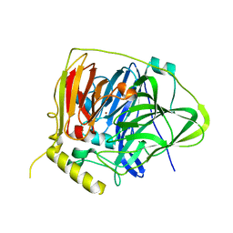 | | Crystal structure of Multicopper Oxidase CueO G304K mutant | | 分子名称: | Blue copper oxidase CueO, COPPER (II) ION | | 著者 | Wang, H.Q, Liu, X.Q, Zhao, J.T, Yue, Q.X, Yan, Y.H, Dong, Y.H, Fan, Y.L, Tian, J, Wu, N.F, Gong, Y. | | 登録日 | 2017-11-12 | | 公開日 | 2018-10-17 | | 最終更新日 | 2023-11-22 | | 実験手法 | X-RAY DIFFRACTION (1.49 Å) | | 主引用文献 | Crystal structures of multicopper oxidase CueO G304K mutant: structural basis of the increased laccase activity
Sci Rep, 8, 2018
|
|
5YS5
 | | Crystal structure of Multicopper Oxidase CueO G304K mutant with seven copper ions | | 分子名称: | Blue copper oxidase CueO, COPPER (II) ION | | 著者 | Wang, H.Q, Liu, X.Q, Zhao, J.T, Yue, Q.X, Yan, Y.H, Dong, Y.H, Fan, Y.L, Tian, J, Wu, N.F, Gong, Y. | | 登録日 | 2017-11-13 | | 公開日 | 2018-10-17 | | 最終更新日 | 2023-11-22 | | 実験手法 | X-RAY DIFFRACTION (2.2 Å) | | 主引用文献 | Crystal structures of multicopper oxidase CueO G304K mutant: structural basis of the increased laccase activity
Sci Rep, 8, 2018
|
|


