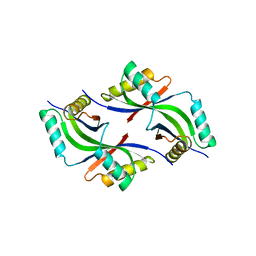
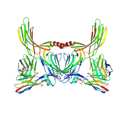
3B77
 
 | |
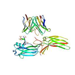
3BYQ
 | |
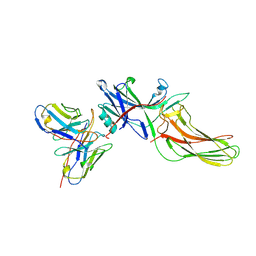
3BY7
 | |
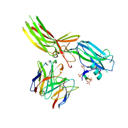
1VR0
 | |
1VQ3
 | |
1ZCZ
 | |
2A6A
 | |
2ETS
 | |
2F46
 | |
2FG0
 | |
2FEA
 | |
2EVR
 | |
1VK9
 | |
5V7Q
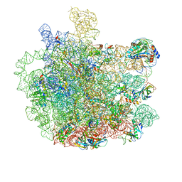 | | Cryo-EM structure of the large ribosomal subunit from Mycobacterium tuberculosis bound with a potent linezolid analog | | 分子名称: | 23S rRNA, 50S ribosomal protein L13, 50S ribosomal protein L14, ... | | 著者 | Yang, K, Chang, J.-Y, Cui, Z, Zhang, J. | | 登録日 | 2017-03-20 | | 公開日 | 2017-09-20 | | 最終更新日 | 2024-03-13 | | 実験手法 | ELECTRON MICROSCOPY (3.7 Å) | | 主引用文献 | Structural insights into species-specific features of the ribosome from the human pathogen Mycobacterium tuberculosis.
Nucleic Acids Res., 45, 2017
|
|
3IRB
 | |
2OOK
 | |
2OOC
 | |
2PV7
 | |
2Q3L
 | |
2Q8U
 | |
8J8Z
 | | Structure of beta-arrestin1 in complex with D6Rpp | | 分子名称: | Atypical chemokine receptor 2, Beta-arrestin-1, Fab30 Heavy Chain, ... | | 著者 | Maharana, J, Sarma, P, Yadav, M.K, Chami, M, Banerjee, R, Shukla, A.K. | | 登録日 | 2023-05-02 | | 公開日 | 2023-12-27 | | 最終更新日 | 2024-01-17 | | 実験手法 | ELECTRON MICROSCOPY (3.4 Å) | | 主引用文献 | Molecular insights into atypical modes of beta-arrestin interaction with seven transmembrane receptors.
Science, 383, 2024
|
|
8J8V
 | | Structure of beta-arrestin2 in complex with D6Rpp (Local Refine) | | 分子名称: | Atypical chemokine receptor 2, Beta-arrestin-2, Fab30 Heavy Chain, ... | | 著者 | Maharana, J, Sarma, P, Yadav, M.K, Chami, M, Banerjee, R, Shukla, A.K. | | 登録日 | 2023-05-02 | | 公開日 | 2023-12-27 | | 最終更新日 | 2024-01-17 | | 実験手法 | ELECTRON MICROSCOPY (3.22 Å) | | 主引用文献 | Molecular insights into atypical modes of beta-arrestin interaction with seven transmembrane receptors.
Science, 383, 2024
|
|
8J97
 | | Structure of Muscarinic receptor (M2R) in complex with beta-arrestin1 (Local refine, cross-linked) | | 分子名称: | Beta-arrestin-1, Fab30 Heavy Chain, Fab30 Light Chain, ... | | 著者 | Maharana, J, Sano, F.K, Shihoya, W, Banerjee, R, Nureki, O, Shukla, A.K. | | 登録日 | 2023-05-02 | | 公開日 | 2023-12-27 | | 最終更新日 | 2024-10-09 | | 実験手法 | ELECTRON MICROSCOPY (3.2 Å) | | 主引用文献 | Molecular insights into atypical modes of beta-arrestin interaction with seven transmembrane receptors.
Science, 383, 2024
|
|
8J9K
 | | Structure of basal beta-arrestin2 | | 分子名称: | Beta-arrestin-2, Fab6 heavy chain, Fab6 light chain | | 著者 | Maharana, J, Sarma, P, Yadav, M.K, Chami, M, Banerjee, R, Shukla, A.K. | | 登録日 | 2023-05-03 | | 公開日 | 2023-12-27 | | 最終更新日 | 2024-01-17 | | 実験手法 | ELECTRON MICROSCOPY (3.5 Å) | | 主引用文献 | Molecular insights into atypical modes of beta-arrestin interaction with seven transmembrane receptors.
Science, 383, 2024
|
|
8JAF
 | | Structure of Muscarinic receptor (M2R) in complex with beta-arrestin1 (Local Refine, non-cross linked) | | 分子名称: | Beta-arrestin-1, Fab30 heavy chain, Fab30 light chain, ... | | 著者 | Maharana, J, Sano, F.K, Shihoya, W, Banerjee, R, Nureki, O, Shukla, A.K. | | 登録日 | 2023-05-05 | | 公開日 | 2023-12-27 | | 最終更新日 | 2024-01-17 | | 実験手法 | ELECTRON MICROSCOPY (3.1 Å) | | 主引用文献 | Molecular insights into atypical modes of beta-arrestin interaction with seven transmembrane receptors.
Science, 383, 2024
|
|


