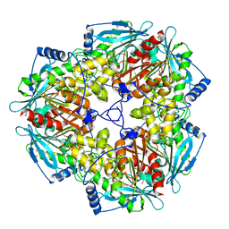
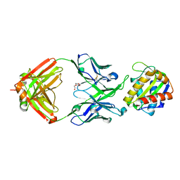
4JM4
 
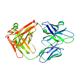 | | Crystal Structure of PGT 135 Fab | | 分子名称: | PGT 135 Heavy Chain, PGT 135 Light Chain | | 著者 | Kong, L, Wilson, I.A. | | 登録日 | 2013-03-13 | | 公開日 | 2013-05-29 | | 最終更新日 | 2013-07-17 | | 実験手法 | X-RAY DIFFRACTION (1.751 Å) | | 主引用文献 | Supersite of immune vulnerability on the glycosylated face of HIV-1 envelope glycoprotein gp120.
Nat.Struct.Mol.Biol., 20, 2013
|
|
2PV7
 | |
2Q3L
 | |
2OOK
 | |
2OOC
 | |
2Q8U
 | |
3L5O
 | |
3H50
 | |
3HBZ
 | |
3O0F
 | |
3M82
 | |
3IRB
 | |
3M81
 | |
3M83
 | |
3NL9
 | |
3HSA
 | |
3K5J
 | |
3H0N
 | |
3KK7
 | |
5SY8
 | |
5T5B
 | |
5T6L
 | |
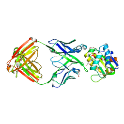
5TFW
 | | Crystal structure of 10E8 Fab light chain mutant2 against the MPER region of the HIV-1 Env, in complex with T117v2 epitope scaffold | | 分子名称: | 1,2-ETHANEDIOL, 10E8 EPITOPE SCAFFOLD T117V2, Antibody 10E8 FAB HEAVY CHAIN, ... | | 著者 | Irimia, A, Wilson, I.A. | | 登録日 | 2016-09-26 | | 公開日 | 2017-03-08 | | 最終更新日 | 2023-10-04 | | 実験手法 | X-RAY DIFFRACTION (2.168 Å) | | 主引用文献 | Lipid interactions and angle of approach to the HIV-1 viral membrane of broadly neutralizing antibody 10E8: Insights for vaccine and therapeutic design.
PLoS Pathog., 13, 2017
|
|
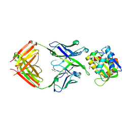
5T29
 | | Crystal structure of 10E8 Fab light chain mutant3, against the MPER region of the HIV-1 Env, in complex with the MPER epitope scaffold T117v2 | | 分子名称: | 10E8 EPITOPE SCAFFOLD T117V2, Antibody 10E8 FAB HEAVY CHAIN, Antibody 10E8 FAB LIGHT CHAIN, ... | | 著者 | Irimia, A, Wilson, I.A. | | 登録日 | 2016-08-23 | | 公開日 | 2017-03-08 | | 最終更新日 | 2023-10-04 | | 実験手法 | X-RAY DIFFRACTION (2.03 Å) | | 主引用文献 | Lipid interactions and angle of approach to the HIV-1 viral membrane of broadly neutralizing antibody 10E8: Insights for vaccine and therapeutic design.
PLoS Pathog., 13, 2017
|
|
5T80
 | |


