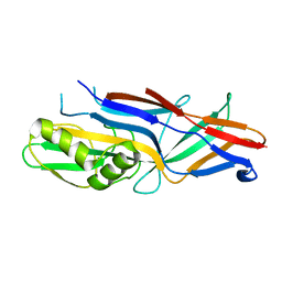
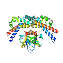
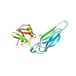
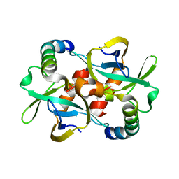
1N3Q
 
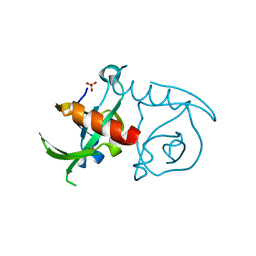
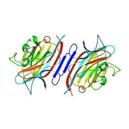 | | Pterocarpus angolensis lectin complexed with turanose | | 分子名称: | CALCIUM ION, MANGANESE (II) ION, alpha-D-glucopyranose-(1-3)-beta-D-fructofuranose, ... | | 著者 | Loris, R, Imberty, A, Beeckmans, S, Van Driessche, E, Read, J.S, Bouckaert, J, De Greve, H, Buts, L, Wyns, L. | | 登録日 | 2002-10-29 | | 公開日 | 2002-11-20 | | 最終更新日 | 2024-03-13 | | 実験手法 | X-RAY DIFFRACTION (2.2 Å) | | 主引用文献 | Crystal structure of Pterocarpus angolensis lectin in complex with glucose, sucrose, and turanose
J.BIOL.CHEM., 278, 2003
|
|
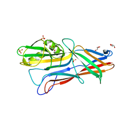
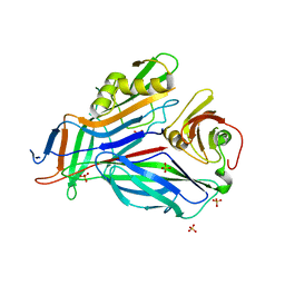
1N3O
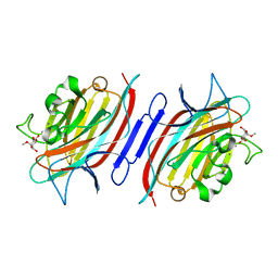 | | Pterocarcpus angolensis lectin in complex with alpha-methyl glucose | | 分子名称: | CALCIUM ION, MANGANESE (II) ION, lectin PAL, ... | | 著者 | Loris, R, Imberty, A, Beeckmans, S, Van Driessche, E, Read, J.S, Bouckaert, J, De Greve, H, Buts, L, Wyns, L. | | 登録日 | 2002-10-29 | | 公開日 | 2002-11-20 | | 最終更新日 | 2024-03-13 | | 実験手法 | X-RAY DIFFRACTION (2 Å) | | 主引用文献 | Crystal structure of Pterocarpus angolensis lectin in complex with glucose, sucrose, and turanose
J.BIOL.CHEM., 278, 2003
|
|
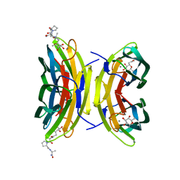
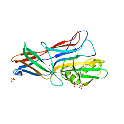
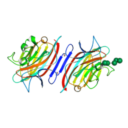
1N3P
 | | Pterocarpus angolensis lectin in complex with sucrose | | 分子名称: | CALCIUM ION, MANGANESE (II) ION, beta-D-fructofuranose-(2-1)-alpha-D-glucopyranose, ... | | 著者 | Loris, R, Imberty, A, Beeckmans, S, Van Driessche, E, Read, J.S, Bouckaert, J, De Greve, H, Buts, L, Wyns, L. | | 登録日 | 2002-10-29 | | 公開日 | 2002-11-20 | | 最終更新日 | 2024-03-13 | | 実験手法 | X-RAY DIFFRACTION (2.1 Å) | | 主引用文献 | Crystal structure of Pterocarpus angolensis lectin in complex with glucose, sucrose, and turanose
J.BIOL.CHEM., 278, 2003
|
|
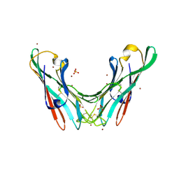
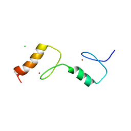
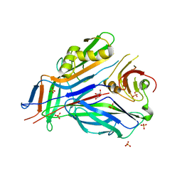
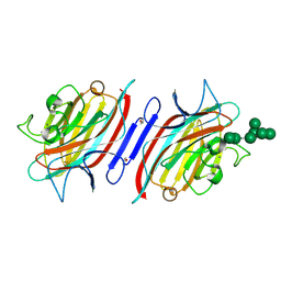
3DCQ
 | | LECB (PA-LII) in complex with the synthetic ligand 2G0 | | 分子名称: | (2S)-1-[(2S)-6-amino-2-({[(2S,3S,4R,5S,6S)-3,4,5-trihydroxy-6-methyltetrahydro-2H-pyran-2-yl]acetyl}amino)hexanoyl]-N-[(1S)-1-carbamoyl-3-methylbutyl]pyrrolidine-2-carboxamide, CALCIUM ION, Fucose-binding lectin PA-IIL | | 著者 | Johansson, E.M, Crusz, S.A, Kolomiets, E, Buts, L, Kadam, R.U, Cacciarini, M, Bartels, K.M, Diggle, S.P, Camara, M, Williams, P, Loris, R, Nativi, C, Rosenau, F, Jaeger, K.E, Darbre, T, Reymond, J.L. | | 登録日 | 2008-06-04 | | 公開日 | 2009-01-13 | | 最終更新日 | 2023-11-01 | | 実験手法 | X-RAY DIFFRACTION (1.8 Å) | | 主引用文献 | Inhibition and dispersion of Pseudomonas aeruginosa biofilms by glycopeptide dendrimers targeting the fucose-specific lectin LecB.
Chem.Biol., 15, 2008
|
|
3JRZ
 | |
3JSC
 | | CcdBVfi-FormI-pH7.0 | | 分子名称: | CcdB, SULFATE ION | | 著者 | De Jonge, N, Buts, L, Loris, R. | | 登録日 | 2009-09-10 | | 公開日 | 2009-12-01 | | 最終更新日 | 2023-11-01 | | 実験手法 | X-RAY DIFFRACTION (1.5 Å) | | 主引用文献 | Structural and thermodynamic characterization of vibrio fischeri CCDB
J.Biol.Chem., 285, 2010
|
|
6TR8
 | | Corynebacterium diphtheriae methionine sulfoxide reductase B (MsrB) solution structure - reduced form | | 分子名称: | Peptide-methionine (R)-S-oxide reductase, ZINC ION | | 著者 | Volkov, A.N, Tossounian, M.A, Buts, L, Messens, J. | | 登録日 | 2019-12-18 | | 公開日 | 2020-02-05 | | 最終更新日 | 2024-05-15 | | 実験手法 | SOLUTION NMR | | 主引用文献 | Methionine sulfoxide reductase B fromCorynebacterium diphtheriaecatalyzes sulfoxide reduction via an intramolecular disulfide cascade.
J.Biol.Chem., 295, 2020
|
|
4BWO
 | | The FedF adhesin from entrrotoxigenic Escherichia coli is a sulfate- binding lectin | | 分子名称: | BROMIDE ION, F18 FIMBRIAL ADHESIN AC, SULFATE ION | | 著者 | Lonardi, E, Moonens, K, Buts, L, de Boer, A.R, Olsson, J.D.M, Weiss, M.S, Fabre, E, Guerardel, Y, Deelder, A.M, Oscarson, S, Wuhrer, M, Bouckaert, J. | | 登録日 | 2013-07-03 | | 公開日 | 2013-08-28 | | 最終更新日 | 2014-05-28 | | 実験手法 | X-RAY DIFFRACTION (1.8 Å) | | 主引用文献 | Structural Sampling of Glycan Interaction Profiles Reveals Mucosal Receptors for Fimbrial Adhesins of Enterotoxigenic Escherichia Coli
Biology, 2, 2013
|
|
3ZPD
 | |
3HLR
 | | Donor strand complemented FaeG of F4ad fimbriae | | 分子名称: | K88 fimbrial protein AD | | 著者 | Van Molle, I, Moonens, K, Garcia-Pino, A, Buts, L, Bouckaert, J, De Greve, H. | | 登録日 | 2009-05-28 | | 公開日 | 2009-10-20 | | 最終更新日 | 2024-03-20 | | 実験手法 | X-RAY DIFFRACTION (2.3 Å) | | 主引用文献 | Structural and thermodynamic characterization of pre- and postpolymerization states in the F4 fimbrial subunit FaeG
J.Mol.Biol., 394, 2009
|
|
3HPW
 | | CcdB dimer in complex with one C-terminal CcdA domain | | 分子名称: | Cytotoxic protein ccdB, DI(HYDROXYETHYL)ETHER, Protein ccdA, ... | | 著者 | De Jonge, N, Loris, R, Garcia-Pino, A, Buts, L. | | 登録日 | 2009-06-05 | | 公開日 | 2009-08-11 | | 最終更新日 | 2023-09-06 | | 実験手法 | X-RAY DIFFRACTION (1.452 Å) | | 主引用文献 | Rejuvenation of CcdB-Poisoned Gyrase by an Intrinsically Disordered Protein Domain.
Mol.Cell, 35, 2009
|
|
2FXI
 | |
4ELY
 | | CCDBVFI:GYRA14EC | | 分子名称: | CHLORIDE ION, CcdB, DNA gyrase subunit A, ... | | 著者 | De Jonge, N, Simic, R, Buts, L, Haesaerts, S, Roelants, K, Garcia-Pino, A, Sterckx, Y, De Greve, H, Lah, J, Loris, R. | | 登録日 | 2012-04-11 | | 公開日 | 2012-05-30 | | 最終更新日 | 2023-09-13 | | 実験手法 | X-RAY DIFFRACTION (1.932 Å) | | 主引用文献 | Alternative interactions define gyrase specificity in the CcdB family.
Mol.Microbiol., 84, 2012
|
|
4ELZ
 | | CCDBVFI:GYRA14VFI | | 分子名称: | CcdB, DNA gyrase subunit A, GLYCEROL | | 著者 | De Jonge, N, Simic, M, Buts, L, Haesaerts, S, Roelants, K, Garcia-Pino, A, Sterckx, Y, De Greve, H, Lah, J, Loris, R. | | 登録日 | 2012-04-11 | | 公開日 | 2012-05-30 | | 最終更新日 | 2023-09-13 | | 実験手法 | X-RAY DIFFRACTION (2.2 Å) | | 主引用文献 | Alternative interactions define gyrase specificity in the CcdB family.
Mol.Microbiol., 84, 2012
|
|
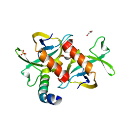
4BMJ
 | | Structure of the UBZ1and2 tandem of the ubiquitin-binding adaptor protein TAX1BP1 | | 分子名称: | CHLORIDE ION, TAX1-BINDING PROTEIN 1, ZINC ION | | 著者 | Ceregido, M.A, Spinola-Amilibia, M, Buts, L, Rivera, J, Bravo, J, van Nuland, N.A.J. | | 登録日 | 2013-05-09 | | 公開日 | 2013-11-20 | | 最終更新日 | 2024-05-08 | | 実験手法 | X-RAY DIFFRACTION (2.75 Å) | | 主引用文献 | The Structure of Tax1BP1 Ubz1 + 2 Provides Insight Into Target Specificity and Adaptability
J.Mol.Biol., 426, 2014
|
|
3F6L
 | | Structure of the F4 fimbrial chaperone FaeE | | 分子名称: | Chaperone protein faeE | | 著者 | Van Molle, I, Moonens, K, Buts, L, Garcia-Pino, A, Wyns, L, De Greve, H, Bouckaert, J. | | 登録日 | 2008-11-06 | | 公開日 | 2009-05-19 | | 最終更新日 | 2023-11-01 | | 実験手法 | X-RAY DIFFRACTION (2.801 Å) | | 主引用文献 | The F4 fimbrial chaperone FaeE is stable as a monomer that does not require self-capping of its pilin-interactive surfaces
Acta Crystallogr.,Sect.D, 65, 2009
|
|
3F6I
 | | Structure of the SeMet labeled F4 fibrial chaperone FaeE | | 分子名称: | Chaperone protein faeE | | 著者 | Van Molle, I, Moonens, K, Buts, L, Garcia-Pino, A, Wyns, L, De Greve, H, Bouckaert, J. | | 登録日 | 2008-11-06 | | 公開日 | 2009-05-19 | | 最終更新日 | 2023-12-27 | | 実験手法 | X-RAY DIFFRACTION (2.788 Å) | | 主引用文献 | The F4 fimbrial chaperone FaeE is stable as a monomer that does not require self-capping of its pilin-interactive surfaces
Acta Crystallogr.,Sect.D, 65, 2009
|
|
3F65
 | | The F4 fimbrial chaperone FaeE does not self-cap its interactive surfaces | | 分子名称: | (4S)-2-METHYL-2,4-PENTANEDIOL, Chaperone protein faeE, PHOSPHATE ION | | 著者 | Van Molle, I, Moonens, K, Buts, L, Garcia-Pino, A, Wyns, L, De Greve, H, Bouckaert, J. | | 登録日 | 2008-11-05 | | 公開日 | 2009-05-19 | | 最終更新日 | 2023-11-01 | | 実験手法 | X-RAY DIFFRACTION (2.29 Å) | | 主引用文献 | The F4 fimbrial chaperone FaeE is stable as a monomer that does not require self-capping of its pilin-interactive surfaces
Acta Crystallogr.,Sect.D, 65, 2009
|
|
3GEA
 | | Donor strand complemented FaeG monomer of F4 variant ad | | 分子名称: | GLYCEROL, K88 fimbrial protein AD, SULFATE ION | | 著者 | Van Molle, I, Moonens, K, Garcia-Pino, A, Buts, L, Bouckaert, J, De Greve, H. | | 登録日 | 2009-02-25 | | 公開日 | 2009-10-20 | | 最終更新日 | 2023-11-01 | | 実験手法 | X-RAY DIFFRACTION (1.699 Å) | | 主引用文献 | Structural and thermodynamic characterization of pre- and postpolymerization states in the F4 fimbrial subunit FaeG
J.Mol.Biol., 394, 2009
|
|
3GGH
 | | Donor strand complemented FaeG of F4ad fimbriae | | 分子名称: | K88 fimbrial protein AD, SULFATE ION | | 著者 | Van Molle, I, Moonens, K, Garcia-Pino, A, Buts, L, Bouckaert, J, De Greve, H. | | 登録日 | 2009-02-28 | | 公開日 | 2009-10-20 | | 最終更新日 | 2023-11-01 | | 実験手法 | X-RAY DIFFRACTION (1.639 Å) | | 主引用文献 | Structural and thermodynamic characterization of pre- and postpolymerization states in the F4 fimbrial subunit FaeG
J.Mol.Biol., 394, 2009
|
|
3GEW
 | | FaeE-FaeG chaperone-major pilin complex of F4 ad fimbriae | | 分子名称: | Chaperone protein faeE, GLYCEROL, K88 fimbrial protein AD, ... | | 著者 | Van Molle, I, Moonens, K, Garcia-Pino, A, Buts, L, Bouckaert, J, De Greve, H. | | 登録日 | 2009-02-26 | | 公開日 | 2009-10-20 | | 最終更新日 | 2023-11-01 | | 実験手法 | X-RAY DIFFRACTION (2 Å) | | 主引用文献 | Structural and thermodynamic characterization of pre- and postpolymerization states in the F4 fimbrial subunit FaeG
J.Mol.Biol., 394, 2009
|
|
3G7Z
 | | CcdB dimer in complex with two C-terminal CcdA domains | | 分子名称: | Cytotoxic protein ccdB, Protein ccdA | | 著者 | De Jonge, N, Loris, R, Garcia-Pino, A, Buts, L. | | 登録日 | 2009-02-11 | | 公開日 | 2009-08-11 | | 最終更新日 | 2023-09-06 | | 実験手法 | X-RAY DIFFRACTION (2.351 Å) | | 主引用文献 | Rejuvenation of CcdB-Poisoned Gyrase by an Intrinsically Disordered Protein Domain.
Mol.Cell, 35, 2009
|
|
3GFU
 | | FaeE-FaeG chaperone-major pilin complex of F4 ac 5/95 fimbriae | | 分子名称: | Chaperone protein faeE, FaeG, SULFATE ION | | 著者 | Van Molle, I, Moonens, K, Garcia-Pino, A, Buts, L, Bouckaert, J, De Greve, H. | | 登録日 | 2009-02-27 | | 公開日 | 2009-10-20 | | 最終更新日 | 2023-11-01 | | 実験手法 | X-RAY DIFFRACTION (1.991 Å) | | 主引用文献 | Structural and thermodynamic characterization of pre- and postpolymerization states in the F4 fimbrial subunit FaeG
J.Mol.Biol., 394, 2009
|
|
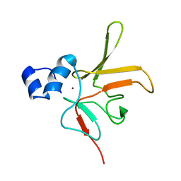
2PHT
 | | Pterocarpus angolensis lectin (P L) in complex with Man-7D3 | | 分子名称: | CALCIUM ION, Lectin, MANGANESE (II) ION, ... | | 著者 | Garcia-Pino, A, Buts, L, Wyns, L, Imberty, A, Loris, R. | | 登録日 | 2007-04-12 | | 公開日 | 2007-07-10 | | 最終更新日 | 2023-08-30 | | 実験手法 | X-RAY DIFFRACTION (2.1 Å) | | 主引用文献 | How a plant lectin recognizes high mannose oligosaccharides
Plant Physiol., 144, 2007
|
|
2PHW
 | | Pterocarpus angolensis lectin (PAL) in complex with Man-9 | | 分子名称: | CALCIUM ION, Lectin, MANGANESE (II) ION, ... | | 著者 | Garcia-Pino, A, Buts, L, Wyns, L, Imberty, A, Loris, R. | | 登録日 | 2007-04-12 | | 公開日 | 2007-07-10 | | 最終更新日 | 2023-08-30 | | 実験手法 | X-RAY DIFFRACTION (1.8 Å) | | 主引用文献 | How a plant lectin recognizes high mannose oligosaccharides
Plant Physiol., 144, 2007
|
|


