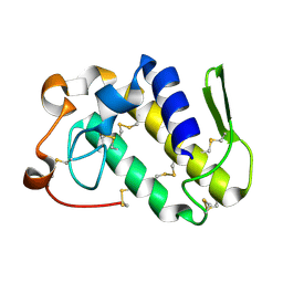
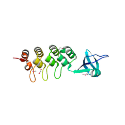
1TDN
 
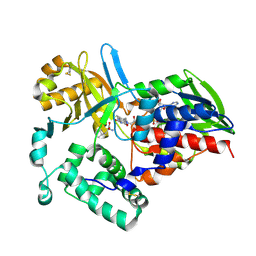 | |
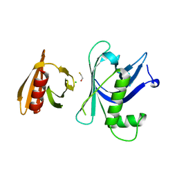
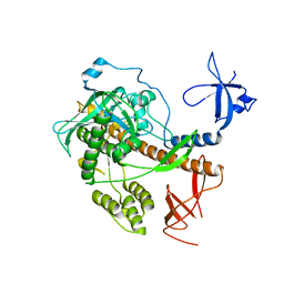
6IZO
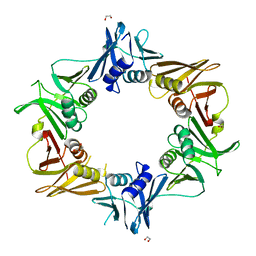 | | Crystal structure of DNA polymerase sliding clamp from Caulobacter crescentus | | 分子名称: | 1,2-ETHANEDIOL, Beta sliding clamp, DI(HYDROXYETHYL)ETHER | | 著者 | Jiang, X, Zhang, L, Teng, M, Li, X. | | 登録日 | 2018-12-20 | | 公開日 | 2019-11-27 | | 最終更新日 | 2023-11-22 | | 実験手法 | X-RAY DIFFRACTION (1.94 Å) | | 主引用文献 | Caulobacter crescentus beta sliding clamp employs a noncanonical regulatory model of DNA replication.
Febs J., 287, 2020
|
|
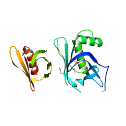
6JIR
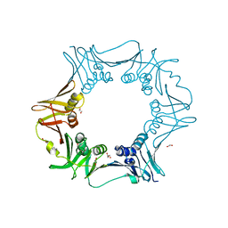 | | Crystal structure of C. crescentus beta sliding clamp with PEG bound to putative beta-motif tethering region | | 分子名称: | 1,2-ETHANEDIOL, Beta sliding clamp, DI(HYDROXYETHYL)ETHER, ... | | 著者 | Jiang, X, Teng, M, Li, X. | | 登録日 | 2019-02-23 | | 公開日 | 2019-11-27 | | 最終更新日 | 2023-11-22 | | 実験手法 | X-RAY DIFFRACTION (1.95 Å) | | 主引用文献 | Caulobacter crescentus beta sliding clamp employs a noncanonical regulatory model of DNA replication.
Febs J., 287, 2020
|
|
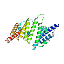
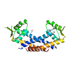
1VB0
 | | Atomic resolution structure of atratoxin-b, one short-chain neurotoxin from Naja atra | | 分子名称: | 2-AMINO-2-HYDROXYMETHYL-PROPANE-1,3-DIOL, Cobrotoxin b, SULFATE ION | | 著者 | Lou, X, Liu, Q, Teng, M, Niu, L, Huang, Q, Hao, Q. | | 登録日 | 2004-02-20 | | 公開日 | 2004-12-21 | | 最終更新日 | 2024-11-13 | | 実験手法 | X-RAY DIFFRACTION (0.92 Å) | | 主引用文献 | The atomic resolution crystal structure of atratoxin determined by single wavelength anomalous diffraction phasing
J.Biol.Chem., 279, 2004
|
|
4NQ0
 | | Structural insights into yeast histone chaperone Hif1: a scaffold protein recruiting protein complexes to core histones | | 分子名称: | HAT1-interacting factor 1 | | 著者 | Liu, H, Zhang, M, He, W, Zhu, Z, Teng, M, Gao, Y, Niu, L. | | 登録日 | 2013-11-23 | | 公開日 | 2014-07-16 | | 最終更新日 | 2024-03-20 | | 実験手法 | X-RAY DIFFRACTION (2.1 Å) | | 主引用文献 | Structural insights into yeast histone chaperone Hif1: a scaffold protein recruiting protein complexes to core histones
Biochem.J., 462, 2014
|
|
1WT9
 | | crystal structure of Aa-X-bp-I, a snake venom protein with the activity of binding to coagulation factor X from Agkistrodon acutus | | 分子名称: | CALCIUM ION, agkisacutacin A chain, anticoagulant protein-B | | 著者 | Zhu, Z, Liu, S, Mo, X, Yu, X, Liang, Z, Zang, J, Zhao, W, Teng, M, Niu, L. | | 登録日 | 2004-11-18 | | 公開日 | 2006-03-07 | | 最終更新日 | 2024-10-16 | | 実験手法 | X-RAY DIFFRACTION (2.01 Å) | | 主引用文献 | Characterizations and Crystal structures of two snake venom proteins with the activity of binding coagulation factor X from Agkistrodon acutus
To be Published
|
|
1MG6
 | |
1V6P
 | | Crystal structure of Cobrotoxin | | 分子名称: | CHLORIDE ION, COPPER (II) ION, Cobrotoxin, ... | | 著者 | Lou, X, Tu, X, Wang, J, Teng, M, Niu, L, Liu, Q, Huang, Q, Hao, Q. | | 登録日 | 2003-12-03 | | 公開日 | 2004-12-21 | | 最終更新日 | 2024-11-20 | | 実験手法 | X-RAY DIFFRACTION (0.87 Å) | | 主引用文献 | The atomic resolution crystal structure of atratoxin determined by single wavelength anomalous diffraction phasing
J.Biol.Chem., 279, 2004
|
|
1ONJ
 | | Crystal structure of Atratoxin-b from Chinese cobra venom of Naja atra | | 分子名称: | Cobrotoxin b, SULFATE ION | | 著者 | Lou, X, Tu, X, Pan, G, Xu, C, Fan, R, Lu, W, Deng, W, Rao, P, Teng, M, Niu, L. | | 登録日 | 2003-02-28 | | 公開日 | 2004-02-28 | | 最終更新日 | 2024-11-06 | | 実験手法 | X-RAY DIFFRACTION (1.555 Å) | | 主引用文献 | Purification, N-terminal sequencing, crystallization and preliminary structural determination of atratoxin-b, a short-chain alpha-neurotoxin from Naja atra venom.
Acta Crystallogr.,Sect.D, 59, 2003
|
|
1Y17
 | | crystal structure of Aa-X-bp-II, a snake venom protein with the activity of binding to coagulation factor X from Agkistrodon acutus | | 分子名称: | CALCIUM ION, anticoagulant protein A, anticoagulant protein-B | | 著者 | Zhu, Z, Liu, S, Mo, X, Yu, X, Liang, Z, Zang, J, Zhao, W, Teng, M, Niu, L. | | 登録日 | 2004-11-17 | | 公開日 | 2006-03-07 | | 最終更新日 | 2024-11-13 | | 実験手法 | X-RAY DIFFRACTION (2.4 Å) | | 主引用文献 | Characterizations and Crystal structures of two snake venom proteins with the activity of binding coagulation factor X from Agkistrodon acutus
To be Published
|
|
4LZO
 | | Crystal structure of human PRS1 A87T mutant | | 分子名称: | Ribose-phosphate pyrophosphokinase 1, SULFATE ION | | 著者 | Chen, P, Teng, M, Li, X. | | 登録日 | 2013-07-31 | | 公開日 | 2015-02-04 | | 最終更新日 | 2024-03-20 | | 実験手法 | X-RAY DIFFRACTION (3.31 Å) | | 主引用文献 | Crystal and EM Structures of Human Phosphoribosyl Pyrophosphate Synthase I (PRS1) Provide Novel Insights into the Disease-Associated Mutations
Plos One, 10, 2015
|
|
4LYG
 | | Crystal structure of human PRS1 E43T mutant | | 分子名称: | Ribose-phosphate pyrophosphokinase 1, SULFATE ION | | 著者 | Chen, P, Teng, M, Li, X. | | 登録日 | 2013-07-31 | | 公開日 | 2015-02-04 | | 最終更新日 | 2024-03-20 | | 実験手法 | X-RAY DIFFRACTION (3 Å) | | 主引用文献 | Crystal and EM Structures of Human Phosphoribosyl Pyrophosphate Synthase I (PRS1) Provide Novel Insights into the Disease-Associated Mutations
Plos One, 10, 2015
|
|
4M0U
 | | crystal structure of human PRS1 Q133P mutant | | 分子名称: | Ribose-phosphate pyrophosphokinase 1, SULFATE ION | | 著者 | Chen, P, Teng, M, Li, X. | | 登録日 | 2013-08-02 | | 公開日 | 2015-02-04 | | 最終更新日 | 2023-11-08 | | 実験手法 | X-RAY DIFFRACTION (2.74 Å) | | 主引用文献 | Crystal and EM Structures of Human Phosphoribosyl Pyrophosphate Synthase I (PRS1) Provide Novel Insights into the Disease-Associated Mutations
Plos One, 10, 2015
|
|
4LZN
 | | Crystal structure of human PRS1 D65N mutant | | 分子名称: | Ribose-phosphate pyrophosphokinase 1, SULFATE ION | | 著者 | Chen, P, Teng, M, Li, X. | | 登録日 | 2013-07-31 | | 公開日 | 2015-02-04 | | 最終更新日 | 2023-11-08 | | 実験手法 | X-RAY DIFFRACTION (2.14 Å) | | 主引用文献 | Crystal and EM Structures of Human Phosphoribosyl Pyrophosphate Synthase I (PRS1) Provide Novel Insights into the Disease-Associated Mutations
Plos One, 10, 2015
|
|
4M0P
 | | Crystal structure of human PRS1 M115T mutant | | 分子名称: | Ribose-phosphate pyrophosphokinase 1, SULFATE ION | | 著者 | Chen, P, Teng, M, Li, X. | | 登録日 | 2013-08-01 | | 公開日 | 2015-02-04 | | 最終更新日 | 2024-03-20 | | 実験手法 | X-RAY DIFFRACTION (2.11 Å) | | 主引用文献 | Crystal and EM Structures of Human Phosphoribosyl Pyrophosphate Synthase I (PRS1) Provide Novel Insights into the Disease-Associated Mutations
Plos One, 10, 2015
|
|
3GYP
 | | Rtt106p | | 分子名称: | BETA-MERCAPTOETHANOL, CHLORIDE ION, Histone chaperone RTT106 | | 著者 | Liu, Y, Huang, H, Shi, Y, Teng, M. | | 登録日 | 2009-04-04 | | 公開日 | 2009-12-08 | | 最終更新日 | 2024-05-29 | | 実験手法 | X-RAY DIFFRACTION (2.406 Å) | | 主引用文献 | Structural analysis of Rtt106p reveals a DNA-binding role required for heterochromatin silencing
J.Biol.Chem., 285, 2010
|
|
3H3B
 | | Crystal structure of the single-chain Fv (scFv) fragment of an anti-ErbB2 antibody chA21 in complex with residues 1-192 of ErbB2 extracellular domain | | 分子名称: | Receptor tyrosine-protein kinase erbB-2, anti-ErbB2 antibody chA21 | | 著者 | Zhou, H, Liu, Y, Niu, L, Zhu, J, Teng, M. | | 登録日 | 2009-04-16 | | 公開日 | 2010-04-28 | | 最終更新日 | 2024-10-30 | | 実験手法 | X-RAY DIFFRACTION (2.45 Å) | | 主引用文献 | Structural Insights into the Down-regulation of Overexpressed p185her2/neu Protein of Transformed Cells by the Antibody chA21.
J.Biol.Chem., 286, 2011
|
|
3GYO
 | | Se-Met Rtt106p | | 分子名称: | Histone chaperone RTT106 | | 著者 | Liu, Y, Huang, H, Shi, Y, Teng, M. | | 登録日 | 2009-04-04 | | 公開日 | 2009-12-08 | | 最終更新日 | 2024-10-30 | | 実験手法 | X-RAY DIFFRACTION (3.1 Å) | | 主引用文献 | Structural analysis of Rtt106p reveals a DNA-binding role required for heterochromatin silencing
J.Biol.Chem., 285, 2010
|
|
4Q77
 | | Crystal structure of Rot, a global regulator of virulence genes in Staphylococcus aureus | | 分子名称: | GLYCEROL, HTH-type transcriptional regulator rot | | 著者 | Zhu, Y, Fan, X, Li, X, Teng, M. | | 登録日 | 2014-04-24 | | 公開日 | 2014-09-17 | | 最終更新日 | 2024-03-20 | | 実験手法 | X-RAY DIFFRACTION (1.77 Å) | | 主引用文献 | Structure of Rot, a global regulator of virulence genes in Staphylococcus aureus.
Acta Crystallogr.,Sect.D, 70, 2014
|
|
4QWQ
 | | Crystal structure of the DNA-binding domain of the response regulator SaeR from Staphylococcus aureus | | 分子名称: | Response regulator SaeR | | 著者 | Fan, X, Zhu, Y, Zhang, X, Teng, M, Li, X. | | 登録日 | 2014-07-17 | | 公開日 | 2015-08-19 | | 最終更新日 | 2024-03-20 | | 実験手法 | X-RAY DIFFRACTION (2.501 Å) | | 主引用文献 | Structure of the DNA-binding domain of the response regulator SaeR from Staphylococcus aureus.
Acta Crystallogr.,Sect.D, 71, 2015
|
|
3EHQ
 | | Crystal Structure of Human Osteoclast Stimulating Factor | | 分子名称: | 1,2-ETHANEDIOL, Osteoclast-stimulating factor 1 | | 著者 | Tong, S, Zhou, H, Gao, Y, Zhu, Z, Zhang, X, Teng, M, Niu, L. | | 登録日 | 2008-09-14 | | 公開日 | 2009-08-04 | | 最終更新日 | 2024-11-20 | | 実験手法 | X-RAY DIFFRACTION (2.57 Å) | | 主引用文献 | Crystal structure of human osteoclast stimulating factor
Proteins, 75, 2009
|
|
3EHR
 | | Crystal Structure of Human Osteoclast Stimulating Factor | | 分子名称: | Osteoclast-stimulating factor 1 | | 著者 | Tong, S, Zhou, H, Gao, Y, Zhu, Z, Zhang, X, Teng, M, Niu, L. | | 登録日 | 2008-09-14 | | 公開日 | 2009-08-04 | | 最終更新日 | 2024-10-30 | | 実験手法 | X-RAY DIFFRACTION (1.95 Å) | | 主引用文献 | Crystal structure of human osteoclast stimulating factor
Proteins, 75, 2009
|
|
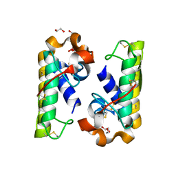
4RO1
 | |
4OU7
 | | Crystal structure of DnaT84-153-dT10 ssDNA complex reveals a novel single-stranded DNA binding mode | | 分子名称: | DNA (5'-D(P*TP*TP*TP*TP*TP*TP*TP*TP*TP*T)-3'), Primosomal protein 1 | | 著者 | Liu, Z, Chen, P, Niu, L, Teng, M, Li, X. | | 登録日 | 2014-02-15 | | 公開日 | 2014-08-13 | | 最終更新日 | 2024-05-29 | | 実験手法 | X-RAY DIFFRACTION (2.83 Å) | | 主引用文献 | Crystal structure of DnaT84-153-dT10 ssDNA complex reveals a novel single-stranded DNA binding mode.
Nucleic Acids Res., 42, 2014
|
|
4RFP
 | |


