3CSC
 
 | |
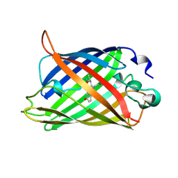
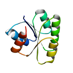
2YFP
 | | STRUCTURE OF YELLOW-EMISSION VARIANT OF GFP | | 分子名称: | PROTEIN (GREEN FLUORESCENT PROTEIN) | | 著者 | Wachter, R.M, Elsliger, M.A, Kallio, K, Hanson, G.T, Remington, S.J. | | 登録日 | 1998-08-17 | | 公開日 | 1999-01-13 | | 最終更新日 | 2023-11-15 | | 実験手法 | X-RAY DIFFRACTION (2.6 Å) | | 主引用文献 | Structural basis of spectral shifts in the yellow-emission variants of green fluorescent protein.
Structure, 6, 1998
|
|
1AMZ
 | |
1AL6
 | |
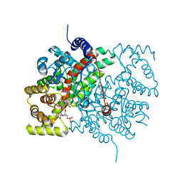
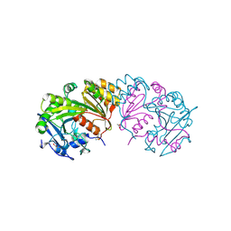
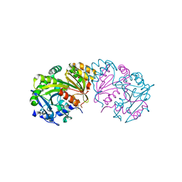
3BLS
 | | AMPC BETA-LACTAMASE FROM ESCHERICHIA COLI | | 分子名称: | AMPC BETA-LACTAMASE, M-AMINOPHENYLBORONIC ACID | | 著者 | Usher, K.C, Shoichet, B.K, Remington, S.J. | | 登録日 | 1998-06-04 | | 公開日 | 1998-08-12 | | 最終更新日 | 2023-08-09 | | 実験手法 | X-RAY DIFFRACTION (2.3 Å) | | 主引用文献 | Three-dimensional structure of AmpC beta-lactamase from Escherichia coli bound to a transition-state analogue: possible implications for the oxyanion hypothesis and for inhibitor design.
Biochemistry, 37, 1998
|
|
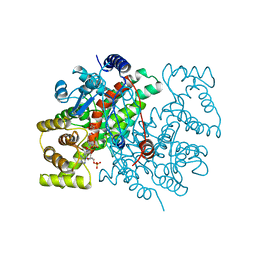
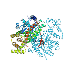
1BU6
 | | CRYSTAL STRUCTURES OF ESCHERICHIA COLI GLYCEROL KINASE AND THE MUTANT A65T IN AN INACTIVE TETRAMER: CONFORMATIONAL CHANGES AND IMPLICATIONS FOR ALLOSTERIC REGULATION | | 分子名称: | GLYCEROL, PROTEIN (GLYCEROL KINASE), SULFATE ION | | 著者 | Feese, M.D, Faber, H.R, Bystrom, C.E, Pettigrew, D.W, Remington, S.J. | | 登録日 | 1998-08-30 | | 公開日 | 1998-09-16 | | 最終更新日 | 2023-08-09 | | 実験手法 | X-RAY DIFFRACTION (2.37 Å) | | 主引用文献 | Glycerol kinase from Escherichia coli and an Ala65-->Thr mutant: the crystal structures reveal conformational changes with implications for allosteric regulation.
Structure, 6, 1998
|
|
1C4F
 | | GREEN FLUORESCENT PROTEIN S65T AT PH 4.6 | | 分子名称: | GREEN FLUORESCENT PROTEIN | | 著者 | Elsliger, M.A, Wachter, R.M, Kallio, K, Hanson, G.T, Remington, S.J. | | 登録日 | 1999-08-21 | | 公開日 | 1999-08-31 | | 最終更新日 | 2023-11-15 | | 実験手法 | X-RAY DIFFRACTION (2.25 Å) | | 主引用文献 | Structural and spectral response of green fluorescent protein variants to changes in pH.
Biochemistry, 38, 1999
|
|
1XMZ
 | | Crystal structure of the dark state of kindling fluorescent protein kfp from anemonia sulcata | | 分子名称: | BETA-MERCAPTOETHANOL, GFP-like non-fluorescent chromoprotein FP595 chain 1, GFP-like non-fluorescent chromoprotein FP595 chain 2 | | 著者 | Quillin, M.L, Anstrom, D.M, Shu, X, O'Leary, S, Kallio, K, Chudakov, D.M, Remington, S.J. | | 登録日 | 2004-10-04 | | 公開日 | 2005-04-19 | | 最終更新日 | 2024-07-10 | | 実験手法 | X-RAY DIFFRACTION (1.38 Å) | | 主引用文献 | Kindling Fluorescent Protein from Anemonia sulcata: Dark-State Structure at 1.38 Resolution
Biochemistry, 44, 2005
|
|
1CSS
 | |
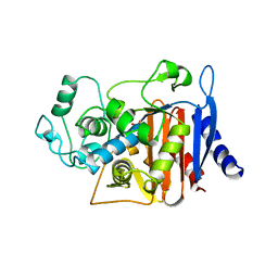
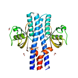
3TMY
 | | CHEY FROM THERMOTOGA MARITIMA (MN-III) | | 分子名称: | CHEY PROTEIN, MANGANESE (II) ION | | 著者 | Usher, K.C, De La Cruz, A, Dahlquist, F.W, Remington, S.J. | | 登録日 | 1997-06-04 | | 公開日 | 1997-12-03 | | 最終更新日 | 2024-05-22 | | 実験手法 | X-RAY DIFFRACTION (2.2 Å) | | 主引用文献 | Crystal structures of CheY from Thermotoga maritima do not support conventional explanations for the structural basis of enhanced thermostability.
Protein Sci., 7, 1998
|
|
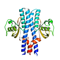
3UB9
 | | Periplasmic portion of the Helicobacter pylori chemoreceptor TlpB with hydroxyurea bound | | 分子名称: | GLYCEROL, N-HYDROXYUREA, SULFATE ION, ... | | 著者 | Henderson, J.N, Sweeney, E.G, Goers, J, Wreden, C, Hicks, K.G, Parthasarathy, R, Guillemin, K.J, Remington, S.J. | | 登録日 | 2011-10-23 | | 公開日 | 2012-06-27 | | 最終更新日 | 2024-02-28 | | 実験手法 | X-RAY DIFFRACTION (1.42 Å) | | 主引用文献 | Structure and proposed mechanism for the pH-sensing Helicobacter pylori chemoreceptor TlpB.
Structure, 20, 2012
|
|
1BCS
 | | COMPLEX OF THE WHEAT SERINE CARBOXYPEPTIDASE, CPDW-II, WITH THE MICROBIAL PEPTIDE ALDEHYDE INHIBITOR, CHYMOSTATIN, AND ARGININE AT 100 DEGREES KELVIN | | 分子名称: | 2-acetamido-2-deoxy-beta-D-glucopyranose, 2-acetamido-2-deoxy-beta-D-glucopyranose-(1-4)-2-acetamido-2-deoxy-beta-D-glucopyranose, ACETATE ION, ... | | 著者 | Bullock, T.L, Remington, S.J. | | 登録日 | 1995-11-03 | | 公開日 | 1996-03-08 | | 最終更新日 | 2020-07-29 | | 実験手法 | X-RAY DIFFRACTION (2.08 Å) | | 主引用文献 | Peptide aldehyde complexes with wheat serine carboxypeptidase II: implications for the catalytic mechanism and substrate specificity.
J.Mol.Biol., 255, 1996
|
|
1BCR
 | | COMPLEX OF THE WHEAT SERINE CARBOXYPEPTIDASE, CPDW-II, WITH THE MICROBIAL PEPTIDE ALDEHYDE INHIBITOR, ANTIPAIN, AND ARGININE AT ROOM TEMPERATURE | | 分子名称: | 2-acetamido-2-deoxy-beta-D-glucopyranose, 2-acetamido-2-deoxy-beta-D-glucopyranose-(1-4)-2-acetamido-2-deoxy-beta-D-glucopyranose, ANTIPAIN, ... | | 著者 | Bullock, T.L, Remington, S.J. | | 登録日 | 1995-11-03 | | 公開日 | 1996-03-08 | | 最終更新日 | 2023-11-15 | | 実験手法 | X-RAY DIFFRACTION (2.5 Å) | | 主引用文献 | Peptide aldehyde complexes with wheat serine carboxypeptidase II: implications for the catalytic mechanism and substrate specificity.
J.Mol.Biol., 255, 1996
|
|
1CSR
 | |
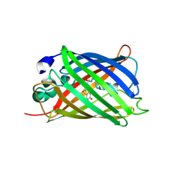
1P7T
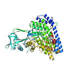 | | Structure of Escherichia coli malate synthase G:pyruvate:acetyl-Coenzyme A abortive ternary complex at 1.95 angstrom resolution | | 分子名称: | ACETYL COENZYME *A, DI(HYDROXYETHYL)ETHER, MAGNESIUM ION, ... | | 著者 | Anstrom, D.M, Kallio, K, Remington, S.J. | | 登録日 | 2003-05-05 | | 公開日 | 2003-09-09 | | 最終更新日 | 2023-11-15 | | 実験手法 | X-RAY DIFFRACTION (1.95 Å) | | 主引用文献 | Structure of the Escherichia Coli Malate Synthase G:pyruvate:acetyl-coenzyme A Abortive Ternary Complex at 1.95 Angstrom Resolution
Protein Sci., 12, 2003
|
|
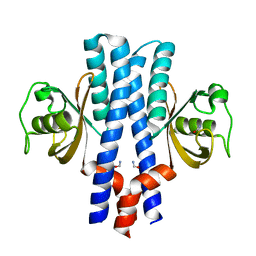
3UB6
 | | Periplasmic portion of the Helicobacter pylori chemoreceptor TlpB with urea bound | | 分子名称: | DI(HYDROXYETHYL)ETHER, GLYCEROL, SULFATE ION, ... | | 著者 | Henderson, J.N, Sweeney, E.G, Goers, J, Wreden, C, Hicks, K.G, Parthasarathy, R, Guillemin, K.J, Remington, S.J. | | 登録日 | 2011-10-23 | | 公開日 | 2012-06-27 | | 最終更新日 | 2024-02-28 | | 実験手法 | X-RAY DIFFRACTION (1.38 Å) | | 主引用文献 | Structure and proposed mechanism for the pH-sensing Helicobacter pylori chemoreceptor TlpB.
Structure, 20, 2012
|
|
3UB7
 | | Periplasmic portion of the Helicobacter pylori chemoreceptor TlpB with acetamide bound | | 分子名称: | ACETAMIDE, GLYCEROL, SULFATE ION, ... | | 著者 | Henderson, J.N, Sweeney, E.G, Goers, J, Wreden, C, Hicks, K.G, Parthasarathy, R, Guillemin, K.J, Remington, S.J. | | 登録日 | 2011-10-23 | | 公開日 | 2012-06-27 | | 最終更新日 | 2024-02-28 | | 実験手法 | X-RAY DIFFRACTION (1.4 Å) | | 主引用文献 | Structure and proposed mechanism for the pH-sensing Helicobacter pylori chemoreceptor TlpB.
Structure, 20, 2012
|
|
3UB8
 | | Periplasmic portion of the Helicobacter pylori chemoreceptor TlpB with formamide bound | | 分子名称: | FORMAMIDE, GLYCEROL, SULFATE ION, ... | | 著者 | Henderson, J.N, Sweeney, E.G, Goers, J, Wreden, C, Hicks, K.G, Parthasarathy, R, Guillemin, K.J, Remington, S.J. | | 登録日 | 2011-10-23 | | 公開日 | 2012-06-27 | | 最終更新日 | 2024-02-28 | | 実験手法 | X-RAY DIFFRACTION (1.42 Å) | | 主引用文献 | Structure and proposed mechanism for the pH-sensing Helicobacter pylori chemoreceptor TlpB.
Structure, 20, 2012
|
|
2QLH
 | | mPlum I65L mutant | | 分子名称: | Fluorescent protein plum | | 著者 | Shu, X, Remington, S.J. | | 登録日 | 2007-07-12 | | 公開日 | 2008-07-22 | | 最終更新日 | 2023-11-15 | | 実験手法 | X-RAY DIFFRACTION (1.9 Å) | | 主引用文献 | Structural Studies of Far-Red Emission in mPlum, a Monomeric Red Fluorescent Protein
To be Published
|
|
2QLI
 | | mPlum E16Q mutant | | 分子名称: | Fluorescent protein plum | | 著者 | Shu, X, Remington, S.J. | | 登録日 | 2007-07-12 | | 公開日 | 2008-07-22 | | 最終更新日 | 2023-11-15 | | 実験手法 | X-RAY DIFFRACTION (1.34 Å) | | 主引用文献 | Structural Studies of Far-Red Emission in mPlum, a Monomeric Red Fluorescent Protein
To be Published
|
|
2QLE
 | | GFP/S205V mutant | | 分子名称: | Green fluorescent protein, IMIDAZOLE | | 著者 | Shu, X, Remington, S.J. | | 登録日 | 2007-07-12 | | 公開日 | 2008-02-12 | | 最終更新日 | 2023-11-15 | | 実験手法 | X-RAY DIFFRACTION (1.59 Å) | | 主引用文献 | An alternative excited-state proton transfer pathway in green fluorescent protein variant S205V.
Protein Sci., 16, 2007
|
|
2TMY
 | |
2OTB
 | |
2OTE
 | | Crystal structure of a monomeric cyan fluorescent protein in the photobleached state | | 分子名称: | ACETATE ION, GFP-like fluorescent chromoprotein cFP484 | | 著者 | Henderson, J.N, Ai, H, Campbell, R.E, Remington, S.J. | | 登録日 | 2007-02-07 | | 公開日 | 2007-04-03 | | 最終更新日 | 2023-11-15 | | 実験手法 | X-RAY DIFFRACTION (1.47 Å) | | 主引用文献 | Structural basis for reversible photobleaching of a green fluorescent protein homologue.
Proc.Natl.Acad.Sci.Usa, 104, 2007
|
|
2QLG
 | | mPlum | | 分子名称: | Fluorescent protein plum | | 著者 | Shu, X, Remington, S.J. | | 登録日 | 2007-07-12 | | 公開日 | 2008-07-22 | | 最終更新日 | 2023-11-15 | | 実験手法 | X-RAY DIFFRACTION (1.8 Å) | | 主引用文献 | Structural Studies of Far-Red Emission in mPlum, a Monomeric Red Fluorescent Protein
To be Published
|
|


