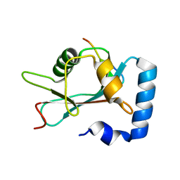
3WE2
 
 | |
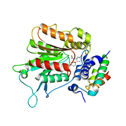
3WIM
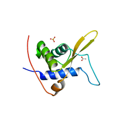
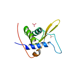
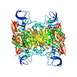
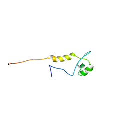
 | | GABARAP-LIR peptide complex | | 分子名称: | Gamma-aminobutyric acid receptor-associated protein, WD repeat and FYVE domain-containing protein 3 | | 著者 | Lystad, A, Ichimura, Y, Takagi, K, Yang, Y, Pankiv, S, Mizushima, T, Komatsu, M, Simonsen, A. | | 登録日 | 2013-09-19 | | 公開日 | 2014-03-12 | | 最終更新日 | 2023-11-08 | | 実験手法 | X-RAY DIFFRACTION (2.6 Å) | | 主引用文献 | GABARAP-LIR peptide complex
To be Published, 2014
|
|
3WQO
 | | Crystal structure of D-tagatose 3-epimerase-like protein | | 分子名称: | MANGANESE (II) ION, Uncharacterized protein MJ1311 | | 著者 | Uechi, K, Takata, G, Yoneda, K, Ohshima, T, Sakuraba, H. | | 登録日 | 2014-01-28 | | 公開日 | 2014-07-09 | | 最終更新日 | 2024-03-20 | | 実験手法 | X-RAY DIFFRACTION (2.64 Å) | | 主引用文献 | Structure of D-tagatose 3-epimerase-like protein from Methanocaldococcus jannaschii
Acta Crystallogr.,Sect.F, 70, 2014
|
|
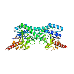
3WE3
 | |
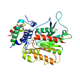
3WIC
 | | Structure of a substrate/cofactor-unbound glucose dehydrogenase | | 分子名称: | Glucose 1-dehydrogenase, PENTAETHYLENE GLYCOL, S-1,2-PROPANEDIOL, ... | | 著者 | Sakuraba, H, Kanoh, Y, Yoneda, K, Ohshima, T. | | 登録日 | 2013-09-10 | | 公開日 | 2014-05-14 | | 最終更新日 | 2023-11-08 | | 実験手法 | X-RAY DIFFRACTION (2.6 Å) | | 主引用文献 | Structural insight into glucose dehydrogenase from the thermoacidophilic archaeon Thermoplasma volcanium.
Acta Crystallogr.,Sect.D, 70, 2014
|
|
2YSJ
 | |
2YSL
 | |
2ZC0
 | | Crystal structure of an archaeal alanine:glyoxylate aminotransferase | | 分子名称: | 4'-DEOXY-4'-AMINOPYRIDOXAL-5'-PHOSPHATE, Alanine glyoxylate transaminase, IMIDAZOLE, ... | | 著者 | Sakuraba, H, Yoneda, K, Tsuge, H, Ohshima, T. | | 登録日 | 2007-10-31 | | 公開日 | 2008-06-17 | | 最終更新日 | 2024-03-13 | | 実験手法 | X-RAY DIFFRACTION (2.3 Å) | | 主引用文献 | Structure of an archaeal alanine:glyoxylate aminotransferase
Acta Crystallogr.,Sect.D, 64, 2008
|
|
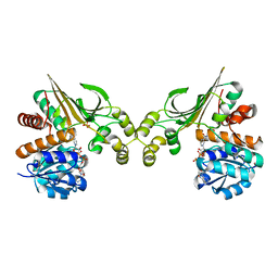
3A4U
 | | Crystal structure of MCFD2 in complex with carbohydrate recognition domain of ERGIC-53 | | 分子名称: | CALCIUM ION, GLYCEROL, Multiple coagulation factor deficiency protein 2, ... | | 著者 | Nishio, M, Kamiya, Y, Mizushima, T, Wakatsuki, S, Sasakawa, H, Yamamoto, K, Uchiyama, S, Noda, M, McKay, A.R, Fukui, K, Hauri, H.P, Kato, K. | | 登録日 | 2009-07-17 | | 公開日 | 2010-01-05 | | 最終更新日 | 2023-11-01 | | 実験手法 | X-RAY DIFFRACTION (1.84 Å) | | 主引用文献 | Structural basis for the cooperative interplay between the two causative gene products of combined factor V and factor VIII deficiency.
Proc.Natl.Acad.Sci.USA, 107, 2010
|
|
3AAF
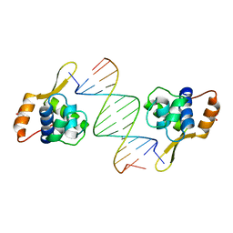 | | Structure of WRN RQC domain bound to double-stranded DNA | | 分子名称: | ACETATE ION, DNA (5'-D(*AP*CP*CP*CP*TP*AP*AP*TP*TP*AP*GP*GP*GP*T)-3'), Werner syndrome ATP-dependent helicase | | 著者 | Kitano, K, Hakoshima, T. | | 登録日 | 2009-11-16 | | 公開日 | 2010-02-16 | | 最終更新日 | 2024-03-13 | | 実験手法 | X-RAY DIFFRACTION (1.9 Å) | | 主引用文献 | Structural basis for DNA strand separation by the unconventional winged-helix domain of RecQ helicase WRN
Structure, 18, 2010
|
|
336D
 | | INTERACTION BETWEEN LEFT-HANDED Z-DNA AND POLYAMINE-3 THE CRYSTAL STRUCTURE OF THE D(CG)3 AND THERMOSPERMINE COMPLEX | | 分子名称: | DNA (5'-D(*CP*GP*CP*GP*CP*G)-3'), MAGNESIUM ION, N-(3-AMINO-PROPYL)-N-(5-AMINOPROPYL)-1,4-DIAMINOBUTANE | | 著者 | Ohishi, H, Terasoma, N, Nakanishi, I, Van Der Marel, G, Van Boom, J.H, Rich, A, Wang, A.H.-J, Hakoshima, T, Tomita, K.-I. | | 登録日 | 1997-06-24 | | 公開日 | 1998-04-10 | | 最終更新日 | 2024-02-21 | | 実験手法 | X-RAY DIFFRACTION (1 Å) | | 主引用文献 | Interaction between left-handed Z-DNA and polyamine - 3. The crystal structure of the d(CG)3 and thermospermine complex.
FEBS Lett., 398, 1996
|
|
2ZDJ
 | | Crystal Structure of TTMA177, a Hypothetical Protein from Thermus thermophilus phage TMA | | 分子名称: | hypothetical protein TTMA177 | | 著者 | Agari, Y, Tamakoshi, M, Yamagishi, A, Shinkai, A, Ebihara, A, Yokoyama, S, Kuramitsu, S, Oshima, T, RIKEN Structural Genomics/Proteomics Initiative (RSGI) | | 登録日 | 2007-11-26 | | 公開日 | 2008-12-02 | | 最終更新日 | 2011-07-13 | | 実験手法 | X-RAY DIFFRACTION (2.2 Å) | | 主引用文献 | Crystal Structure of TTMA177, a Hypothetical Protein from Thermus thermophilus phage TMA
To be Published
|
|
3AY8
 | | Glutathione S-transferase unclassified 2 from Bombyx mori | | 分子名称: | GLYCEROL, Glutathione S-transferase | | 著者 | Kakuta, Y, Usuda, K, Nakashima, T, Kimura, M, Aso, Y, Yamamoto, K. | | 登録日 | 2011-05-02 | | 公開日 | 2011-09-07 | | 最終更新日 | 2023-11-01 | | 実験手法 | X-RAY DIFFRACTION (2.1 Å) | | 主引用文献 | Crystallographic survey of active sites of an unclassified glutathione transferase from Bombyx mori
Biochim.Biophys.Acta, 1810, 2011
|
|
2YVS
 | |
2ZSH
 | | Structural basis of gibberellin(GA3)-induced DELLA recognition by the gibberellin receptor | | 分子名称: | DELLA protein GAI, GIBBERELLIN A3, Probable gibberellin receptor GID1L1 | | 著者 | Murase, K, Hirano, Y, Sun, T.P, Hakoshima, T. | | 登録日 | 2008-09-10 | | 公開日 | 2008-11-25 | | 最終更新日 | 2024-03-13 | | 実験手法 | X-RAY DIFFRACTION (1.8 Å) | | 主引用文献 | Gibberellin-induced DELLA recognition by the gibberellin receptor GID1
Nature, 456, 2008
|
|
2ZSI
 | | Structural basis of gibberellin(GA4)-induced DELLA recognition by the gibberellin receptor | | 分子名称: | DELLA protein GAI, GIBBERELLIN A4, Probable gibberellin receptor GID1L1 | | 著者 | Murase, K, Hirano, Y, Sun, T.P, Hakoshima, T. | | 登録日 | 2008-09-10 | | 公開日 | 2008-11-25 | | 最終更新日 | 2023-11-01 | | 実験手法 | X-RAY DIFFRACTION (1.8 Å) | | 主引用文献 | Gibberellin-induced DELLA recognition by the gibberellin receptor GID1
Nature, 456, 2008
|
|
3ABI
 | | Crystal Structure of L-Lysine Dehydrogenase from Hyperthermophilic Archaeon Pyrococcus horikoshii | | 分子名称: | NICOTINAMIDE-ADENINE-DINUCLEOTIDE, Putative uncharacterized protein PH1688, SULFATE ION | | 著者 | Yoneda, K, Sakuraba, H, Fukuda, J, Ohshima, T. | | 登録日 | 2009-12-14 | | 公開日 | 2009-12-29 | | 最終更新日 | 2024-03-13 | | 実験手法 | X-RAY DIFFRACTION (2.44 Å) | | 主引用文献 | First crystal structure of L-lysine 6-dehydrogenase as an NAD-dependent amine dehydrogenase.
J.Biol.Chem., 285, 2010
|
|
3AH8
 | | Structure of heterotrimeric G protein Galpha-q beta gamma in complex with an inhibitor YM-254890 | | 分子名称: | GUANOSINE-5'-DIPHOSPHATE, Guanine nucleotide-binding protein G(I)/G(S)/G(O) subunit gamma-2, Guanine nucleotide-binding protein G(I)/G(S)/G(T) subunit beta-1, ... | | 著者 | Nishimura, A, Kitano, K, Takasaki, J, Taniguchi, M, Mizuno, N, Tago, K, Hakoshima, T, Itoh, H. | | 登録日 | 2010-04-20 | | 公開日 | 2010-07-21 | | 最終更新日 | 2023-11-15 | | 実験手法 | X-RAY DIFFRACTION (2.9 Å) | | 主引用文献 | Structural basis for the specific inhibition of heterotrimeric Gq protein by a small molecule.
Proc.Natl.Acad.Sci.USA, 107, 2010
|
|
2D11
 | |
2D10
 | |
2CS8
 | |
2DL6
 | |
2DCH
 | | Crystal structure of archaeal intron-encoded homing endonuclease I-Tsp061I | | 分子名称: | CHLORIDE ION, SULFATE ION, putative homing endonuclease | | 著者 | Nakayama, H, Tsuge, H, Shimamura, T, Miyano, M, Nomura, N, Sako, Y. | | 登録日 | 2006-01-06 | | 公開日 | 2006-07-06 | | 最終更新日 | 2024-03-13 | | 実験手法 | X-RAY DIFFRACTION (2.06 Å) | | 主引用文献 | Structure of a hyperthermophilic archaeal homing endonuclease, I-Tsp061I: contribution of cross-domain polar networks to thermostability.
J.Mol.Biol., 365, 2007
|
|
2E4H
 | |
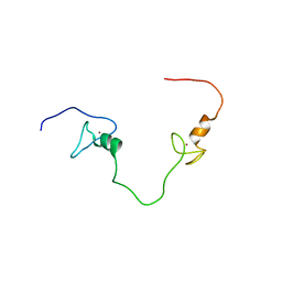
2EO1
 | | Solution structure of the ig domain of human OBSCN protein | | 分子名称: | CDNA FLJ14124 fis, clone MAMMA1002498 | | 著者 | Nagashima, K, Nagashima, T, Yoshida, M, Hayashi, F, Yokoyama, S, RIKEN Structural Genomics/Proteomics Initiative (RSGI) | | 登録日 | 2007-03-28 | | 公開日 | 2007-10-02 | | 最終更新日 | 2024-05-29 | | 実験手法 | SOLUTION NMR | | 主引用文献 | Solution structure of the ig domain of human OBSCN protein
To be Published
|
|


