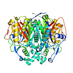
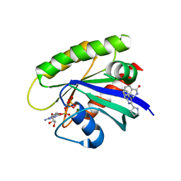
8ADG
 
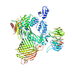 | | Cryo-EM structure of Darobactin 22 bound BAM complex | | 分子名称: | Darobactin 22, Outer membrane protein assembly factor BamA, Outer membrane protein assembly factor BamB, ... | | 著者 | Yuan, B, Marlovits, T.C. | | 登録日 | 2022-07-08 | | 公開日 | 2023-01-11 | | 実験手法 | ELECTRON MICROSCOPY (3 Å) | | 主引用文献 | Darobactins Exhibiting Superior Antibiotic Activity by Cryo-EM Structure Guided Biosynthetic Engineering.
Angew.Chem.Int.Ed.Engl., 62, 2023
|
|
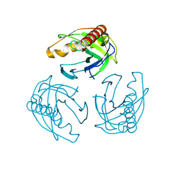
8ADI
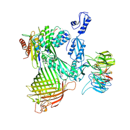 | | Cryo-EM structure of Darobactin 9 bound BAM complex | | 分子名称: | Darobactin 9, Outer membrane protein assembly factor BamA, Outer membrane protein assembly factor BamB, ... | | 著者 | Yuan, B, Marlovits, T.C. | | 登録日 | 2022-07-08 | | 公開日 | 2023-01-11 | | 実験手法 | ELECTRON MICROSCOPY (3.4 Å) | | 主引用文献 | Darobactins Exhibiting Superior Antibiotic Activity by Cryo-EM Structure Guided Biosynthetic Engineering.
Angew.Chem.Int.Ed.Engl., 62, 2023
|
|
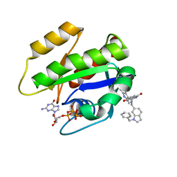
5MJ1
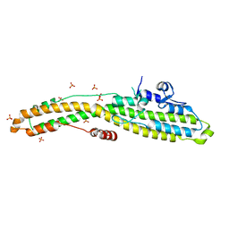
 | | Extracellular domain of human CD83 - rhombohedral crystal form | | 分子名称: | CD83 antigen, DI(HYDROXYETHYL)ETHER | | 著者 | Klingl, S, Egerer-Sieber, C, Schmid, B, Weiler, S, Muller, Y.A. | | 登録日 | 2016-11-29 | | 公開日 | 2017-03-29 | | 最終更新日 | 2018-03-07 | | 実験手法 | X-RAY DIFFRACTION (1.8 Å) | | 主引用文献 | Crystal Structure of the Extracellular Domain of the Human Dendritic Cell Surface Marker CD83.
J. Mol. Biol., 429, 2017
|
|
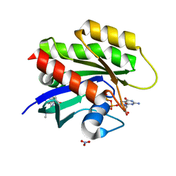
2BHO
 | |
6MHJ
 | | Structure of BoNT mutant | | 分子名称: | Botulinum neurotoxin type A, PHOSPHATE ION | | 著者 | Lam, K, Jin, R. | | 登録日 | 2018-09-18 | | 公開日 | 2018-12-26 | | 最終更新日 | 2023-10-11 | | 実験手法 | X-RAY DIFFRACTION (3.019 Å) | | 主引用文献 | A viral-fusion-peptide-like molecular switch drives membrane insertion of botulinum neurotoxin A1.
Nat Commun, 9, 2018
|
|
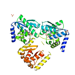
3L0N
 | | Human orotidyl-5'-monophosphate decarboxylase in complex with 6-mercapto-UMP | | 分子名称: | 6-sulfanyluridine-5'-phosphate, Uridine 5'-monophosphate synthase | | 著者 | Heinrich, D, Wittmann, J, Diederichsen, U, Rudolph, M. | | 登録日 | 2009-12-10 | | 公開日 | 2010-01-26 | | 最終更新日 | 2024-03-20 | | 実験手法 | X-RAY DIFFRACTION (1.74 Å) | | 主引用文献 | Lys314 is a nucleophile in non-classical reactions of orotidine-5'-monophosphate decarboxylase
Chemistry, 15, 2009
|
|
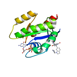
5MAV
 | |
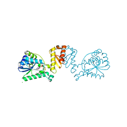
2IWZ
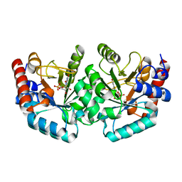
 | | Human mitochondrial beta-ketoacyl ACP synthase complexed with hexanoic acid | | 分子名称: | 3-OXOACYL-[ACYL-CARRIER-PROTEIN] SYNTHASE, AMMONIUM ION, HEXANOIC ACID | | 著者 | Christensen, C.E, Kragelund, B.B, von Wettstein-Knowles, P, Henriksen, A. | | 登録日 | 2006-07-05 | | 公開日 | 2007-02-06 | | 最終更新日 | 2023-12-13 | | 実験手法 | X-RAY DIFFRACTION (1.65 Å) | | 主引用文献 | Structure of the Human Beta-Ketoacyl [Acp] Synthase from the Mitochondrial Type II Fatty Acid Synthase.
Protein Sci., 16, 2007
|
|
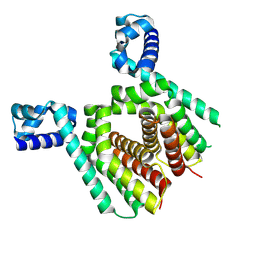
2IWY
 | | Human mitochondrial beta-ketoacyl ACP synthase | | 分子名称: | 3-OXOACYL-[ACYL-CARRIER-PROTEIN] SYNTHASE, AMMONIUM ION | | 著者 | Christensen, C.E, Kragelund, B.B, von Wettstein-Knowles, P, Henriksen, A. | | 登録日 | 2006-07-05 | | 公開日 | 2007-02-06 | | 最終更新日 | 2023-12-13 | | 実験手法 | X-RAY DIFFRACTION (2.06 Å) | | 主引用文献 | Structure of the Human Beta-Ketoacyl [Acp] Synthase from the Mitochondrial Type II Fatty Acid Synthase.
Protein Sci., 16, 2007
|
|
2JVF
 | | Solution structure of M7, a computationally-designed artificial protein | | 分子名称: | de novo protein M7 | | 著者 | Stordeur, C, Dalluege, R, Birkenmeier, O, Wienk, H, Rudolph, R, Lange, C, Luecke, C. | | 登録日 | 2007-09-19 | | 公開日 | 2008-08-12 | | 最終更新日 | 2024-05-01 | | 実験手法 | SOLUTION NMR | | 主引用文献 | The NMR solution structure of the artificial protein M7 matches the computationally designed model
Proteins, 72, 2008
|
|
7OU8
 | | Human O-GlcNAc hydrolase in complex with DNJNAc-thiazolidines | | 分子名称: | 1,2-ETHANEDIOL, CALCIUM ION, O-GlcNAcase BT_4395, ... | | 著者 | Males, A, Davies, G.J, Gonzalez-Cuesta, M, Mellet, C.O, Fernandez, J.M.G, Sidhu, P, Ashmus, R, Busmann, J, Vocadlo, D.J, Foster, L. | | 登録日 | 2021-06-11 | | 公開日 | 2022-04-13 | | 最終更新日 | 2024-01-31 | | 実験手法 | X-RAY DIFFRACTION (1.5 Å) | | 主引用文献 | Bicyclic Picomolar OGA Inhibitors Enable Chemoproteomic Mapping of Its Endogenous Post-translational Modifications
J.Am.Chem.Soc., 144, 2022
|
|
7OU6
 | | Human O-GlcNAc hydrolase in complex with DNJNAc-thiazolidines | | 分子名称: | Protein O-GlcNAcase, ~{N}-[(3~{Z},6~{S},7~{R},8~{R},8~{a}~{S})-7,8-bis(oxidanyl)-3-(phenylmethyl)imino-1,5,6,7,8,8~{a}-hexahydro-[1,3]thiazolo[3,4-a]pyridin-6-yl]ethanamide | | 著者 | Males, A, Davies, G.J, Gonzalez-Cuesta, M, Mellet, C.O, Fernandez, J.M.G, Sidhu, P, Ashmus, R, Busmann, J, Vocadlo, D.J, Foster, L. | | 登録日 | 2021-06-11 | | 公開日 | 2022-04-13 | | 最終更新日 | 2024-01-31 | | 実験手法 | X-RAY DIFFRACTION (2.41 Å) | | 主引用文献 | Bicyclic Picomolar OGA Inhibitors Enable Chemoproteomic Mapping of Its Endogenous Post-translational Modifications
J.Am.Chem.Soc., 144, 2022
|
|
8CP3
 | | Apo-LarE in complex with AMP-PNP | | 分子名称: | BETA-MERCAPTOETHANOL, GLYCEROL, NAD_synthase domain-containing protein, ... | | 著者 | Zecchin, P, Pecqueur, L, Golinelli-Pimpaneau, B. | | 登録日 | 2023-03-01 | | 公開日 | 2024-01-31 | | 最終更新日 | 2024-02-07 | | 実験手法 | X-RAY DIFFRACTION (2.35 Å) | | 主引用文献 | Structure-based insights into the mechanism of [4Fe-4S]-dependent sulfur insertase LarE.
Protein Sci., 33, 2024
|
|
8CNZ
 | | mmLarE-[4Fe-4S] phased by Fe-SAD | | 分子名称: | CHLORIDE ION, IODIDE ION, IRON/SULFUR CLUSTER, ... | | 著者 | Pecqueur, L, Zecchin, P, Golinelli-Pimpaneau, B. | | 登録日 | 2023-02-24 | | 公開日 | 2024-01-31 | | 最終更新日 | 2024-02-07 | | 実験手法 | X-RAY DIFFRACTION (3.6 Å) | | 主引用文献 | Structure-based insights into the mechanism of [4Fe-4S]-dependent sulfur insertase LarE.
Protein Sci., 33, 2024
|
|
8CP4
 | | [4Fe-4S] cluster containing LarE in complex with AMP | | 分子名称: | ADENOSINE MONOPHOSPHATE, CHLORIDE ION, IRON/SULFUR CLUSTER, ... | | 著者 | Zecchin, P, Pecqueur, L, Golinelli-Pimpaneau, B. | | 登録日 | 2023-03-01 | | 公開日 | 2024-01-31 | | 最終更新日 | 2024-02-07 | | 実験手法 | X-RAY DIFFRACTION (3.19 Å) | | 主引用文献 | Structure-based insights into the mechanism of [4Fe-4S]-dependent sulfur insertase LarE.
Protein Sci., 33, 2024
|
|
5N1I
 | |
5N1C
 | |
6GJ5
 | | CRYSTAL STRUCTURE OF KRAS G12D (GPPCP) IN COMPLEX WITH 15 | | 分子名称: | (3~{S})-3-[2-[(2~{R})-pyrrolidin-2-yl]-1~{H}-indol-3-yl]-2,3-dihydroisoindol-1-one, GTPase KRas, MAGNESIUM ION, ... | | 著者 | Kessler, D, Mcconnell, D.M, Mantoulidis, A. | | 登録日 | 2018-05-16 | | 公開日 | 2019-07-31 | | 最終更新日 | 2024-01-17 | | 実験手法 | X-RAY DIFFRACTION (1.499 Å) | | 主引用文献 | Drugging an undruggable pocket on KRAS.
Proc.Natl.Acad.Sci.USA, 116, 2019
|
|
4LUG
 | |
6GJ7
 | | CRYSTAL STRUCTURE OF KRAS G12D (GPPCP) IN COMPLEX WITH 22 | | 分子名称: | (3~{S})-5-oxidanyl-3-[2-[[[1-(phenylmethyl)indol-6-yl]methylamino]methyl]-1~{H}-indol-3-yl]-2,3-dihydroisoindol-1-one, GTPase KRas, MAGNESIUM ION, ... | | 著者 | Kessler, D, Mcconnell, D.M, Mantoulidis, A. | | 登録日 | 2018-05-16 | | 公開日 | 2019-07-31 | | 最終更新日 | 2024-01-17 | | 実験手法 | X-RAY DIFFRACTION (1.67 Å) | | 主引用文献 | Drugging an undruggable pocket on KRAS.
Proc.Natl.Acad.Sci.USA, 116, 2019
|
|
5N7O
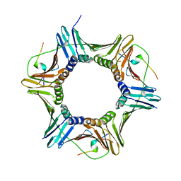
 | | EthR2 in complex with SMARt-420 compound | | 分子名称: | 4,4,4-trifluoro-1-(3-phenyl-1-oxa-2,8-diazaspiro[4.5]dec-2-en-8-yl)butan-1-one, Probable transcriptional regulatory protein | | 著者 | Wohlkonig, A, Wintjens, R. | | 登録日 | 2017-02-21 | | 公開日 | 2017-04-26 | | 最終更新日 | 2024-01-17 | | 実験手法 | X-RAY DIFFRACTION (2.3 Å) | | 主引用文献 | Structural analysis of the interaction between spiroisoxazoline SMARt-420 and the Mycobacterium tuberculosis repressor EthR2.
Biochem. Biophys. Res. Commun., 487, 2017
|
|
6GJ6
 | | CRYSTAL STRUCTURE OF KRAS G12D (GPPCP) IN COMPLEX WITH 18 | | 分子名称: | (3~{S})-3-[2-[(dimethylamino)methyl]-1~{H}-indol-3-yl]-5-oxidanyl-2,3-dihydroisoindol-1-one, GTPase KRas, MAGNESIUM ION, ... | | 著者 | Kessler, D, Mcconnell, D.M, Mantoulidis, A, Phan, J. | | 登録日 | 2018-05-16 | | 公開日 | 2019-07-31 | | 最終更新日 | 2024-05-15 | | 実験手法 | X-RAY DIFFRACTION (1.761 Å) | | 主引用文献 | Drugging an undruggable pocket on KRAS.
Proc.Natl.Acad.Sci.USA, 116, 2019
|
|
6GJ8
 | | CRYSTAL STRUCTURE OF KRAS G12D (GPPCP) IN COMPLEX WITH BI 2852 | | 分子名称: | (3~{S})-3-[2-[[[1-[(1-methylimidazol-4-yl)methyl]indol-6-yl]methylamino]methyl]-1~{H}-indol-3-yl]-5-oxidanyl-2,3-dihydroisoindol-1-one, GTPase KRas, MAGNESIUM ION, ... | | 著者 | Kessler, D, Mcconnell, D.M, Mantoulidis, A. | | 登録日 | 2018-05-16 | | 公開日 | 2019-07-31 | | 最終更新日 | 2024-01-17 | | 実験手法 | X-RAY DIFFRACTION (1.65 Å) | | 主引用文献 | Drugging an undruggable pocket on KRAS.
Proc.Natl.Acad.Sci.USA, 116, 2019
|
|
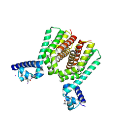
4N22
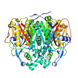
 | | Crystal structure of Protein Arginine Deiminase 2 (50 uM Ca2+) | | 分子名称: | (4S)-2-METHYL-2,4-PENTANEDIOL, ACETATE ION, CALCIUM ION, ... | | 著者 | Slade, D.J, Zhang, X, Fang, P, Dreyton, C.J, Zhang, Y, Gross, M.L, Guo, M, Coonrod, S.A, Thompson, P.R. | | 登録日 | 2013-10-04 | | 公開日 | 2015-02-04 | | 最終更新日 | 2023-09-20 | | 実験手法 | X-RAY DIFFRACTION (1.889 Å) | | 主引用文献 | Protein arginine deiminase 2 binds calcium in an ordered fashion: implications for inhibitor design.
Acs Chem.Biol., 10, 2015
|
|
4N2I
 | | Crystal structure of Protein Arginine Deiminase 2 (D177A, 10 mM Ca2+) | | 分子名称: | (4S)-2-METHYL-2,4-PENTANEDIOL, ACETATE ION, CALCIUM ION, ... | | 著者 | Slade, D.J, Zhang, X, Fang, P, Dreyton, C.J, Zhang, Y, Gross, M.L, Guo, M, Coonrod, S.A, Thompson, P.R. | | 登録日 | 2013-10-04 | | 公開日 | 2015-02-04 | | 最終更新日 | 2023-09-20 | | 実験手法 | X-RAY DIFFRACTION (1.9 Å) | | 主引用文献 | Protein arginine deiminase 2 binds calcium in an ordered fashion: implications for inhibitor design.
Acs Chem.Biol., 10, 2015
|
|


