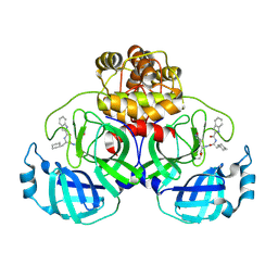
5DEU
 
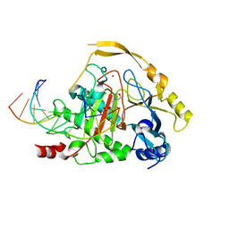 | | Crystal structure of TET2-5hmC complex | | 分子名称: | 2-(N-MORPHOLINO)-ETHANESULFONIC ACID, CHLORIDE ION, DNA (5'-D(*AP*CP*CP*AP*CP*(5HC)P*GP*GP*TP*GP*GP*T)-3'), ... | | 著者 | Hu, L, Cheng, J, Rao, Q, Li, Z, Li, J, Xu, Y. | | 登録日 | 2015-08-26 | | 公開日 | 2015-11-04 | | 最終更新日 | 2023-09-27 | | 実験手法 | X-RAY DIFFRACTION (1.801 Å) | | 主引用文献 | Structural insight into substrate preference for TET-mediated oxidation.
Nature, 527, 2015
|
|
6E83
 | |
5XNV
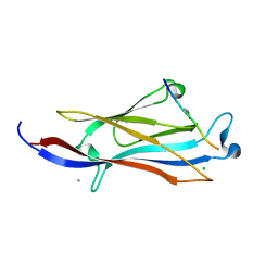 | | Crystal structure of YEATS2 YEATS bound to H3K27ac peptide | | 分子名称: | ALA-ALA-ARG-ALY-SER-ALA-PRO-ALA, AMMONIUM ION, CHLORIDE ION, ... | | 著者 | Li, H.T, Guan, H.P, Zhao, D. | | 登録日 | 2017-05-24 | | 公開日 | 2017-11-01 | | 最終更新日 | 2024-10-16 | | 実験手法 | X-RAY DIFFRACTION (2.696 Å) | | 主引用文献 | YEATS2 links histone acetylation to tumorigenesis of non-small cell lung cancer.
Nat Commun, 8, 2017
|
|
8HRD
 | | Crystal structure of the receptor binding domain of SARS-CoV-2 Delta variant in complex with IMCAS74 Fab and W14 Fab | | 分子名称: | 2-acetamido-2-deoxy-beta-D-glucopyranose, IMCAS74 Fab heavy chain, IMCAS74 Fab light chain, ... | | 著者 | Zhao, R.C, Wu, L.L, Han, P. | | 登録日 | 2022-12-15 | | 公開日 | 2023-12-20 | | 最終更新日 | 2024-10-23 | | 実験手法 | X-RAY DIFFRACTION (2.86 Å) | | 主引用文献 | Defining a de novo non-RBM antibody as RBD-8 and its synergistic rescue of immune-evaded antibodies to neutralize Omicron SARS-CoV-2.
Proc.Natl.Acad.Sci.USA, 120, 2023
|
|
9LUG
 | |
9LUF
 | |
7NAM
 | | LRP6_E1 in complex with Lr-EET-3.5 | | 分子名称: | Low-density lipoprotein receptor-related protein 6, SODIUM ION, Trypsin inhibitor 2, ... | | 著者 | Hansen, S, Hannoush, R.N. | | 登録日 | 2021-06-21 | | 公開日 | 2022-06-29 | | 最終更新日 | 2024-10-23 | | 実験手法 | X-RAY DIFFRACTION (1.6 Å) | | 主引用文献 | Directed evolution identifies high-affinity cystine-knot peptide agonists and antagonists of Wnt/ beta-catenin signaling.
Proc.Natl.Acad.Sci.USA, 119, 2022
|
|
7FIV
 | | Crystal structure of the complex formed by Wolbachia cytoplasmic incompatibility factors CidA and CidBND1-ND2 from wPip(Tunis) | | 分子名称: | CidA_I gamma/2 protein, CidB_I b/2 protein | | 著者 | Xiao, Y.J, Wang, W, Chen, X, Ji, X.Y, Yang, H.T. | | 登録日 | 2021-08-01 | | 公開日 | 2022-04-06 | | 最終更新日 | 2023-11-29 | | 実験手法 | X-RAY DIFFRACTION (2.59 Å) | | 主引用文献 | Crystal Structures of Wolbachia CidA and CidB Reveal Determinants of Bacteria-induced Cytoplasmic Incompatibility and Rescue.
Nat Commun, 13, 2022
|
|
7FIW
 | | Crystal structure of the complex formed by Wolbachia cytoplasmic incompatibility factors CidAwMel(ST) and CidBND1-ND2 from wPip(Pel) | | 分子名称: | ULP_PROTEASE domain-containing protein, bacteria factor 4,CidA I(Zeta/1) protein | | 著者 | Xiao, Y.J, Wang, W, Chen, X, Ji, X.Y, Yang, H.T. | | 登録日 | 2021-08-01 | | 公開日 | 2022-04-06 | | 最終更新日 | 2023-11-29 | | 実験手法 | X-RAY DIFFRACTION (2.16 Å) | | 主引用文献 | Crystal Structures of Wolbachia CidA and CidB Reveal Determinants of Bacteria-induced Cytoplasmic Incompatibility and Rescue.
Nat Commun, 13, 2022
|
|
7FIU
 | | Crystal structure of the DUB domain of Wolbachia cytoplasmic incompatibility factor CidB from wMel | | 分子名称: | ULP_PROTEASE domain-containing protein | | 著者 | Xiao, Y.J, Wang, W, Chen, X, Ji, X.Y, Yang, H.T. | | 登録日 | 2021-08-01 | | 公開日 | 2022-04-06 | | 最終更新日 | 2024-05-29 | | 実験手法 | X-RAY DIFFRACTION (1.84 Å) | | 主引用文献 | Crystal Structures of Wolbachia CidA and CidB Reveal Determinants of Bacteria-induced Cytoplasmic Incompatibility and Rescue.
Nat Commun, 13, 2022
|
|
7FIT
 | | Crystal structure of Wolbachia cytoplasmic incompatibility factor CidA from wMel | | 分子名称: | bacteria factor 1 | | 著者 | Xiao, Y.J, Wang, W, Chen, X, Ji, X.Y, Yang, H.T. | | 登録日 | 2021-08-01 | | 公開日 | 2022-04-06 | | 最終更新日 | 2024-05-29 | | 実験手法 | X-RAY DIFFRACTION (2.75 Å) | | 主引用文献 | Crystal Structures of Wolbachia CidA and CidB Reveal Determinants of Bacteria-induced Cytoplasmic Incompatibility and Rescue.
Nat Commun, 13, 2022
|
|
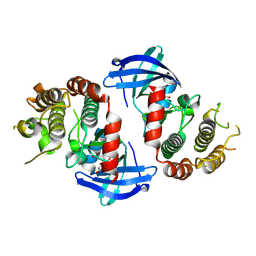
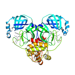
9LFU
 | | Crystal structure of human RIP3 kinase domain complexed with LK01003 | | 分子名称: | 2-cyclopentyl-~{N}-(6-propan-2-ylsulfonylquinolin-4-yl)-1,3-benzothiazol-5-amine, Receptor-interacting serine/threonine-protein kinase 3 | | 著者 | Xie, H, Su, H.X, Li, M.J, Xu, Y.C. | | 登録日 | 2025-01-09 | | 公開日 | 2025-05-07 | | 最終更新日 | 2025-05-21 | | 実験手法 | X-RAY DIFFRACTION (2.93 Å) | | 主引用文献 | Structure-based design of potent and selective inhibitors targeting RIPK3 for eliminating on-target toxicity in vitro.
Nat Commun, 16, 2025
|
|
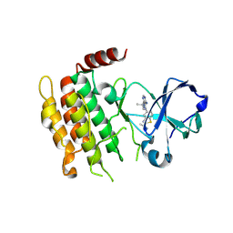
9LVG
 | | Crystal structure of the mouse RIP3 kinase domain in complexed with Robinetin | | 分子名称: | 3,7-bis(oxidanyl)-2-[3,4,5-tris(oxidanyl)phenyl]chromen-4-one, Receptor-interacting serine/threonine-protein kinase 3 | | 著者 | Xie, H, Su, H.X, Li, M.J, Xu, Y.C. | | 登録日 | 2025-02-12 | | 公開日 | 2025-05-07 | | 最終更新日 | 2025-05-21 | | 実験手法 | X-RAY DIFFRACTION (2.03 Å) | | 主引用文献 | Identification of RIPK3 as a target of flavonoids for anti-necroptosis in vitro.
Bioorg.Chem., 161, 2025
|
|
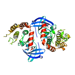
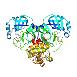
9LFW
 | | Crystal structure of mouse RIP3 kinase domain(R69H) complexed with LK01003 | | 分子名称: | 2-cyclopentyl-~{N}-(6-propan-2-ylsulfonylquinolin-4-yl)-1,3-benzothiazol-5-amine, Receptor-interacting serine/threonine-protein kinase 3 | | 著者 | Xie, H, Su, H.X, Li, M.J, Xu, Y.C. | | 登録日 | 2025-01-09 | | 公開日 | 2025-05-07 | | 最終更新日 | 2025-05-21 | | 実験手法 | X-RAY DIFFRACTION (1.59 Å) | | 主引用文献 | Structure-based design of potent and selective inhibitors targeting RIPK3 for eliminating on-target toxicity in vitro.
Nat Commun, 16, 2025
|
|
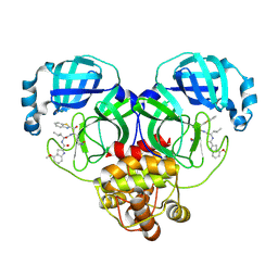
9LFV
 | | Crystal structure of mouse RIP3 kinase domain complexed with LK01004 | | 分子名称: | 3-[2-[(2~{R})-butan-2-yl]-1,3-benzothiazol-5-yl]-1-~{tert}-butyl-pyrazolo[3,4-d]pyrimidin-4-amine, 3-[2-[(2~{S})-butan-2-yl]-1,3-benzothiazol-5-yl]-1-~{tert}-butyl-pyrazolo[3,4-d]pyrimidin-4-amine, Receptor-interacting serine/threonine-protein kinase 3 | | 著者 | Xie, H, Su, H.X, Li, M.J, Xu, Y.C. | | 登録日 | 2025-01-09 | | 公開日 | 2025-05-07 | | 最終更新日 | 2025-05-21 | | 実験手法 | X-RAY DIFFRACTION (2.78 Å) | | 主引用文献 | Structure-based design of potent and selective inhibitors targeting RIPK3 for eliminating on-target toxicity in vitro.
Nat Commun, 16, 2025
|
|
9LVH
 | | Crystal structure of the mouse RIP3 kinase domain in complexed with Tricetin | | 分子名称: | 5,7-DIHYDROXY-2-(3,4,5-TRIHYDROXYPHENYL)-4H-CHROMEN-4-ONE, Receptor-interacting serine/threonine-protein kinase 3 | | 著者 | Xie, H, Su, H.X, Li, M.J, Xu, Y.C. | | 登録日 | 2025-02-12 | | 公開日 | 2025-05-07 | | 最終更新日 | 2025-05-21 | | 実験手法 | X-RAY DIFFRACTION (2.38 Å) | | 主引用文献 | Identification of RIPK3 as a target of flavonoids for anti-necroptosis in vitro.
Bioorg.Chem., 161, 2025
|
|
8HQI
 | |
8HQG
 | |
8HQH
 | |
8HQJ
 | |
8HQF
 | |
5XTZ
 | | Crystal structure of GAS41 YEATS bound to H3K27ac peptide | | 分子名称: | ACETATE ION, THR-LYS-ALA-ALA-ARG-ALY-SER-ALA-PRO-ALA, YEATS domain-containing protein 4 | | 著者 | Li, H.T, Zhao, D. | | 登録日 | 2017-06-21 | | 公開日 | 2018-06-27 | | 最終更新日 | 2024-10-30 | | 実験手法 | X-RAY DIFFRACTION (2.105 Å) | | 主引用文献 | Gas41 links histone acetylation to H2A.Z deposition and maintenance of embryonic stem cell identity.
Cell Discov, 4, 2018
|
|
5XA6
 | |
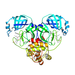
1TY4
 | | Crystal structure of a CED-9/EGL-1 complex | | 分子名称: | Apoptosis regulator ced-9, EGg Laying defective EGL-1, programmed cell death activator | | 著者 | Yan, N, Gu, L, Kokel, D, Xue, D, Shi, Y. | | 登録日 | 2004-07-07 | | 公開日 | 2004-09-28 | | 最終更新日 | 2024-11-20 | | 実験手法 | X-RAY DIFFRACTION (2.2 Å) | | 主引用文献 | Structural, Biochemical, and Functional Analyses of CED-9 Recognition by the Proapoptotic Proteins EGL-1 and CED-4
Mol.Cell, 15, 2004
|
|
9E9H
 | | Crystal structure of human KRAS G12C covalently bound to DEL triazine compound 5 | | 分子名称: | (3S)-N,5-dimethyl-3-({4-[3-(morpholin-4-yl)phenyl]-6-(2-propanoyl-2,6-diazaspiro[3.4]octan-6-yl)-1,3,5-triazin-2-yl}amino)hexanamide, CALCIUM ION, GTPase KRas, ... | | 著者 | Mohr, C. | | 登録日 | 2024-11-08 | | 公開日 | 2025-02-26 | | 最終更新日 | 2025-03-05 | | 実験手法 | X-RAY DIFFRACTION (1.65 Å) | | 主引用文献 | Identification of Structurally Novel KRAS G12C Inhibitors through Covalent DNA-Encoded Library Screening.
J.Med.Chem., 68, 2025
|
|


