9J52
 
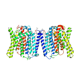 | | CryoEM structure of human XPR1 in complex with phosphate in state B | | 分子名称: | 1,2-DIACYL-SN-GLYCERO-3-PHOSPHOCHOLINE, PHOSPHATE ION, Solute carrier family 53 member 1 | | 著者 | Zhang, W.H, Chen, Y.K, Guan, Z.Y, Liu, Z. | | 登録日 | 2024-08-11 | | 公開日 | 2025-01-15 | | 実験手法 | ELECTRON MICROSCOPY (3.1 Å) | | 主引用文献 | Structural insights into the mechanism of phosphate recognition and transport by XPR1.
Nat Commun, 16, 2025
|
|
9J51
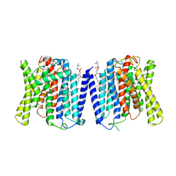 | | CryoEM structure of human XPR1 in complex with phosphate in state A | | 分子名称: | 1,2-DIACYL-SN-GLYCERO-3-PHOSPHOCHOLINE, PHOSPHATE ION, Solute carrier family 53 member 1 | | 著者 | Zhang, W.H, Chen, Y.K, Guan, Z.Y, Liu, Z. | | 登録日 | 2024-08-11 | | 公開日 | 2025-01-15 | | 実験手法 | ELECTRON MICROSCOPY (3.1 Å) | | 主引用文献 | Structural insights into the mechanism of phosphate recognition and transport by XPR1.
Nat Commun, 16, 2025
|
|
9J53
 | | CryoEM structure of human XPR1 in complex with phosphate in state C | | 分子名称: | 1,2-DIACYL-SN-GLYCERO-3-PHOSPHOCHOLINE, PHOSPHATE ION, Solute carrier family 53 member 1 | | 著者 | Zhang, W.H, Chen, Y.K, Guan, Z.Y, Liu, Z. | | 登録日 | 2024-08-11 | | 公開日 | 2025-01-15 | | 実験手法 | ELECTRON MICROSCOPY (3.3 Å) | | 主引用文献 | Structural insights into the mechanism of phosphate recognition and transport by XPR1.
Nat Commun, 16, 2025
|
|
6RTI
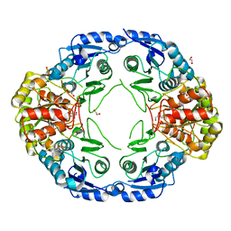
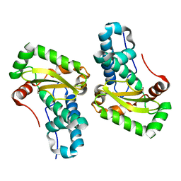
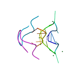
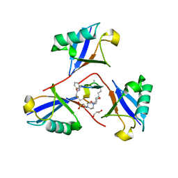
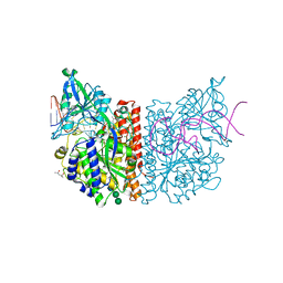 | | X-ray structure of human glutamate carboxypeptidase II (GCPII) in complex with aptamer A9g | | 分子名称: | (2S)-2-(PHOSPHONOMETHYL)PENTANEDIOIC ACID, (4S)-2-METHYL-2,4-PENTANEDIOL, 2-acetamido-2-deoxy-beta-D-glucopyranose-(1-4)-2-acetamido-2-deoxy-beta-D-glucopyranose, ... | | 著者 | Motlova, L, Kolenko, P, Barinka, C. | | 登録日 | 2019-05-24 | | 公開日 | 2020-06-10 | | 最終更新日 | 2024-11-13 | | 実験手法 | X-RAY DIFFRACTION (2.2 Å) | | 主引用文献 | Structural basis of prostate-specific membrane antigen recognition by the A9g RNA aptamer.
Nucleic Acids Res., 48, 2020
|
|
7R6A
 | |
7R69
 | |
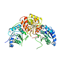
7R6B
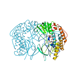
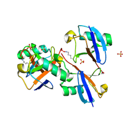
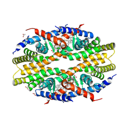
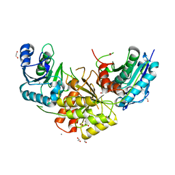 | | Crystal structure of mutant R43D/L124D/R125A/C273S of L-Asparaginase I from Yersinia pestis | | 分子名称: | 1,2-ETHANEDIOL, CALCIUM ION, L-asparaginase I | | 著者 | Strzelczyk, P, Wlodawer, A, Lubkowski, J. | | 登録日 | 2021-06-22 | | 公開日 | 2022-07-06 | | 最終更新日 | 2023-10-25 | | 実験手法 | X-RAY DIFFRACTION (2.03 Å) | | 主引用文献 | The dimeric form of bacterial l-asparaginase YpAI is fully active.
Febs J., 290, 2023
|
|
5HX6
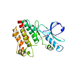 | | Crystal structure of RIP1 kinase with a benzo[b][1,4]oxazepin-4-one | | 分子名称: | 5-benzyl-N-[(3S)-5-methyl-4-oxo-2,3,4,5-tetrahydro-1,5-benzoxazepin-3-yl]-1,2-oxazole-3-carboxamide, Receptor-interacting serine/threonine-protein kinase 1 | | 著者 | Campobasso, N, Ward, P. | | 登録日 | 2016-01-29 | | 公開日 | 2016-03-02 | | 最終更新日 | 2024-03-06 | | 実験手法 | X-RAY DIFFRACTION (2.23 Å) | | 主引用文献 | DNA-Encoded Library Screening Identifies Benzo[b][1,4]oxazepin-4-ones as Highly Potent and Monoselective Receptor Interacting Protein 1 Kinase Inhibitors.
J.Med.Chem., 59, 2016
|
|
8S9G
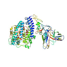 | | SARS-CoV-2 BN.1 spike RBD bound to the human ACE2 ectodomain and the S309 neutralizing antibody Fab fragment | | 分子名称: | 2-acetamido-2-deoxy-beta-D-glucopyranose, Processed angiotensin-converting enzyme 2, S309 Fab Heavy chain, ... | | 著者 | Park, Y.J, Seattle Structural Genomics Center for Infectious Disease (SSGCID), Veesler, D. | | 登録日 | 2023-03-28 | | 公開日 | 2023-10-04 | | 最終更新日 | 2024-11-20 | | 実験手法 | ELECTRON MICROSCOPY (3 Å) | | 主引用文献 | Neutralization, effector function and immune imprinting of Omicron variants.
Nature, 621, 2023
|
|
4YET
 | | X-ray crystal structure of superoxide dismutase from Babesia bovis solved by Sulfur SAD | | 分子名称: | FE (III) ION, Superoxide dismutase | | 著者 | Fairman, J.W, Clifton, M.C, Abendroth, J, Edwards, T.E, Lorimer, D, Seattle Structural Genomics Center for Infectious Disease (SSGCID) | | 登録日 | 2015-02-24 | | 公開日 | 2015-04-15 | | 最終更新日 | 2024-02-28 | | 実験手法 | X-RAY DIFFRACTION (1.75 Å) | | 主引用文献 | Iron superoxide dismutases in eukaryotic pathogens: new insights from Apicomplexa and Trypanosoma structures.
Acta Crystallogr.,Sect.F, 71, 2015
|
|
1ZGK
 | | 1.35 angstrom structure of the Kelch domain of Keap1 | | 分子名称: | Kelch-like ECH-associated protein 1 | | 著者 | Li, X, Bottoms, C.A, Hannink, M, Beamer, L.J. | | 登録日 | 2005-04-21 | | 公開日 | 2005-10-04 | | 最終更新日 | 2024-11-20 | | 実験手法 | X-RAY DIFFRACTION (1.35 Å) | | 主引用文献 | Conserved solvent and side-chain interactions in the 1.35 Angstrom structure of the Kelch domain of Keap1.
Acta Crystallogr.,Sect.D, 61, 2005
|
|
6MC2
 | |
5GSN
 | | Tmm in complex with methimazole | | 分子名称: | 1-METHYL-1,3-DIHYDRO-2H-IMIDAZOLE-2-THIONE, FLAVIN-ADENINE DINUCLEOTIDE, Flavin-containing monooxygenase, ... | | 著者 | Zhang, Y.Z, Li, C.Y. | | 登録日 | 2016-08-16 | | 公開日 | 2017-01-18 | | 最終更新日 | 2023-11-08 | | 実験手法 | X-RAY DIFFRACTION (2.203 Å) | | 主引用文献 | Structural mechanism for bacterial oxidation of oceanic trimethylamine into trimethylamine N-oxide
Mol. Microbiol., 103, 2017
|
|
8E7O
 | |
6N4G
 | |
5IQ4
 | | Crystal structure of RnTmm mutant Y207S soaking | | 分子名称: | FLAVIN-ADENINE DINUCLEOTIDE, Flavin-containing monooxygenase, NADP NICOTINAMIDE-ADENINE-DINUCLEOTIDE PHOSPHATE | | 著者 | Zhang, Y.Z, Li, C.Y. | | 登録日 | 2016-03-10 | | 公開日 | 2017-01-18 | | 最終更新日 | 2023-11-08 | | 実験手法 | X-RAY DIFFRACTION (1.5 Å) | | 主引用文献 | Structural mechanism for bacterial oxidation of oceanic trimethylamine into trimethylamine N-oxide
Mol. Microbiol., 103, 2017
|
|
5IPY
 | | Crystal structure of WT RnTmm | | 分子名称: | FLAVIN-ADENINE DINUCLEOTIDE, Flavin-containing monooxygenase, NADP NICOTINAMIDE-ADENINE-DINUCLEOTIDE PHOSPHATE | | 著者 | Li, C.Y, Zhang, Y.Z. | | 登録日 | 2016-03-10 | | 公開日 | 2017-01-18 | | 最終更新日 | 2023-11-08 | | 実験手法 | X-RAY DIFFRACTION (1.5 Å) | | 主引用文献 | Structural mechanism for bacterial oxidation of oceanic trimethylamine into trimethylamine N-oxide
Mol. Microbiol., 103, 2017
|
|
5IQ1
 | | Crystal structure of RnTmm mutant Y207S | | 分子名称: | FLAVIN-ADENINE DINUCLEOTIDE, Flavin-containing monooxygenase, NADP NICOTINAMIDE-ADENINE-DINUCLEOTIDE PHOSPHATE | | 著者 | Li, C.Y, Zhang, Y.Z. | | 登録日 | 2016-03-10 | | 公開日 | 2017-01-18 | | 最終更新日 | 2023-11-08 | | 実験手法 | X-RAY DIFFRACTION (1.75 Å) | | 主引用文献 | Structural mechanism for bacterial oxidation of oceanic trimethylamine into trimethylamine N-oxide
Mol. Microbiol., 103, 2017
|
|
8F1F
 | | Structure of K48-linked tri-ubiquitin in complex with cyclic peptide | | 分子名称: | 4-(2-HYDROXYETHYL)-1-PIPERAZINE ETHANESULFONIC ACID, GLYCEROL, Non-proteinogenic cyclic peptide (inhibitor), ... | | 著者 | Lubkowski, J, Fushman, D, Lemma, B. | | 登録日 | 2022-11-05 | | 公開日 | 2023-11-01 | | 最終更新日 | 2024-11-20 | | 実験手法 | X-RAY DIFFRACTION (1.85 Å) | | 主引用文献 | Mechanism of selective recognition of Lys48-linked polyubiquitin by macrocyclic peptide inhibitors of proteasomal degradation.
Nat Commun, 14, 2023
|
|
5TBP
 | | Crystal Structure of RXR-alpha ligand binding domain complexed with synthetic modulator K8003 | | 分子名称: | ACETATE ION, DIMETHYL SULFOXIDE, GLYCEROL, ... | | 著者 | Aleshin, A.E, Liddington, R.C, Su, Y, Zhang, X. | | 登録日 | 2016-09-12 | | 公開日 | 2017-08-09 | | 最終更新日 | 2024-10-09 | | 実験手法 | X-RAY DIFFRACTION (2.6 Å) | | 主引用文献 | Modulation of nongenomic activation of PI3K signalling by tetramerization of N-terminally-cleaved RXR alpha.
Nat Commun, 8, 2017
|
|
7UD7
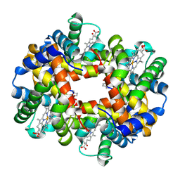 | | Crystal structure of deoxygenated hemoglobin in complex with 5HMF-NO at 1.8 Angstrom | | 分子名称: | Hemoglobin subunit alpha, Hemoglobin subunit beta, PROTOPORPHYRIN IX CONTAINING FE, ... | | 著者 | Donkor, A.K, Musayev, F.N, Safo, M.K. | | 登録日 | 2022-03-18 | | 公開日 | 2022-03-30 | | 最終更新日 | 2024-11-06 | | 実験手法 | X-RAY DIFFRACTION (1.8 Å) | | 主引用文献 | Design, Synthesis, and Antisickling Investigation of a Nitric Oxide-Releasing Prodrug of 5HMF for the Treatment of Sickle Cell Disease.
Biomolecules, 12, 2022
|
|
7UD8
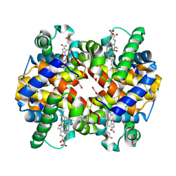 | | Crystal structure of carbon monoxy Hemoglobin in complex with 5HMF at 1.8 Angstrom | | 分子名称: | (5-methylfuran-2-yl)methanol, Hemoglobin subunit alpha, Hemoglobin subunit beta, ... | | 著者 | Donkor, A.K, Musayev, F.N, Safo, M.S. | | 登録日 | 2022-03-18 | | 公開日 | 2022-03-30 | | 最終更新日 | 2024-11-20 | | 実験手法 | X-RAY DIFFRACTION (1.8 Å) | | 主引用文献 | Design, Synthesis, and Antisickling Investigation of a Nitric Oxide-Releasing Prodrug of 5HMF for the Treatment of Sickle Cell Disease.
Biomolecules, 12, 2022
|
|
4JBA
 | |
6G76
 | |
6G78
 | |


