1GKG
 
 | | Structure Determination and Rational Mutagenesis reveal binding surface of immune adherence receptor, CR1 (CD35) | | 分子名称: | COMPLEMENT RECEPTOR TYPE 1 | | 著者 | Smith, B.O, Mallin, R.L, Krych-Goldberg, M, Wang, X, Hauhart, R.E, Bromek, K, Uhrin, D, Atkinson, J.P, Barlow, P.N. | | 登録日 | 2001-08-14 | | 公開日 | 2002-04-18 | | 最終更新日 | 2011-07-13 | | 実験手法 | SOLUTION NMR | | 主引用文献 | Structure of the C3B Binding Site of Cr1 (Cd35), the Immune Adherence Receptor
Cell(Cambridge,Mass.), 108, 2002
|
|
4EQ3
 | | Crystal Structure Analysis of Selenomethionine (Se-Met) Substituted Chicken Interferon Gamma Receptor Alpha Chain | | 分子名称: | Interferon gamma receptor 1 | | 著者 | Ping, Z, Qi, J, Lu, G, Shi, Y, Wang, X, Gao, G.F, Wang, M. | | 登録日 | 2012-04-18 | | 公開日 | 2013-04-24 | | 最終更新日 | 2014-05-21 | | 実験手法 | X-RAY DIFFRACTION (2.001 Å) | | 主引用文献 | Crystal structure of the interferon gamma receptor alpha chain from chicken reveals an undetected extra helix compared with the human counterparts.
J.Interferon Cytokine Res., 34, 2014
|
|
2ACV
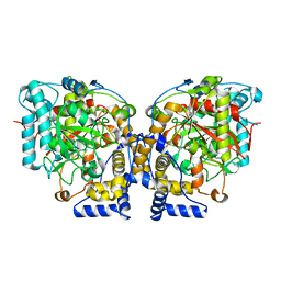
 | | Crystal Structure of Medicago truncatula UGT71G1 | | 分子名称: | URIDINE-5'-DIPHOSPHATE, triterpene UDP-glucosyl transferase UGT71G1 | | 著者 | Shao, H, He, X, Achnine, L, Blount, J.W, Dixon, R.A, Wang, X. | | 登録日 | 2005-07-19 | | 公開日 | 2005-11-15 | | 最終更新日 | 2024-02-14 | | 実験手法 | X-RAY DIFFRACTION (2 Å) | | 主引用文献 | Crystal Structures of a Multifunctional Triterpene/Flavonoid Glycosyltransferase from Medicago truncatula.
Plant Cell, 17, 2005
|
|
2ACW
 | | Crystal Structure of Medicago truncatula UGT71G1 complexed with UDP-glucose | | 分子名称: | URIDINE-5'-DIPHOSPHATE-GLUCOSE, triterpene UDP-glucosyl transferase UGT71G1 | | 著者 | Shao, H, He, X, Achnine, L, Blount, J.W, Dixon, R.A, Wang, X. | | 登録日 | 2005-07-19 | | 公開日 | 2005-11-15 | | 最終更新日 | 2024-02-14 | | 実験手法 | X-RAY DIFFRACTION (2.6 Å) | | 主引用文献 | Crystal Structures of a Multifunctional Triterpene/Flavonoid Glycosyltransferase from Medicago truncatula.
Plant Cell, 17, 2005
|
|
3V32
 | | Crystal structure of MCPIP1 N-terminal conserved domain | | 分子名称: | Ribonuclease ZC3H12A | | 著者 | Xu, J, Peng, W, Sun, Y, Wang, X, Xu, Y, Li, X, Gao, G, Rao, Z. | | 登録日 | 2011-12-12 | | 公開日 | 2012-05-23 | | 最終更新日 | 2024-03-20 | | 実験手法 | X-RAY DIFFRACTION (2 Å) | | 主引用文献 | Structural study of MCPIP1 N-terminal conserved domain reveals a PIN-like RNase
Nucleic Acids Res., 40, 2012
|
|
5H4R
 | | the complex of Glycoside Hydrolase 5 Lichenase from Caldicellulosiruptor sp. F32 E188Q mutant and cellotetraose | | 分子名称: | Beta-1,3-1,4-glucanase, GLYCEROL, beta-D-glucopyranose-(1-4)-beta-D-glucopyranose-(1-4)-beta-D-glucopyranose-(1-4)-beta-D-glucopyranose | | 著者 | Dong, S, Zhou, H, Liu, X, Wang, X, Feng, Y. | | 登録日 | 2016-11-02 | | 公開日 | 2017-09-13 | | 最終更新日 | 2023-11-08 | | 実験手法 | X-RAY DIFFRACTION (1.70310438 Å) | | 主引用文献 | Structural insights into the substrate specificity of a glycoside hydrolase family 5 lichenase from Caldicellulosiruptor sp. F32
Biochem. J., 474, 2017
|
|
3V34
 | | Crystal structure of MCPIP1 conserved domain with magnesium ion in the catalytic center | | 分子名称: | MAGNESIUM ION, Ribonuclease ZC3H12A | | 著者 | Xu, J, Peng, W, Sun, Y, Wang, X, Xu, Y, Li, X, Gao, G, Rao, Z. | | 登録日 | 2011-12-12 | | 公開日 | 2012-05-23 | | 最終更新日 | 2024-03-20 | | 実験手法 | X-RAY DIFFRACTION (2.003 Å) | | 主引用文献 | Structural study of MCPIP1 N-terminal conserved domain reveals a PIN-like RNase
Nucleic Acids Res., 40, 2012
|
|
3V33
 | | Crystal structure of MCPIP1 conserved domain with zinc-finger motif | | 分子名称: | Ribonuclease ZC3H12A | | 著者 | Xu, J, Peng, W, Sun, Y, Wang, X, Xu, Y, Li, X, Gao, G, Rao, Z. | | 登録日 | 2011-12-12 | | 公開日 | 2012-05-23 | | 最終更新日 | 2024-03-20 | | 実験手法 | X-RAY DIFFRACTION (2.005 Å) | | 主引用文献 | Structural study of MCPIP1 N-terminal conserved domain reveals a PIN-like RNase
Nucleic Acids Res., 40, 2012
|
|
4JGZ
 | | Crystal structure of human coxsackievirus A16 uncoating intermediate (space group I222) | | 分子名称: | Polyprotein, capsid protein VP1, capsid protein VP2, ... | | 著者 | Ren, J, Wang, X, Hu, Z, Gao, Q, Sun, Y, Li, X, Porta, C, Walter, T.S, Gilbert, R.J, Zhao, Y, Axford, D, Williams, M, McAuley, K, Rowlands, D.J, Yin, W, Wang, J, Stuart, D.I, Rao, Z, Fry, E.E. | | 登録日 | 2013-03-04 | | 公開日 | 2013-06-05 | | 最終更新日 | 2023-09-20 | | 実験手法 | X-RAY DIFFRACTION (3 Å) | | 主引用文献 | Picornavirus uncoating intermediate captured in atomic detail.
Nat Commun, 4, 2013
|
|
4JGY
 | | Crystal structure of human coxsackievirus A16 uncoating intermediate (space group P4232) | | 分子名称: | Polyprotein, capsid protein VP1, capsid protein VP2, ... | | 著者 | Ren, J, Wang, X, Hu, Z, Gao, Q, Sun, Y, Li, X, Porta, C, Walter, T.S, Gilbert, R.J, Zhao, Y, Axford, D, Williams, M, Mcauley, K, Rowlands, D.J, Yin, W, Wang, J, Stuart, D.I, Rao, Z, Fry, E.E. | | 登録日 | 2013-03-04 | | 公開日 | 2013-06-05 | | 最終更新日 | 2023-09-20 | | 実験手法 | X-RAY DIFFRACTION (3 Å) | | 主引用文献 | Picornavirus uncoating intermediate captured in atomic detail.
Nat Commun, 4, 2013
|
|
6KG3
 | |
6KH2
 | |
8GXS
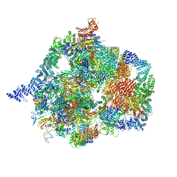
 | | PIC-Mediator in complex with +1 nucleosome (T40N) in H-binding state | | 分子名称: | CDK-activating kinase assembly factor MAT1, Cyclin-H, Cyclin-dependent kinase 7, ... | | 著者 | Chen, X, Wang, X, Liu, W, Ren, Y, Qu, X, Li, J, Yin, X. | | 登録日 | 2022-09-21 | | 公開日 | 2022-11-02 | | 実験手法 | ELECTRON MICROSCOPY (4.16 Å) | | 主引用文献 | Structures of +1 nucleosome-bound PIC-Mediator complex.
Science, 378, 2022
|
|
8GXQ
 | | PIC-Mediator in complex with +1 nucleosome (T40N) in MH-binding state | | 分子名称: | CDK-activating kinase assembly factor MAT1, Cyclin-H, Cyclin-dependent kinase 7, ... | | 著者 | Chen, X, Wang, X, Liu, W, Ren, Y, Qu, X, Li, J, Yin, X, Xu, Y. | | 登録日 | 2022-09-21 | | 公開日 | 2022-11-02 | | 実験手法 | ELECTRON MICROSCOPY (5.04 Å) | | 主引用文献 | Structures of +1 nucleosome-bound PIC-Mediator complex.
Science, 378, 2022
|
|
6DHV
 | | Structure of Arabidopsis Fatty Acid Amide Hydrolase | | 分子名称: | Fatty acid amide hydrolase | | 著者 | Aziz, M, Wang, X, Tripathi, A, Bankaitis, V, Chapman, K.D. | | 登録日 | 2018-05-21 | | 公開日 | 2019-03-27 | | 最終更新日 | 2023-10-11 | | 実験手法 | X-RAY DIFFRACTION (2.099 Å) | | 主引用文献 | Structural analysis of a plant fatty acid amide hydrolase provides insights into the evolutionary diversity of bioactive acylethanolamides.
J.Biol.Chem., 294, 2019
|
|
2RO1
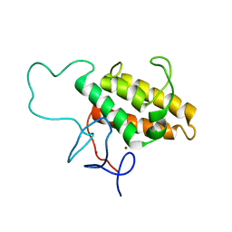 | | NMR Solution Structures of Human KAP1 PHD finger-bromodomain | | 分子名称: | Transcription intermediary factor 1-beta, ZINC ION | | 著者 | Zeng, L, Yap, K.L, Ivanov, A.V, Wang, X, Mujtaba, S, Plotnikova, O, Rauscher, F.J. | | 登録日 | 2008-03-04 | | 公開日 | 2008-05-20 | | 最終更新日 | 2024-05-29 | | 実験手法 | SOLUTION NMR | | 主引用文献 | Structural insights into human KAP1 PHD finger-bromodomain and its role in gene silencing
Nat.Struct.Mol.Biol., 15, 2008
|
|
7YCX
 | | The structure of INTAC-PEC complex | | 分子名称: | DNA-directed RNA polymerase II subunit E, DNA-directed RNA polymerase II subunit F, DNA-directed RNA polymerase II subunit RPB1,DNA-directed RNA polymerase II subunit RPB1, ... | | 著者 | Zheng, H, Jin, Q, Wang, X, Qi, Y, Liu, W, Ren, Y, Zhao, D, Chen, F.X, Cheng, J, Chen, X, Xu, Y. | | 登録日 | 2022-07-02 | | 公開日 | 2023-03-15 | | 最終更新日 | 2023-09-27 | | 実験手法 | ELECTRON MICROSCOPY (4.18 Å) | | 主引用文献 | Structural basis of INTAC-regulated transcription.
Protein Cell, 14, 2023
|
|
1B8W
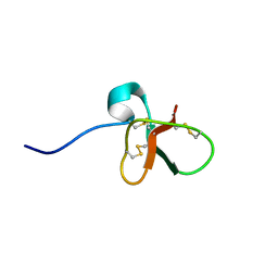 | | DEFENSIN-LIKE PEPTIDE 1 | | 分子名称: | PROTEIN (DEFENSIN-LIKE PEPTIDE 1) | | 著者 | Torres, A.M, Wang, X, Fletcher, J.I, Alewood, D, Alewood, P.F, Smith, R, Simpson, R.J, Nicholson, G.M, Sutherland, S.K, Gallagher, C.H, King, G.F, Kuchel, P.W. | | 登録日 | 1999-02-02 | | 公開日 | 1999-09-15 | | 最終更新日 | 2023-12-27 | | 実験手法 | SOLUTION NMR | | 主引用文献 | Solution structure of a defensin-like peptide from platypus venom.
Biochem.J., 341, 1999
|
|
9FT0
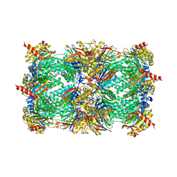 | | Yeast 20S proteasome in complex with epoxyketone inhibitor 42 | | 分子名称: | 2-(N-MORPHOLINO)-ETHANESULFONIC ACID, 3-PYRIDIN-4-YL-2,4-DIHYDRO-INDENO[1,2-.C.]PYRAZOLE, CHLORIDE ION, ... | | 著者 | Maurits, E, Huber, E.M, Dekker, P.M, Wang, X, Heinemeyer, W, Florea, B.I, Groll, M, Overkleeft, H.S. | | 登録日 | 2024-06-23 | | 公開日 | 2024-07-10 | | 実験手法 | X-RAY DIFFRACTION (2.75 Å) | | 主引用文献 | Structure-based design of peptide epoxyketones selectively targeting the three human immunoproteasome active sites
to be published
|
|
9FT1
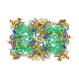 | | Yeast 20S proteasome in complex with epoxyketone inhibitor 13 | | 分子名称: | 2-(N-MORPHOLINO)-ETHANESULFONIC ACID, 3-PYRIDIN-4-YL-2,4-DIHYDRO-INDENO[1,2-.C.]PYRAZOLE, MAGNESIUM ION, ... | | 著者 | Maurits, E, Huber, E.M, Dekker, P.M, Wang, X, Heinemeyer, W, Florea, B.I, Groll, M, Overkleeft, H.S. | | 登録日 | 2024-06-23 | | 公開日 | 2024-07-10 | | 実験手法 | X-RAY DIFFRACTION (2.6 Å) | | 主引用文献 | Structure-based design of peptide epoxyketones selectively targeting the three human immunoproteasome active sites
to be published
|
|
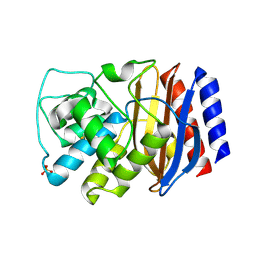
1BK9
 | | PHOSPHOLIPASE A2 MODIFIED BY PBPB | | 分子名称: | 1,4-BUTANEDIOL, CALCIUM ION, PHOSPHOLIPASE A2, ... | | 著者 | Zhao, H, Tang, L, Wang, X, Lin, Z, Zhou, Y. | | 登録日 | 1998-07-16 | | 公開日 | 1999-03-02 | | 最終更新日 | 2023-08-02 | | 実験手法 | X-RAY DIFFRACTION (2 Å) | | 主引用文献 | Structure of a snake venom phospholipase A2 modified by p-bromo-phenacyl-bromide.
Toxicon, 36, 1998
|
|
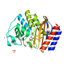
3CMZ
 | | TEM-1 Class-A beta-lactamase L201P mutant apo structure | | 分子名称: | Beta-lactamase TEM, PHOSPHATE ION | | 著者 | Marciano, D.C, Wang, X, Wang, J, Chen, Y, Thomas, V.L, Shoichet, B.K, Palzkill, T. | | 登録日 | 2008-03-24 | | 公開日 | 2008-11-25 | | 最終更新日 | 2023-08-30 | | 実験手法 | X-RAY DIFFRACTION (1.92 Å) | | 主引用文献 | Genetic and structural characterization of an L201P global suppressor substitution in TEM-1 beta-lactamase
J.Mol.Biol., 384, 2008
|
|
1M40
 | | ULTRA HIGH RESOLUTION CRYSTAL STRUCTURE OF TEM-1 | | 分子名称: | BETA-LACTAMASE TEM, PHOSPHATE ION, PINACOL[[2-AMINO-ALPHA-(1-CARBOXY-1-METHYLETHOXYIMINO)-4-THIAZOLEACETYL]AMINO]METHANEBORONATE, ... | | 著者 | Minasov, G, Wang, X, Shoichet, B.K. | | 登録日 | 2002-07-01 | | 公開日 | 2002-07-17 | | 最終更新日 | 2021-10-27 | | 実験手法 | X-RAY DIFFRACTION (0.85 Å) | | 主引用文献 | An ultrahigh resolution structure of TEM-1 beta-lactamase suggests a role for Glu166 as the general base in acylation.
J.Am.Chem.Soc., 124, 2002
|
|
7RST
 | | The Crystal Structure of Recombinant Chloroperoxidase Expressed in Aspergillus niger | | 分子名称: | 2-acetamido-2-deoxy-beta-D-glucopyranose, 2-acetamido-2-deoxy-beta-D-glucopyranose-(1-4)-2-acetamido-2-deoxy-beta-D-glucopyranose, Chloroperoxidase, ... | | 著者 | Tang, X, Venkadesh, S, Zhou, J, Rosen, B, Wang, X. | | 登録日 | 2021-08-11 | | 公開日 | 2022-08-24 | | 最終更新日 | 2023-10-18 | | 実験手法 | X-RAY DIFFRACTION (1.69 Å) | | 主引用文献 | The Crystal Structure of Recombinant Chloroperoxidase Expressed in Aspergillus niger
To Be Published
|
|
1T5K
 | | Crystal structure of amicyanin substituted with cobalt | | 分子名称: | Amicyanin, COBALT (II) ION, PHOSPHATE ION | | 著者 | Carrell, C.J, Wang, X, Jones, L, Jarrett, W.L, Davidson, V.L, Mathews, F.S. | | 登録日 | 2004-05-04 | | 公開日 | 2004-07-27 | | 最終更新日 | 2024-02-14 | | 実験手法 | X-RAY DIFFRACTION (1.4 Å) | | 主引用文献 | Crystallographic and NMR Investigation of Cobalt-Substituted Amicyanin.
Biochemistry, 43, 2004
|
|


