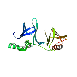
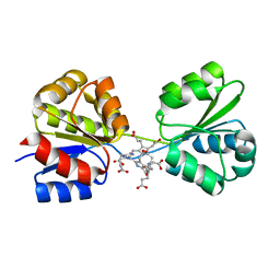
2ECS
 
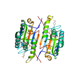
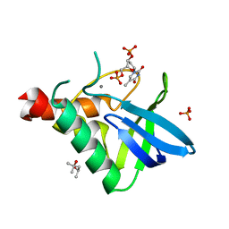
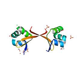 | | Lambda Cro mutant Q27P/A29S/K32Q at 1.4 A in space group C2 | | 分子名称: | ACETATE ION, CHLORIDE ION, LITHIUM ION, ... | | 著者 | Hall, B.M, Roberts, S.A, Cordes, M.H. | | 登録日 | 2007-02-14 | | 公開日 | 2008-01-08 | | 最終更新日 | 2024-04-03 | | 実験手法 | X-RAY DIFFRACTION (1.4 Å) | | 主引用文献 | Two structures of a lambda Cro variant highlight dimer flexibility but disfavor major dimer distortions upon specific binding of cognate DNA.
J.Mol.Biol., 375, 2008
|
|
1RE1
 | |
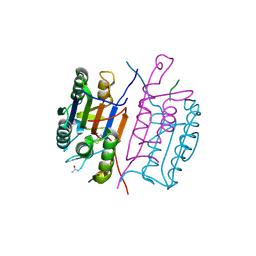
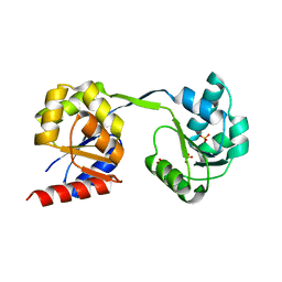
2GCL
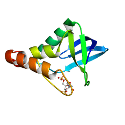
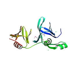 | | Structure of the Pob3 Middle domain | | 分子名称: | CHLORIDE ION, Hypothetical 63.0 kDa protein in DAK1-ORC1 intergenic region | | 著者 | VanDemark, A.P. | | 登録日 | 2006-03-14 | | 公開日 | 2006-05-23 | | 最終更新日 | 2021-10-20 | | 実験手法 | X-RAY DIFFRACTION (2.21 Å) | | 主引用文献 | The Structure of the yFACT Pob3-M Domain, Its Interaction with the DNA Replication Factor RPA, and a Potential Role in Nucleosome Deposition.
Mol.Cell, 22, 2006
|
|
1RHQ
 | |
1RHM
 | |
1RHK
 | |
1RHR
 | |
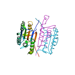
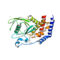
1OMO
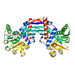 | | alanine dehydrogenase dimer w/bound NAD (archaeal) | | 分子名称: | NICOTINAMIDE-ADENINE-DINUCLEOTIDE, SODIUM ION, alanine dehydrogenase | | 著者 | Gallagher, D.T, Smith, N.N, Holden, M.J, Schroeder, I, Monbouquette, H.G. | | 登録日 | 2003-02-25 | | 公開日 | 2004-07-06 | | 最終更新日 | 2024-02-14 | | 実験手法 | X-RAY DIFFRACTION (2.32 Å) | | 主引用文献 | Structure of alanine dehydrogenase from Archaeoglobus: active site analysis and relation to bacterial cyclodeaminases and mammalian mu crystallin.
J.Mol.Biol., 342, 2004
|
|
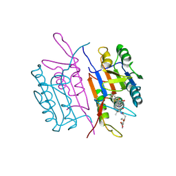
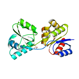
2GCJ
 | | Crystal Structure of the Pob3 middle domain | | 分子名称: | Hypothetical 63.0 kDa protein in DAK1-ORC1 intergenic region | | 著者 | VanDemark, A.P. | | 登録日 | 2006-03-14 | | 公開日 | 2006-05-23 | | 最終更新日 | 2024-04-03 | | 実験手法 | X-RAY DIFFRACTION (2.55 Å) | | 主引用文献 | The Structure of the yFACT Pob3-M Domain, Its Interaction with the DNA Replication Factor RPA, and a Potential Role in Nucleosome Deposition.
Mol.Cell, 22, 2006
|
|
4MLA
 | | Structure of maize cytokinin oxidase/dehydrogenase 2 (ZmCKO2) | | 分子名称: | 1,2-ETHANEDIOL, Cytokinin oxidase 2, FLAVIN-ADENINE DINUCLEOTIDE, ... | | 著者 | Morera, S, Kopecny, D, Briozzo, P, Koncitikova, R. | | 登録日 | 2013-09-06 | | 公開日 | 2015-03-11 | | 最終更新日 | 2016-03-23 | | 実験手法 | X-RAY DIFFRACTION (2.04 Å) | | 主引用文献 | Kinetic and structural investigation of the cytokinin oxidase/dehydrogenase active site.
Febs J., 283, 2016
|
|
4ML8
 | | Structure of maize cytokinin oxidase/dehydrogenase 2 (ZmCKO2) | | 分子名称: | Cytokinin oxidase 2, DI(HYDROXYETHYL)ETHER, FLAVIN-ADENINE DINUCLEOTIDE | | 著者 | Morera, S, Kopecny, D, Briozzo, P, Koncitikova, R. | | 登録日 | 2013-09-06 | | 公開日 | 2015-03-11 | | 最終更新日 | 2016-03-23 | | 実験手法 | X-RAY DIFFRACTION (2.7 Å) | | 主引用文献 | Kinetic and structural investigation of the cytokinin oxidase/dehydrogenase active site.
Febs J., 283, 2016
|
|
3NPQ
 | |
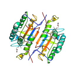
4R8N
 | | Crystal structure of Staphylococcal nuclease variant V23I/V66I/I72V/I92V at cryogenic temperature | | 分子名称: | (4R)-2-METHYLPENTANE-2,4-DIOL, CALCIUM ION, PHOSPHATE ION, ... | | 著者 | Caro, J.A, Flores, E, Schlessman, J.L, Heroux, A, Garcia-Moreno, E.B. | | 登録日 | 2014-09-02 | | 公開日 | 2014-09-17 | | 最終更新日 | 2023-09-20 | | 実験手法 | X-RAY DIFFRACTION (1.65 Å) | | 主引用文献 | Cavities in proteins
To be Published
|
|
3PL6
 | |
4S3S
 | | Crystal structure of Staphylococcal nuclease variant Delta+PHS I92K/V23A at cryogenic temperature | | 分子名称: | CALCIUM ION, THYMIDINE-3',5'-DIPHOSPHATE, Thermonuclease | | 著者 | Caro, J.A, Sue, G, Schlessman, J.L, Heroux, A, Garcia-Moreno E, B. | | 登録日 | 2015-06-19 | | 公開日 | 2015-07-01 | | 最終更新日 | 2023-09-20 | | 実験手法 | X-RAY DIFFRACTION (1.64 Å) | | 主引用文献 | Buried ionizable residues
To be Published
|
|
3CWE
 | | PTP1B in complex with a phosphonic acid inhibitor | | 分子名称: | MAGNESIUM ION, Tyrosine-protein phosphatase non-receptor type 1, [{2-bromo-4-[(2R)-3-oxo-2,3-diphenylpropyl]phenyl}(difluoro)methyl]phosphonic acid | | 著者 | Scapin, G, Han, Y, Kennedy, B.P. | | 登録日 | 2008-04-21 | | 公開日 | 2008-06-10 | | 最終更新日 | 2024-02-21 | | 実験手法 | X-RAY DIFFRACTION (1.6 Å) | | 主引用文献 | Discovery of [(3-bromo-7-cyano-2-naphthyl)(difluoro)methyl]phosphonic acid, a potent and orally active small molecule PTP1B inhibitor
Bioorg.Med.Chem.Lett., 18, 2008
|
|
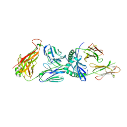
3OSS
 | | The crystal structure of enterotoxigenic Escherichia coli GspC-GspD complex from the type II secretion system | | 分子名称: | CALCIUM ION, CHLORIDE ION, TYPE 2 SECRETION SYSTEM, ... | | 著者 | Korotkov, K.V, Pruneda, J, Hol, W.G.J. | | 登録日 | 2010-09-09 | | 公開日 | 2011-08-03 | | 最終更新日 | 2023-09-06 | | 実験手法 | X-RAY DIFFRACTION (2.63 Å) | | 主引用文献 | Structural and functional studies on the interaction of GspC and GspD in the type II secretion system.
Plos Pathog., 7, 2011
|
|
5UMU
 | | Crystal structure of the middle double PH domain of human FACT complex subunit SPT16 | | 分子名称: | ACETATE ION, FACT complex subunit SPT16, FORMIC ACID | | 著者 | Hu, Q, Thompson, J.R, Heroux, A, Su, D, Botuyan, M.V, Mer, G. | | 登録日 | 2017-01-29 | | 公開日 | 2018-01-31 | | 最終更新日 | 2019-12-04 | | 実験手法 | X-RAY DIFFRACTION (1.903 Å) | | 主引用文献 | Crystal structure of the middle double PH domain of human FACT complex subunit SPT16
To Be Published
|
|
5UMR
 | | Crystal structure of N-terminal domain of human FACT complex subunit SSRP1 | | 分子名称: | FACT complex subunit SSRP1 | | 著者 | Su, D, Hu, Q, Thompson, J.R, Heroux, A, Botuyan, M.V, Mer, G. | | 登録日 | 2017-01-29 | | 公開日 | 2018-01-31 | | 最終更新日 | 2019-12-04 | | 実験手法 | X-RAY DIFFRACTION (1.501 Å) | | 主引用文献 | Crystal structure of N-terminal domain of human FACT complex subunit SSRP1
To Be Published
|
|
3NPN
 | |
3D8N
 | | Uroporphyrinogen III Synthase-Uroporphyringen III Complex | | 分子名称: | 3,3',3'',3'''-[3,8,13,17-tetrakis(carboxymethyl)porphyrin-2,7,12,18-tetrayl]tetrapropanoic acid, Uroporphyrinogen-III synthase | | 著者 | Schubert, H.L. | | 登録日 | 2008-05-23 | | 公開日 | 2008-08-12 | | 最終更新日 | 2024-02-21 | | 実験手法 | X-RAY DIFFRACTION (1.9 Å) | | 主引用文献 | Structure and mechanistic implications of a uroporphyrinogen III synthase-product complex.
Biochemistry, 47, 2008
|
|
3D8R
 | |
3D8S
 | |
3D8T
 | |
3RCP
 | |


