3TSL
 
 | |
2FGO
 | |
2G4R
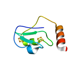
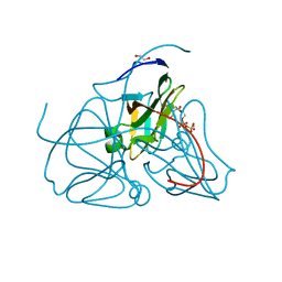
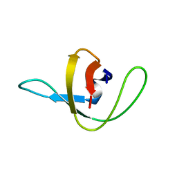
 | | anomalous substructure of MogA | | 分子名称: | 2-AMINO-2-HYDROXYMETHYL-PROPANE-1,3-DIOL, CHLORIDE ION, molybdopterin biosynthesis Mog protein | | 著者 | Mueller-Dieckmann, C, Weiss, M.S. | | 登録日 | 2006-02-22 | | 公開日 | 2007-02-20 | | 最終更新日 | 2024-02-14 | | 実験手法 | X-RAY DIFFRACTION (1.92 Å) | | 主引用文献 | On the routine use of soft X-rays in macromolecular crystallography. Part IV. Efficient determination of anomalous substructures in biomacromolecules using longer X-ray wavelengths.
Acta Crystallogr.,Sect.D, 63, 2007
|
|
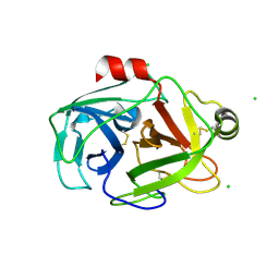
2G4V
 | | anomalous substructure of proteinase K | | 分子名称: | CALCIUM ION, CHLORIDE ION, POTASSIUM ION, ... | | 著者 | Mueller-Dieckmann, C, Weiss, M.S. | | 登録日 | 2006-02-22 | | 公開日 | 2007-02-20 | | 最終更新日 | 2011-07-13 | | 実験手法 | X-RAY DIFFRACTION (2.14 Å) | | 主引用文献 | On the routine use of soft X-rays in macromolecular crystallography. Part IV. Efficient determination of anomalous substructures in biomacromolecules using longer X-ray wavelengths.
Acta Crystallogr.,Sect.D, 63, 2007
|
|
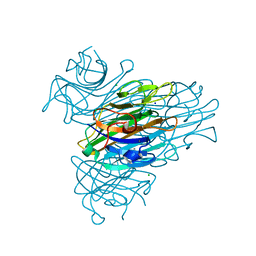
2G51
 | | anomalous substructure of trypsin (p1) | | 分子名称: | CHLORIDE ION, Trypsin | | 著者 | Mueller-Dieckmann, C, Weiss, M.S. | | 登録日 | 2006-02-22 | | 公開日 | 2007-02-20 | | 最終更新日 | 2011-07-13 | | 実験手法 | X-RAY DIFFRACTION (1.84 Å) | | 主引用文献 | On the routine use of soft X-rays in macromolecular crystallography. Part IV. Efficient determination of anomalous substructures in biomacromolecules using longer X-ray wavelengths.
Acta Crystallogr.,Sect.D, 63, 2007
|
|
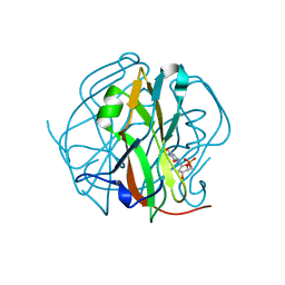
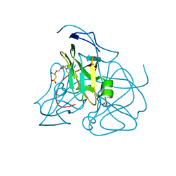
2G4I
 | | Anomalous substructure of Concanavalin A | | 分子名称: | CALCIUM ION, CHLORIDE ION, Concanavalin A, ... | | 著者 | Mueller-Dieckmann, C, Weiss, M.S. | | 登録日 | 2006-02-22 | | 公開日 | 2007-02-20 | | 最終更新日 | 2024-02-14 | | 実験手法 | X-RAY DIFFRACTION (2.4 Å) | | 主引用文献 | On the routine use of soft X-rays in macromolecular crystallography. Part IV. Efficient determination of anomalous substructures in biomacromolecules using longer X-ray wavelengths.
Acta Crystallogr.,Sect.D, 63, 2007
|
|
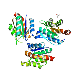
2XZS
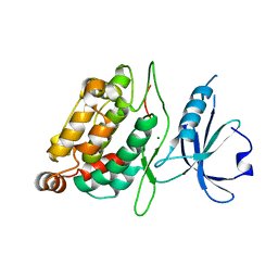 | | Death associated protein kinase 1 residues 1-312 | | 分子名称: | DEATH ASSOCIATED KINASE 1, MAGNESIUM ION | | 著者 | Yumerefendi, H, Mas, P.J, Dordevic, N, McCarthy, A.A, Hart, D.J. | | 登録日 | 2010-11-29 | | 公開日 | 2011-12-07 | | 最終更新日 | 2023-12-20 | | 実験手法 | X-RAY DIFFRACTION (2 Å) | | 主引用文献 | Death-Associated Protein Kinase Activity is Regulated by Coupled Calcium/Calmodulin Binding to Two Distinct Sites.
Structure, 24, 2016
|
|
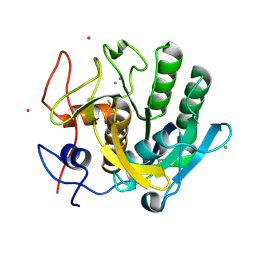
2HRM
 | | Crystal structure of dUTPase complexed with substrate analogue methylene-dUTP | | 分子名称: | 1,2-ETHANEDIOL, 2'-DEOXY-5'-O-[(S)-HYDROXY(PHOSPHONOMETHYL)PHOSPHORYL]URIDINE, Deoxyuridine 5'-triphosphate nucleotidohydrolase | | 著者 | Barabas, O, Kovari, J, Tapai, R, Vertessy, B.G. | | 登録日 | 2006-07-20 | | 公開日 | 2007-07-31 | | 最終更新日 | 2023-08-30 | | 実験手法 | X-RAY DIFFRACTION (1.7 Å) | | 主引用文献 | Methylene substitution at the alpha-beta bridging position within the phosphate chain of dUDP profoundly perturbs ligand accommodation into the dUTPase active site.
Proteins, 71, 2008
|
|
2HR6
 | | Crystal structure of dUTPase in complex with substrate analogue dUDP and manganese | | 分子名称: | 1,2-ETHANEDIOL, DEOXYURIDINE-5'-DIPHOSPHATE, Deoxyuridine 5'-triphosphate nucleotidohydrolase, ... | | 著者 | Barabas, O, Kovari, J, Tapai, R, Vertessy, B.G. | | 登録日 | 2006-07-19 | | 公開日 | 2007-07-31 | | 最終更新日 | 2023-08-30 | | 実験手法 | X-RAY DIFFRACTION (1.84 Å) | | 主引用文献 | Methylene substitution at the alpha-beta bridging position within the phosphate chain of dUDP profoundly perturbs ligand accommodation into the dUTPase active site.
Proteins, 71, 2008
|
|
1TUD
 | |


