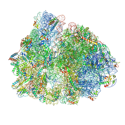
7PAO
 
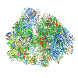
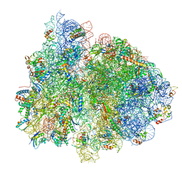
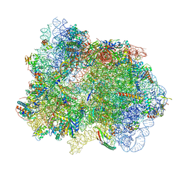 | | 70S ribosome with EF-G, A*- and P/E-site tRNAs in Mycoplasma pneumoniae cells | | 分子名称: | 16S ribosomal RNA, 23S ribosomal RNA, 30S ribosomal protein S10, ... | | 著者 | Xue, L, Lenz, S, Rappsilber, J, Mahamid, J. | | 登録日 | 2021-07-30 | | 公開日 | 2022-05-25 | | 最終更新日 | 2024-10-16 | | 実験手法 | ELECTRON MICROSCOPY (7 Å) | | 主引用文献 | Visualizing translation dynamics at atomic detail inside a bacterial cell.
Nature, 610, 2022
|
|
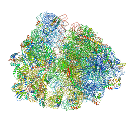
7PAQ
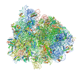
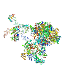
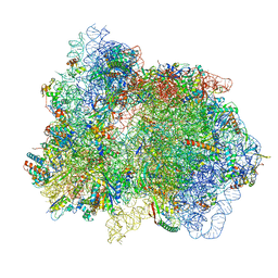 | | 70S ribosome with EF-G, A/P- and P/E-site tRNAs in Mycoplasma pneumoniae cells | | 分子名称: | 16S ribosomal RNA, 23S ribosomal RNA, 30S ribosomal protein S10, ... | | 著者 | Xue, L, Lenz, S, Rappsilber, J, Mahamid, J. | | 登録日 | 2021-07-30 | | 公開日 | 2022-05-25 | | 最終更新日 | 2024-10-23 | | 実験手法 | ELECTRON MICROSCOPY (8.9 Å) | | 主引用文献 | Visualizing translation dynamics at atomic detail inside a bacterial cell.
Nature, 610, 2022
|
|
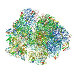
7PIR
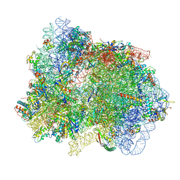
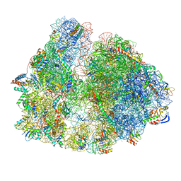
 | | 70S ribosome with A*- and P/E-site tRNAs in pseudouridimycin-treated Mycoplasma pneumoniae cells | | 分子名称: | 16S ribosomal RNA, 23S ribosomal RNA, 30S ribosomal protein S10, ... | | 著者 | Xue, L, Lenz, S, Rappsilber, J, Mahamid, J. | | 登録日 | 2021-08-23 | | 公開日 | 2022-05-25 | | 最終更新日 | 2024-10-16 | | 実験手法 | ELECTRON MICROSCOPY (12.1 Å) | | 主引用文献 | Visualizing translation dynamics at atomic detail inside a bacterial cell.
Nature, 610, 2022
|
|
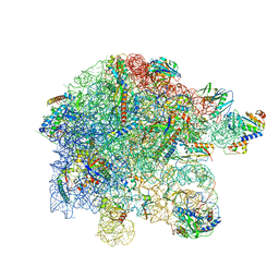
7PIT
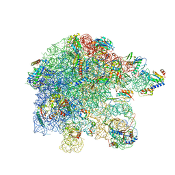
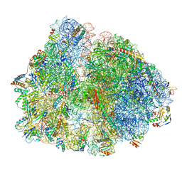
 | | 70S ribosome with EF-G, A/P- and P/E-site tRNAs in pseudouridimycin-treated Mycoplasma pneumoniae cells | | 分子名称: | 16S ribosomal RNA, 23S ribosomal RNA, 30S ribosomal protein S10, ... | | 著者 | Xue, L, Lenz, S, Rappsilber, J, Mahamid, J. | | 登録日 | 2021-08-23 | | 公開日 | 2022-05-25 | | 最終更新日 | 2024-11-06 | | 実験手法 | ELECTRON MICROSCOPY (5.7 Å) | | 主引用文献 | Visualizing translation dynamics at atomic detail inside a bacterial cell.
Nature, 610, 2022
|
|
7PAT
 | | free 50S in untreated Mycoplasma pneumoniae cells | | 分子名称: | 23S ribosomal RNA, 50S ribosomal protein L10, 50S ribosomal protein L11, ... | | 著者 | Xue, L, Lenz, S, Rappsilber, J, Mahamid, J. | | 登録日 | 2021-07-30 | | 公開日 | 2022-05-25 | | 最終更新日 | 2024-10-23 | | 実験手法 | ELECTRON MICROSCOPY (9.2 Å) | | 主引用文献 | Visualizing translation dynamics at atomic detail inside a bacterial cell.
Nature, 610, 2022
|
|
7PHC
 | | 70S ribosome with A*- and P/E-site tRNAs in chloramphenicol-treated Mycoplasma pneumoniae cells | | 分子名称: | 16S ribosomal RNA, 23S ribosomal RNA, 30S ribosomal protein S10, ... | | 著者 | Xue, L, Lenz, S, Rappsilber, J, Mahamid, J. | | 登録日 | 2021-08-16 | | 公開日 | 2022-05-25 | | 最終更新日 | 2024-10-23 | | 実験手法 | ELECTRON MICROSCOPY (9.9 Å) | | 主引用文献 | Visualizing translation dynamics at atomic detail inside a bacterial cell.
Nature, 610, 2022
|
|
7PAJ
 | | 70S ribosome with EF-Tu-tRNA, P- and E-site tRNAs in Mycoplasma pneumoniae cells | | 分子名称: | 16S ribosomal RNA, 23S ribosomal RNA, 30S ribosomal protein S10, ... | | 著者 | Xue, L, Lenz, S, Rappsilber, J, Mahamid, J. | | 登録日 | 2021-07-30 | | 公開日 | 2022-05-25 | | 最終更新日 | 2024-10-16 | | 実験手法 | ELECTRON MICROSCOPY (7.3 Å) | | 主引用文献 | Visualizing translation dynamics at atomic detail inside a bacterial cell.
Nature, 610, 2022
|
|
7PAS
 | | 70S ribosome with P/E-site tRNA in Mycoplasma pneumoniae cells | | 分子名称: | 16S ribosomal RNA, 23S ribosomal RNA, 30S ribosomal protein S10, ... | | 著者 | Xue, L, Lenz, S, Rappsilber, J, Mahamid, J. | | 登録日 | 2021-07-30 | | 公開日 | 2022-05-25 | | 最終更新日 | 2024-10-23 | | 実験手法 | ELECTRON MICROSCOPY (16 Å) | | 主引用文献 | Visualizing translation dynamics at atomic detail inside a bacterial cell.
Nature, 610, 2022
|
|
7PAM
 | | 70S ribosome with A*- and P/E-site tRNAs in Mycoplasma pneumoniae cells | | 分子名称: | 16S ribosomal RNA, 23S ribosomal RNA, 30S ribosomal protein S10, ... | | 著者 | Xue, L, Lenz, S, Rappsilber, J, Mahamid, J. | | 登録日 | 2021-07-30 | | 公開日 | 2022-05-25 | | 最終更新日 | 2024-11-06 | | 実験手法 | ELECTRON MICROSCOPY (6.8 Å) | | 主引用文献 | Visualizing translation dynamics at atomic detail inside a bacterial cell.
Nature, 610, 2022
|
|
7PIC
 | | 70S ribosome with P/E-site tRNA in spectinomycin-treated Mycoplasma pneumoniae cells | | 分子名称: | 16S ribosomal RNA, 23S ribosomal RNA, 30S ribosomal protein S10, ... | | 著者 | Xue, L, Lenz, S, Rappsilber, J, Mahamid, J. | | 登録日 | 2021-08-19 | | 公開日 | 2022-05-25 | | 最終更新日 | 2024-10-09 | | 実験手法 | ELECTRON MICROSCOPY (9.1 Å) | | 主引用文献 | Visualizing translation dynamics at atomic detail inside a bacterial cell.
Nature, 610, 2022
|
|
7PIA
 | | 70S ribosome with A/P- and P/E-site tRNAs in spectinomycin-treated Mycoplasma pneumoniae cells | | 分子名称: | 16S ribosomal RNA, 23S ribosomal RNA, 30S ribosomal protein S10, ... | | 著者 | Xue, L, Lenz, S, Rappsilber, J, Mahamid, J. | | 登録日 | 2021-08-19 | | 公開日 | 2022-05-25 | | 最終更新日 | 2024-11-13 | | 実験手法 | ELECTRON MICROSCOPY (13.6 Å) | | 主引用文献 | Visualizing translation dynamics at atomic detail inside a bacterial cell.
Nature, 610, 2022
|
|
7PIQ
 | | 70S ribosome with A- and P-site tRNAs in pseudouridimycin-treated Mycoplasma pneumoniae cells | | 分子名称: | 16S ribosomal RNA, 23S ribosomal RNA, 30S ribosomal protein S10, ... | | 著者 | Xue, L, Lenz, S, Rappsilber, J, Mahamid, J. | | 登録日 | 2021-08-23 | | 公開日 | 2022-05-25 | | 最終更新日 | 2024-11-06 | | 実験手法 | ELECTRON MICROSCOPY (9.7 Å) | | 主引用文献 | Visualizing translation dynamics at atomic detail inside a bacterial cell.
Nature, 610, 2022
|
|
7PAU
 | | free 50S in complex with ribosome recycling factor in untreated Mycoplasma pneumoniae cells | | 分子名称: | 23S ribosomal RNA, 50S ribosomal protein L10, 50S ribosomal protein L11, ... | | 著者 | Xue, L, Lenz, S, Rappsilber, J, Mahamid, J. | | 登録日 | 2021-07-30 | | 公開日 | 2022-05-25 | | 最終更新日 | 2024-11-13 | | 実験手法 | ELECTRON MICROSCOPY (8.3 Å) | | 主引用文献 | Visualizing translation dynamics at atomic detail inside a bacterial cell.
Nature, 610, 2022
|
|
7PHA
 | | 70S ribosome with EF-Tu-tRNA and P-site tRNA in chloramphenicol-treated Mycoplasma pneumoniae cells | | 分子名称: | 16S ribosomal RNA, 23S ribosomal RNA, 30S ribosomal protein S10, ... | | 著者 | Xue, L, Lenz, S, Rappsilber, J, Mahamid, J. | | 登録日 | 2021-08-16 | | 公開日 | 2022-05-25 | | 最終更新日 | 2024-10-16 | | 実験手法 | ELECTRON MICROSCOPY (8.5 Å) | | 主引用文献 | Visualizing translation dynamics at atomic detail inside a bacterial cell.
Nature, 610, 2022
|
|
9QEQ
 | | Structure of the transcribing Pol II-DSIF-SPT6-U1 snRNP complex | | 分子名称: | DNA-directed RNA polymerase II subunit E, DNA-directed RNA polymerase II subunit RPB11-a, DNA-directed RNA polymerase II subunit RPB3, ... | | 著者 | Zhang, S. | | 登録日 | 2025-03-10 | | 公開日 | 2025-07-09 | | 実験手法 | ELECTRON MICROSCOPY (3.5 Å) | | 主引用文献 | Structure of a transcribing Pol II-DSIF-SPT6-U1 snRNP complex
Nat Commun, 16, 2025
|
|
6X4H
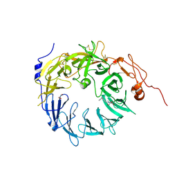 | | Sortilin-Progranulin Interaction With Compound 24 | | 分子名称: | 2-acetamido-2-deoxy-beta-D-glucopyranose-(1-4)-2-acetamido-2-deoxy-beta-D-glucopyranose, 4-methyl-N-(6-phenoxypyridine-3-carbonyl)-L-leucine, GLYCEROL, ... | | 著者 | Parthasarathy, G, Soisson, S.M, Klein, D. | | 登録日 | 2020-05-22 | | 公開日 | 2020-08-12 | | 最終更新日 | 2024-10-23 | | 実験手法 | X-RAY DIFFRACTION (2.9 Å) | | 主引用文献 | Identification of potent inhibitors of the sortilin-progranulin interaction.
Bioorg.Med.Chem.Lett., 30, 2020
|
|
6X3L
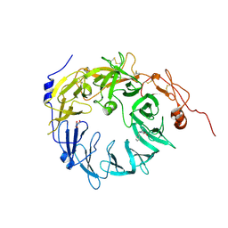 | | Sortilin-Progranulin Interaction With Compound 2 | | 分子名称: | 1-benzyl-3-tert-butyl-1H-pyrazole-5-carboxylic acid, 2-acetamido-2-deoxy-beta-D-glucopyranose-(1-4)-2-acetamido-2-deoxy-beta-D-glucopyranose, GLYCEROL, ... | | 著者 | Parthasarathy, G, Soisson, S.M. | | 登録日 | 2020-05-21 | | 公開日 | 2020-08-12 | | 最終更新日 | 2024-10-23 | | 実験手法 | X-RAY DIFFRACTION (2.7 Å) | | 主引用文献 | Identification of potent inhibitors of the sortilin-progranulin interaction.
Bioorg.Med.Chem.Lett., 30, 2020
|
|
6X48
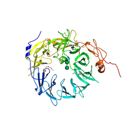 | | Sortilin-Progranulin Interaction With Compound 17 | | 分子名称: | GLYCEROL, N-(3,5-dichlorobenzene-1-carbonyl)-5,5-dimethyl-L-norleucine, Sortilin, ... | | 著者 | Parthasarathy, G, Soisson, S.M. | | 登録日 | 2020-05-22 | | 公開日 | 2020-08-12 | | 最終更新日 | 2024-10-09 | | 実験手法 | X-RAY DIFFRACTION (2.9 Å) | | 主引用文献 | Identification of potent inhibitors of the sortilin-progranulin interaction.
Bioorg.Med.Chem.Lett., 30, 2020
|
|
8TUA
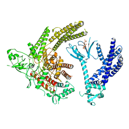 | | Full-length P-Rex1 in complex with inositol 1,3,4,5-tetrakisphosphate (IP4) | | 分子名称: | INOSITOL-(1,3,4,5)-TETRAKISPHOSPHATE, Phosphatidylinositol 3,4,5-trisphosphate-dependent Rac exchanger 1 protein | | 著者 | Cash, J.N, Tesmer, J.J.G. | | 登録日 | 2023-08-15 | | 公開日 | 2024-04-10 | | 実験手法 | ELECTRON MICROSCOPY (4.1 Å) | | 主引用文献 | Full-length P-Rex1 in complex with inositol 1,3,4,5-tetrakisphosphate (IP4)
Elife, 2024
|
|
3QQC
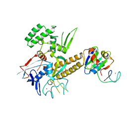 | |
2MOW
 | |
5MDU
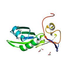 | | Structure of the RNA recognition motif (RRM) of Seb1 from S. pombe. | | 分子名称: | CHLORIDE ION, GLYCEROL, Rpb7-binding protein seb1, ... | | 著者 | Wittmann, S, Renner, M, El Omari, K, Adams, O, Vasiljeva, L, Grimes, J. | | 登録日 | 2016-11-13 | | 公開日 | 2017-04-12 | | 最終更新日 | 2024-05-08 | | 実験手法 | X-RAY DIFFRACTION (1.02 Å) | | 主引用文献 | The conserved protein Seb1 drives transcription termination by binding RNA polymerase II and nascent RNA.
Nat Commun, 8, 2017
|
|
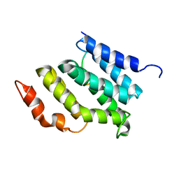
6RFG
 | |
5MDT
 | |
8C8H
 | |


