Loading
PDBj
メニューPDBj@FacebookPDBj@TwitterPDBj@YouTubewwPDB FoundationwwPDB
RCSB PDBPDBeBMRBAdv. SearchSearch help
Search by PDB author
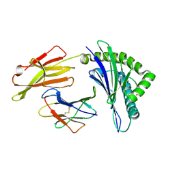
8XES
DownloadVisualize
BU of 8xes by Molmil
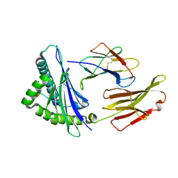
The structure of HLA-A/L1-1
分子名称: Beta-2-microglobulin, HLA class I heavy chain, Major capsid protein L1
著者Zhang, J.N, Yue, C, Liu, J.
登録日2023-12-12
公開日2024-07-10
最終更新日2024-10-16
実験手法X-RAY DIFFRACTION (1.78 Å)
主引用文献Uncommon P1 Anchor-featured Viral T Cell Epitope Preference within HLA-A*2601 and HLA-A*0101 Individuals.
Immunohorizons, 8, 2024
8XKE
DownloadVisualize
BU of 8xke by Molmil
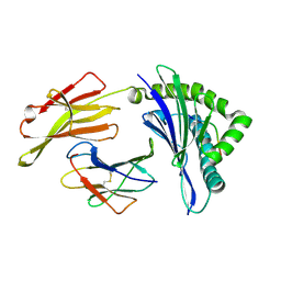
The structure of HLA-A/14-3-D
分子名称: Beta-2-microglobulin, GLU-VAL-ASP-ASN-ALA-THR-ARG-PHE-ALA-SER-VAL-TYR, HLA class I heavy chain
著者Zhang, J.N, Yue, C, Liu, J, Sun, Z.Y.
登録日2023-12-23
公開日2024-07-10
実験手法X-RAY DIFFRACTION (1.92 Å)
主引用文献Uncommon P1 Anchor-featured Viral T Cell Epitope Preference within HLA-A*2601 and HLA-A*0101 Individuals.
Immunohorizons, 8, 2024
8XFZ
DownloadVisualize
BU of 8xfz by Molmil
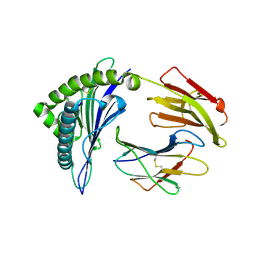
The structure of HLA-A/L1-2
分子名称: Beta-2-microglobulin, HLA class I heavy chain, Major capsid protein L1
著者Zhang, J.N, Yue, C, Liu, J.
登録日2023-12-14
公開日2024-07-10
実験手法X-RAY DIFFRACTION (2.32 Å)
主引用文献Uncommon P1 Anchor-featured Viral T Cell Epitope Preference within HLA-A*2601 and HLA-A*0101 Individuals.
Immunohorizons, 8, 2024
8XKC
DownloadVisualize
BU of 8xkc by Molmil
The structure of HLA-A/Pep16
分子名称: Beta-2-microglobulin, HLA class I heavy chain, Spike protein S1
著者Zhang, J.N, Yue, C, Liu, J.
登録日2023-12-23
公開日2024-07-10
実験手法X-RAY DIFFRACTION (2.18 Å)
主引用文献Uncommon P1 Anchor-featured Viral T Cell Epitope Preference within HLA-A*2601 and HLA-A*0101 Individuals.
Immunohorizons, 8, 2024
8D0A
DownloadVisualize
BU of 8d0a by Molmil
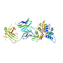
Crystal structure of human USP30 in complex with a covalent inhibitor 829 and a Fab
分子名称: Ubiquitin carboxyl-terminal hydrolase 30, ZINC ION, mouse anti-huUSP30 Fab heavy chain, ...
著者Song, X, Butler, J, Li, C, Zhang, K, Zhang, D, Hao, Y.
登録日2022-05-25
公開日2023-02-01
最終更新日2023-10-25
実験手法X-RAY DIFFRACTION (3.19 Å)
主引用文献TBD
To Be Published
1ZGK
DownloadVisualize
BU of 1zgk by Molmil
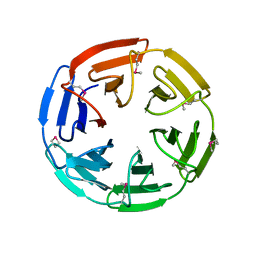
1.35 angstrom structure of the Kelch domain of Keap1
分子名称: Kelch-like ECH-associated protein 1
著者Li, X, Bottoms, C.A, Hannink, M, Beamer, L.J.
登録日2005-04-21
公開日2005-10-04
最終更新日2011-07-13
実験手法X-RAY DIFFRACTION (1.35 Å)
主引用文献Conserved solvent and side-chain interactions in the 1.35 Angstrom structure of the Kelch domain of Keap1.
Acta Crystallogr.,Sect.D, 61, 2005
3W94
DownloadVisualize
BU of 3w94 by Molmil
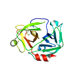
Structure of Oryzias latipes enteropeptidase light chain
分子名称: Enteropeptidase-1
著者Hu, S, Xu, J, Wang, H, Guo, Y.J.
登録日2013-03-26
公開日2014-02-19
最終更新日2024-10-30
実験手法X-RAY DIFFRACTION (2.002 Å)
主引用文献Structure basis for the unique specificity of medaka enteropeptidase light chain.
Protein Cell, 5, 2014
6Z6Y
DownloadVisualize
BU of 6z6y by Molmil
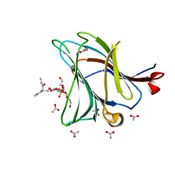
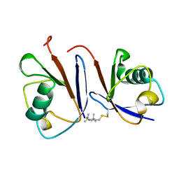
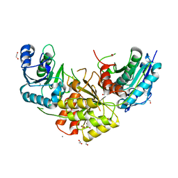
Galectin-8 N-terminal domain in complex with a sulfatide mimicking a sphingolipid
分子名称: ACETATE ION, Galectin-8, TRIETHYLENE GLYCOL, ...
著者Romero, A, Medrano, F.J.
登録日2020-05-29
公開日2020-12-30
最終更新日2024-01-24
実験手法X-RAY DIFFRACTION (1.34 Å)
主引用文献Probing sulfatide-tissue lectin recognition with functionalized glycodendrimersomes.
Iscience, 24, 2021
7VRD
DownloadVisualize
BU of 7vrd by Molmil
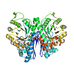
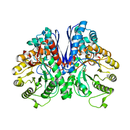
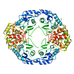
Crystal structure of Enolase1 from Candida albicans complexed with 2'-phosphoglyceric acid sodium
分子名称: 2-PHOSPHOGLYCERIC ACID, Enolase 1, MAGNESIUM ION
著者Zhang, M, Zhang, X.
登録日2021-10-22
公開日2022-07-06
最終更新日2023-11-29
実験手法X-RAY DIFFRACTION (1.7 Å)
主引用文献Baicalein Acts against Candida albicans by Targeting Eno1 and Inhibiting Glycolysis.
Microbiol Spectr, 10, 2022
7V67
DownloadVisualize
BU of 7v67 by Molmil
Crystal Structure of Enolase1 from Candida albicans
分子名称: Enolase 1, SULFATE ION
著者Zhang, M, Zhang, X.
登録日2021-08-19
公開日2022-07-06
最終更新日2023-11-29
実験手法X-RAY DIFFRACTION (2 Å)
主引用文献Baicalein Acts against Candida albicans by Targeting Eno1 and Inhibiting Glycolysis.
Microbiol Spectr, 10, 2022
2MRP
DownloadVisualize
BU of 2mrp by Molmil
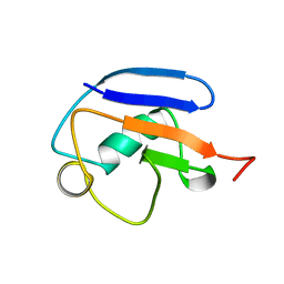
NMR solution structure of the Ubiquitin like domain (UBL) of DNA-damage-inducible 1 protein (Ddi1)
分子名称: DNA damage-inducible protein 1
著者Nowicka, U, Fushman, D, Chen, T.
登録日2014-07-14
公開日2015-03-11
最終更新日2024-05-01
実験手法SOLUTION NMR
主引用文献DNA-Damage-Inducible 1 Protein (Ddi1) Contains an Uncharacteristic Ubiquitin-like Domain that Binds Ubiquitin.
Structure, 23, 2015
2MWS
DownloadVisualize
BU of 2mws by Molmil
Structure of the complex of ubiquitin and the ubiquitin-like (UBL) domain of Ddi1
分子名称: DNA damage-inducible protein 1, Ubiquitin
著者Fushman, D, Nowicka, U, Walker, O.
登録日2014-11-23
公開日2015-03-25
最終更新日2023-06-14
実験手法SOLUTION NMR
主引用文献DNA-Damage-Inducible 1 Protein (Ddi1) Contains an Uncharacteristic Ubiquitin-like Domain that Binds Ubiquitin.
Structure, 23, 2015
3DS6
DownloadVisualize
BU of 3ds6 by Molmil
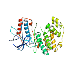
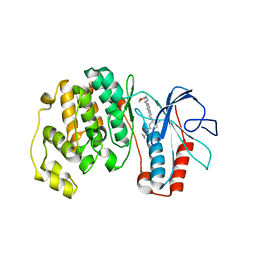
P38 complex with a phthalazine inhibitor
分子名称: Mitogen-activated protein kinase 14, N-cyclopropyl-4-methyl-3-[1-(2-methylphenyl)phthalazin-6-yl]benzamide
著者Herberich, B, Syed, R, Li, V, Grosfeld, D.
登録日2008-07-11
公開日2008-10-07
最終更新日2024-04-03
実験手法X-RAY DIFFRACTION (2.9 Å)
主引用文献Discovery of highly selective and potent p38 inhibitors based on a phthalazine scaffold.
J.Med.Chem., 51, 2008
3DT1
DownloadVisualize
BU of 3dt1 by Molmil
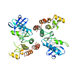
P38 Complexed with a quinazoline inhibitor
分子名称: Mitogen-activated protein kinase 14, N-cyclopropyl-4-methyl-3-{2-[(2-morpholin-4-ylethyl)amino]quinazolin-6-yl}benzamide
著者Herberich, B, Syed, R, Li, V, Tasker, A.S.
登録日2008-07-14
公開日2008-10-14
最終更新日2024-02-21
実験手法X-RAY DIFFRACTION (2.8 Å)
主引用文献Discovery of highly selective and potent p38 inhibitors based on a phthalazine scaffold.
J.Med.Chem., 51, 2008
5TX5
DownloadVisualize
BU of 5tx5 by Molmil
Rip1 Kinase ( flag 1-294, C34A, C127A, C233A, C240A) with GSK772
分子名称: 3-benzyl-N-[(3S)-5-methyl-4-oxo-2,3,4,5-tetrahydro-1,5-benzoxazepin-3-yl]-1H-1,2,4-triazole-5-carboxamide, Receptor-interacting serine/threonine-protein kinase 1
著者Campobasso, N, Ward, P, Thrope, J.
登録日2016-11-15
公開日2017-07-05
最終更新日2024-03-06
実験手法X-RAY DIFFRACTION (2.56 Å)
主引用文献Discovery of a First-in-Class Receptor Interacting Protein 1 (RIP1) Kinase Specific Clinical Candidate (GSK2982772) for the Treatment of Inflammatory Diseases.
J. Med. Chem., 60, 2017
7WBQ
DownloadVisualize
BU of 7wbq by Molmil
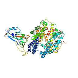
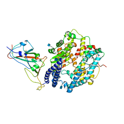
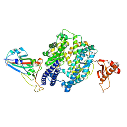
Crystal structure of the receptor binding domain of SARS-CoV-2 Delta variant spike glycoprotein in complex with its receptor human ACE2
分子名称: 2-acetamido-2-deoxy-beta-D-glucopyranose, 2-acetamido-2-deoxy-beta-D-glucopyranose-(1-4)-2-acetamido-2-deoxy-beta-D-glucopyranose, Angiotensin-converting enzyme 2, ...
著者Qi, J, Han, P.
登録日2021-12-17
公開日2022-01-19
最終更新日2023-11-29
実験手法X-RAY DIFFRACTION (3.34 Å)
主引用文献Receptor binding and complex structures of human ACE2 to spike RBD from omicron and delta SARS-CoV-2.
Cell, 185, 2022
7WBP
DownloadVisualize
BU of 7wbp by Molmil
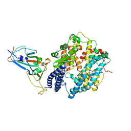
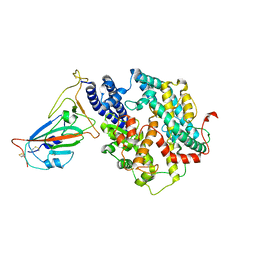
Crystal structure of the receptor binding domain of SARS-CoV-2 Omicron variant spike glycoprotein in complex with its receptor human ACE2
分子名称: 2-acetamido-2-deoxy-beta-D-glucopyranose, 2-acetamido-2-deoxy-beta-D-glucopyranose-(1-4)-2-acetamido-2-deoxy-beta-D-glucopyranose, Angiotensin-converting enzyme 2, ...
著者Qi, J, Han, P.
登録日2021-12-17
公開日2022-01-19
最終更新日2024-10-30
実験手法X-RAY DIFFRACTION (3 Å)
主引用文献Receptor binding and complex structures of human ACE2 to spike RBD from omicron and delta SARS-CoV-2.
Cell, 185, 2022
7WBL
DownloadVisualize
BU of 7wbl by Molmil
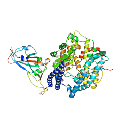
Cryo-EM structure of human ACE2 complexed with SARS-CoV-2 Omicron RBD
分子名称: 2-acetamido-2-deoxy-beta-D-glucopyranose, Angiotensin-converting enzyme 2, Spike protein S1, ...
著者Liu, S, Gao, F.G.
登録日2021-12-17
公開日2022-01-19
最終更新日2022-03-02
実験手法ELECTRON MICROSCOPY (3.4 Å)
主引用文献Receptor binding and complex structures of human ACE2 to spike RBD from omicron and delta SARS-CoV-2.
Cell, 185, 2022
7R6A
DownloadVisualize
BU of 7r6a by Molmil
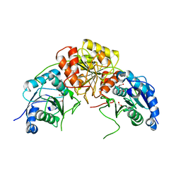
Crystal structure of mutant L124D/R125A/C273S of L-Asparaginase I from Yersinia pestis
分子名称: FORMIC ACID, L-asparaginase I
著者Strzelczyk, P, Wlodawer, A, Lubkowski, J.
登録日2021-06-22
公開日2022-07-06
最終更新日2023-10-25
実験手法X-RAY DIFFRACTION (1.89 Å)
主引用文献The dimeric form of bacterial l-asparaginase YpAI is fully active.
Febs J., 290, 2023
7R69
DownloadVisualize
BU of 7r69 by Molmil
Crystal structure of mutant R43D/C273S of L-Asparaginase I from Yersinia pestis
分子名称: 1,2-ETHANEDIOL, L-asparaginase I
著者Strzelczyk, P, Wlodawer, A, Lubkowski, J.
登録日2021-06-22
公開日2022-07-06
最終更新日2023-10-25
実験手法X-RAY DIFFRACTION (1.8 Å)
主引用文献The dimeric form of bacterial l-asparaginase YpAI is fully active.
Febs J., 290, 2023
7R6B
DownloadVisualize
BU of 7r6b by Molmil
Crystal structure of mutant R43D/L124D/R125A/C273S of L-Asparaginase I from Yersinia pestis
分子名称: 1,2-ETHANEDIOL, CALCIUM ION, L-asparaginase I
著者Strzelczyk, P, Wlodawer, A, Lubkowski, J.
登録日2021-06-22
公開日2022-07-06
最終更新日2023-10-25
実験手法X-RAY DIFFRACTION (2.03 Å)
主引用文献The dimeric form of bacterial l-asparaginase YpAI is fully active.
Febs J., 290, 2023
7WSF
DownloadVisualize
BU of 7wsf by Molmil
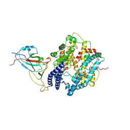
Cryo-EM structure of SARS-CoV spike receptor-binding domain in complex with minke whale ACE2
分子名称: Angiotensin-converting enzyme, Spike protein S1, ZINC ION
著者Li, S, Han, P, Qi, J.
登録日2022-01-29
公開日2022-10-19
実験手法ELECTRON MICROSCOPY (2.87 Å)
主引用文献Cross-species recognition and molecular basis of SARS-CoV-2 and SARS-CoV binding to ACE2s of marine animals.
Natl Sci Rev, 9, 2022
7WSE
DownloadVisualize
BU of 7wse by Molmil
Cryo-EM structure of SARS-CoV-2 spike receptor-binding domain complexed with its receptor minke whale ACE2
分子名称: Angiotensin-converting enzyme, Spike protein S1, ZINC ION
著者Li, S, Han, P.
登録日2022-01-29
公開日2022-10-19
最終更新日2024-10-23
実験手法ELECTRON MICROSCOPY (2.93 Å)
主引用文献Cross-species recognition and molecular basis of SARS-CoV-2 and SARS-CoV binding to ACE2s of marine animals.
Natl Sci Rev, 9, 2022
7WSH
DownloadVisualize
BU of 7wsh by Molmil
Cryo-EM structure of SARS-CoV-2 spike receptor-binding domain in complex with sea lion ACE2
分子名称: 2-acetamido-2-deoxy-beta-D-glucopyranose, Angiotensin-converting enzyme, Spike protein S1, ...
著者Li, S, Han, P, Qi, J.
登録日2022-01-29
公開日2022-11-09
最終更新日2024-10-23
実験手法ELECTRON MICROSCOPY (2.89 Å)
主引用文献Cross-species recognition and molecular basis of SARS-CoV-2 and SARS-CoV binding to ACE2s of marine animals.
Natl Sci Rev, 9, 2022
7WSG
DownloadVisualize
BU of 7wsg by Molmil
Cryo-EM structure of SARS-CoV spike receptor-binding domain in complex with sea lion ACE2
分子名称: Angiotensin-converting enzyme, Spike protein S1, ZINC ION
著者Li, S, Han, P, Qi, J.
登録日2022-01-29
公開日2022-11-09
実験手法ELECTRON MICROSCOPY (3.03 Å)
主引用文献Cross-species recognition and molecular basis of SARS-CoV-2 and SARS-CoV binding to ACE2s of marine animals.
Natl Sci Rev, 9, 2022

226707

件を2024-10-30に公開中

PDB statisticsPDBj update infoContact PDBjnumon