8I94
 
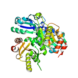
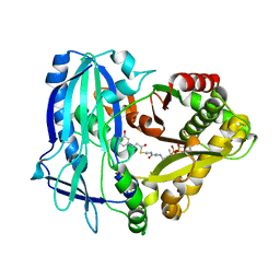
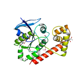
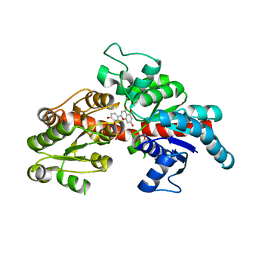 | | Structure of flavone 4'-O-glucoside 7-O-glucosyltransferase from Nemophila menziesii, complex with luteolin | | 分子名称: | 2-(3,4-dihydroxyphenyl)-5,7-dihydroxy-4H-chromen-4-one, Glycosyltransferase, SULFATE ION | | 著者 | Murayama, K, Kato-Murayama, M, Shirouzu, M. | | 登録日 | 2023-02-06 | | 公開日 | 2024-02-14 | | 最終更新日 | 2024-03-27 | | 実験手法 | X-RAY DIFFRACTION (2.43 Å) | | 主引用文献 | Molecular basis of ligand recognition specificity of flavone glucosyltransferases in Nemophila menziesii.
Arch.Biochem.Biophys., 753, 2024
|
|
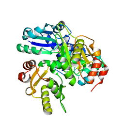
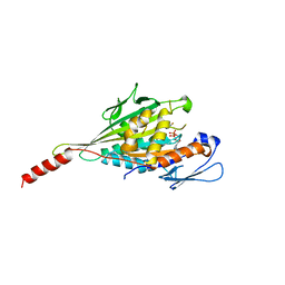
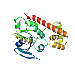
3WO7
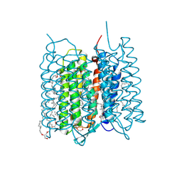
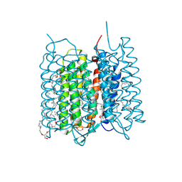
 | | Crystal structure of YidC from Bacillus halodurans (form II) | | 分子名称: | COPPER (II) ION, Membrane protein insertase YidC 2 | | 著者 | Kumazaki, K, Tsukazaki, T, Ishitani, R, Nureki, O. | | 登録日 | 2013-12-20 | | 公開日 | 2014-04-23 | | 最終更新日 | 2024-04-03 | | 実験手法 | X-RAY DIFFRACTION (3.201 Å) | | 主引用文献 | Structural basis of Sec-independent membrane protein insertion by YidC.
Nature, 509, 2014
|
|
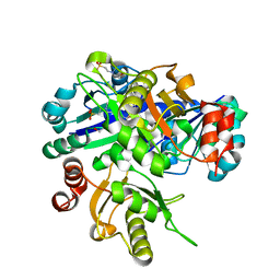
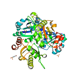
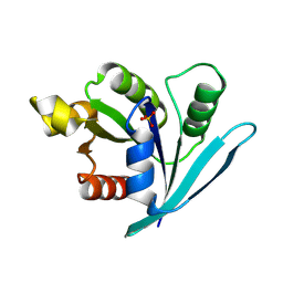
3WO6
 | | Crystal structure of YidC from Bacillus halodurans (form I) | | 分子名称: | (2R)-2,3-dihydroxypropyl (9Z)-octadec-9-enoate, CADMIUM ION, Membrane protein insertase YidC 2 | | 著者 | Kumazaki, K, Tsukazaki, T, Ishitani, R, Nureki, O. | | 登録日 | 2013-12-20 | | 公開日 | 2014-04-23 | | 最終更新日 | 2024-04-03 | | 実験手法 | X-RAY DIFFRACTION (2.403 Å) | | 主引用文献 | Structural basis of Sec-independent membrane protein insertion by YidC.
Nature, 509, 2014
|
|
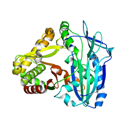
2E4R
 | |
2EJJ
 | |
7XJE
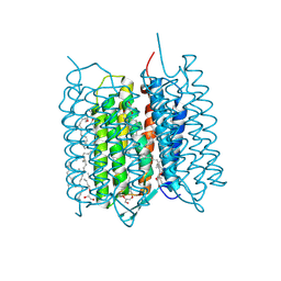 | | Crystal structure of bacteriorhodopsin in the K state refined against the extrapolated dataset | | 分子名称: | 2,3-DI-PHYTANYL-GLYCEROL, Bacteriorhodopsin, RETINAL | | 著者 | Taguchi, S, Niwa, S, Takeda, K. | | 登録日 | 2022-04-16 | | 公開日 | 2023-03-01 | | 最終更新日 | 2024-10-16 | | 実験手法 | X-RAY DIFFRACTION (1.33 Å) | | 主引用文献 | Detailed analysis of distorted retinal and its interaction with surrounding residues in the K intermediate of bacteriorhodopsin
Commun Biol, 6, 2023
|
|
7XJC
 | | Crystal structure of bacteriorhodopsin in the ground and K states after green laser irradiation | | 分子名称: | 2,10,23-TRIMETHYL-TETRACOSANE, 2,3-DI-PHYTANYL-GLYCEROL, Bacteriorhodopsin, ... | | 著者 | Taguchi, S, Niwa, S, Takeda, K. | | 登録日 | 2022-04-16 | | 公開日 | 2023-03-01 | | 最終更新日 | 2023-11-29 | | 実験手法 | X-RAY DIFFRACTION (1.33 Å) | | 主引用文献 | Detailed analysis of distorted retinal and its interaction with surrounding residues in the K intermediate of bacteriorhodopsin
Commun Biol, 6, 2023
|
|
2EJK
 | |
2EMU
 | |
2EN5
 | |
2E4N
 | |
7XJD
 | | Crystal structure of bacteriorhodopsin in the ground state by red laser irradiation | | 分子名称: | 2,10,23-TRIMETHYL-TETRACOSANE, 2,3-DI-PHYTANYL-GLYCEROL, Bacteriorhodopsin, ... | | 著者 | Taguchi, S, Niwa, S, Takeda, K. | | 登録日 | 2022-04-16 | | 公開日 | 2023-03-22 | | 実験手法 | X-RAY DIFFRACTION (1.33 Å) | | 主引用文献 | Detailed analysis of distorted retinal and its interaction with surrounding residues in the K intermediate of bacteriorhodopsin.
Commun Biol, 6, 2023
|
|
2EH2
 | |
7DEV
 | | Crystal Structures of Anthocyanin 5,3'-aromatic acyltransferase from Gentiana triflora | | 分子名称: | Anthocyanin 5-aromatic acyltransferase | | 著者 | Murayama, K, Kato-Murayama, M, Shirouzu, M. | | 登録日 | 2020-11-05 | | 公開日 | 2021-09-15 | | 最終更新日 | 2023-11-29 | | 実験手法 | X-RAY DIFFRACTION (3.1 Å) | | 主引用文献 | Anthocyanin 5,3'-aromatic acyltransferase from Gentiana triflora, a structural insight into biosynthesis of a blue anthocyanin.
Phytochemistry, 186, 2021
|
|
7DEX
 | | Crystal Structures of Anthocyanin 5,3'-aromatic acyltransferase H174A mutant with caffeoyl-CoA | | 分子名称: | Anthocyanin 5-aromatic acyltransferase, S-[2-[3-[[(2R)-4-[[[(2R,3S,4R,5R)-5-(6-aminopurin-9-yl)-4-oxidanyl-3-phosphonooxy-oxolan-2-yl]methoxy-oxidanyl-phosphoryl]oxy-oxidanyl-phosphoryl]oxy-3,3-dimethyl-2-oxidanyl-butanoyl]amino]propanoylamino]ethyl] (E)-3-[3,4-bis(oxidanyl)phenyl]prop-2-enethioate | | 著者 | Murayama, K, Kato-Murayama, M, Shirouzu, M. | | 登録日 | 2020-11-05 | | 公開日 | 2021-09-15 | | 最終更新日 | 2023-11-29 | | 実験手法 | X-RAY DIFFRACTION (2.5 Å) | | 主引用文献 | Anthocyanin 5,3'-aromatic acyltransferase from Gentiana triflora, a structural insight into biosynthesis of a blue anthocyanin.
Phytochemistry, 186, 2021
|
|
7EO9
 | | Crystal structure of KIF1A Motor-Neck domain with ADP-Mg-AlFx | | 分子名称: | ADENOSINE-5'-DIPHOSPHATE, ALUMINUM FLUORIDE, Kinesin-like protein KIF1A, ... | | 著者 | Ogawa, T, Jiang, X, Hirokawa, N. | | 登録日 | 2021-04-21 | | 公開日 | 2021-12-29 | | 最終更新日 | 2023-11-29 | | 実験手法 | X-RAY DIFFRACTION (2.57 Å) | | 主引用文献 | A neuropathy-associated kinesin KIF1A mutation hyper-stabilizes the motor-neck interaction during the ATPase cycle.
Embo J., 41, 2022
|
|
3VHL
 | | Crystal structure of the DHR-2 domain of DOCK8 in complex with Cdc42 (T17N mutant) | | 分子名称: | Cell division control protein 42 homolog, Dedicator of cytokinesis protein 8, PHOSPHATE ION | | 著者 | Hanawa-Suetsugu, K, Kukimoto-Niino, M, Nishizak, T, Terada, T, Shirouzu, M, Fukui, Y, Yokoyama, S. | | 登録日 | 2011-08-26 | | 公開日 | 2012-06-20 | | 最終更新日 | 2023-11-08 | | 実験手法 | X-RAY DIFFRACTION (2.085 Å) | | 主引用文献 | DOCK8 is a Cdc42 activator critical for interstitial dendritic cell migration during immune responses.
Blood, 119, 2012
|
|
2Z4Q
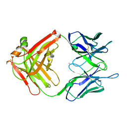 | | Crystal structure of a murine antibody FAB 528 | | 分子名称: | CADMIUM ION, CHLORIDE ION, anti egfr antibody fab, ... | | 著者 | Nakanishi, T, Tsumoto, K, Asano, R, Kondo, H, Kumagai, I. | | 登録日 | 2007-06-22 | | 公開日 | 2007-10-30 | | 最終更新日 | 2023-11-01 | | 実験手法 | X-RAY DIFFRACTION (2.3 Å) | | 主引用文献 | Thermodynamic consequences of mutations in vernier zone residues of a humanized anti-human epidermal growth factor receptor murine antibody, 528
J.Biol.Chem., 283, 2008
|
|
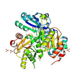
5X32
 | | Crystal structure of D-mannose isomerase | | 分子名称: | N-acylglucosamine 2-epimerase, PHOSPHATE ION | | 著者 | Kato, K, Saburi, W, Yao, M. | | 登録日 | 2017-02-03 | | 公開日 | 2018-02-07 | | 最終更新日 | 2023-11-22 | | 実験手法 | X-RAY DIFFRACTION (2.586 Å) | | 主引用文献 | Biochemical and structural characterization of Marinomonas mediterranead-mannose isomerase Marme_2490 phylogenetically distant from known enzymes
Biochimie, 144, 2018
|
|
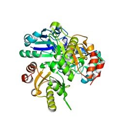
3A1S
 | | Crystal structue of the cytosolic domain of T. maritima FeoB iron iransporter in GDP form I | | 分子名称: | (4R)-2-METHYLPENTANE-2,4-DIOL, (4S)-2-METHYL-2,4-PENTANEDIOL, GUANOSINE-5'-DIPHOSPHATE, ... | | 著者 | Hattori, M, Ishitani, R, Nureki, O. | | 登録日 | 2009-04-22 | | 公開日 | 2009-09-22 | | 最終更新日 | 2024-10-09 | | 実験手法 | X-RAY DIFFRACTION (1.5 Å) | | 主引用文献 | Structural basis of novel interactions between the small-GTPase and GDI-like domains in prokaryotic FeoB iron transporter
Structure, 17, 2009
|
|
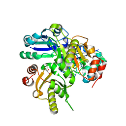
3A1U
 | | Crystal structue of the cytosolic domain of T. maritima FeoB iron iransporter in GMPPNP form | | 分子名称: | (4R)-2-METHYLPENTANE-2,4-DIOL, Iron(II) transport protein B, MAGNESIUM ION, ... | | 著者 | Hattori, M, Ishitani, R, Nureki, O. | | 登録日 | 2009-04-22 | | 公開日 | 2009-09-22 | | 最終更新日 | 2023-11-01 | | 実験手法 | X-RAY DIFFRACTION (1.8 Å) | | 主引用文献 | Structural basis of novel interactions between the small-GTPase and GDI-like domains in prokaryotic FeoB iron transporter
Structure, 17, 2009
|
|
3A1W
 | |
3A1T
 | |
3A1V
 | | Crystal structue of the cytosolic domain of T. maritima FeoB iron iransporter in apo form | | 分子名称: | (4R)-2-METHYLPENTANE-2,4-DIOL, (4S)-2-METHYL-2,4-PENTANEDIOL, 4-(2-HYDROXYETHYL)-1-PIPERAZINE ETHANESULFONIC ACID, ... | | 著者 | Hattori, M, Ishitani, R, Nureki, O. | | 登録日 | 2009-04-22 | | 公開日 | 2009-09-22 | | 最終更新日 | 2023-11-01 | | 実験手法 | X-RAY DIFFRACTION (2.4 Å) | | 主引用文献 | Structural basis of novel interactions between the small-GTPase and GDI-like domains in prokaryotic FeoB iron transporter
Structure, 17, 2009
|
|
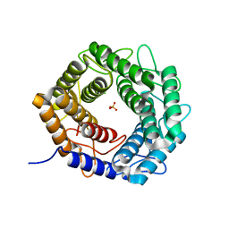
5W49
 | | The crystal structure of human S-adenosylhomocysteine hydrolase (AHCY) bound to oxadiazole inhibitor | | 分子名称: | (4-amino-1,2,5-oxadiazol-3-yl)[(3R)-3-{4-[(3-methoxyphenyl)amino]-6-methylpyridin-2-yl}pyrrolidin-1-yl]methanone, 1,2-ETHANEDIOL, Adenosylhomocysteinase, ... | | 著者 | Dougan, D.R, Lawson, J.D, Lane, W. | | 登録日 | 2017-06-09 | | 公開日 | 2017-06-28 | | 最終更新日 | 2024-03-13 | | 実験手法 | X-RAY DIFFRACTION (2.4 Å) | | 主引用文献 | Identification of AHCY inhibitors using novel high-throughput mass spectrometry.
Biochem. Biophys. Res. Commun., 491, 2017
|
|


