Loading
PDBj
メニューPDBj@FacebookPDBj@TwitterPDBj@YouTubewwPDB FoundationwwPDB
RCSB PDBPDBeBMRBAdv. SearchSearch help
Search by PDB author
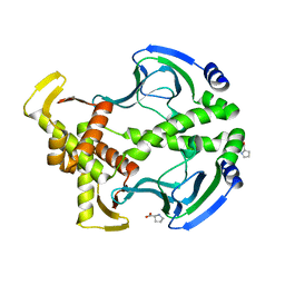
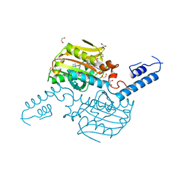
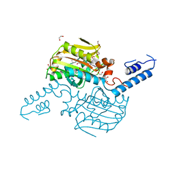
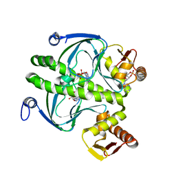
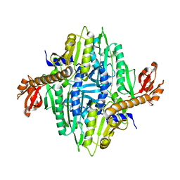
1NJ8
DownloadVisualize
BU of 1nj8 by Molmil
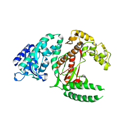
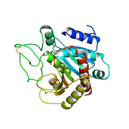
Crystal Structure of Prolyl-tRNA Synthetase from Methanocaldococcus janaschii
分子名称: Proline-tRNA Synthetase
著者Kamtekar, S, Kennedy, W.D, Wang, J, Stathopoulos, C, Soll, D, Steitz, T.A.
登録日2002-12-30
公開日2003-03-04
最終更新日2024-04-03
実験手法X-RAY DIFFRACTION (3.2 Å)
主引用文献The structural basis of cysteine aminoacylation of tRNAPro by prolyl-tRNA synthetases
Proc.Natl.Acad.Sci.USA, 100, 2003
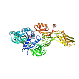
1DPI
DownloadVisualize
BU of 1dpi by Molmil
STRUCTURE OF LARGE FRAGMENT OF ESCHERICHIA COLI DNA POLYMERASE I COMPLEXED WITH D/TMP
分子名称: DNA POLYMERASE I KLENOW FRAGMENT, ZINC ION
著者Beese, L, Ollis, D, Steitz, T.
登録日1987-08-11
公開日1987-10-16
最終更新日2024-02-07
実験手法X-RAY DIFFRACTION (2.8 Å)
主引用文献Structure of large fragment of Escherichia coli DNA polymerase I complexed with dTMP.
Nature, 313, 1985
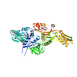
4IQJ
DownloadVisualize
BU of 4iqj by Molmil
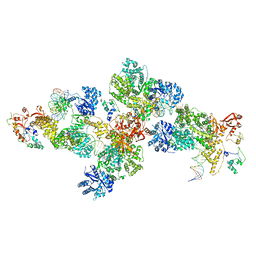
Structure of PolIIIalpha-Tauc-DNA complex suggests an atomic model of the replisome
分子名称: DNA (5'-D(*TP*TP*TP*TP*TP*TP*TP*GP*TP*GP*GP*CP*AP*CP*TP*GP*GP*CP*CP*GP*TP*CP*GP*TP*TP*TP*CP*G)-3'), DNA (5'-D(P*CP*GP*AP*AP*AP*CP*GP*AP*CP*GP*GP*CP*CP*AP*GP*TP*GP*CP*CP*A)-3'), DNA (5'-D(P*CP*GP*AP*AP*AP*CP*GP*AP*CP*GP*GP*CP*CP*AP*GP*TP*GP*CP*CP*AP*(DOC))-3'), ...
著者Liu, B, Lin, J, Steitz, T.
登録日2013-01-11
公開日2013-03-13
最終更新日2023-09-20
実験手法X-RAY DIFFRACTION (3.2 Å)
主引用文献Structure of PolIIIalpha-Tauc-DNA complex suggests an atomic model of the replisome
Structure, 21, 2013
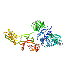
1ARO
DownloadVisualize
BU of 1aro by Molmil
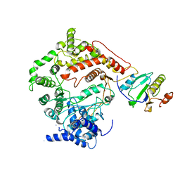
T7 RNA POLYMERASE COMPLEXED WITH T7 LYSOZYME
分子名称: MERCURY (II) ION, T7 LYSOZYME, T7 RNA POLYMERASE
著者Steitz, T, Jeruzalmi, D.
登録日1997-08-08
公開日1998-10-21
最終更新日2024-02-07
実験手法X-RAY DIFFRACTION (2.8 Å)
主引用文献Structure of T7 RNA polymerase complexed to the transcriptional inhibitor T7 lysozyme.
EMBO J., 17, 1998
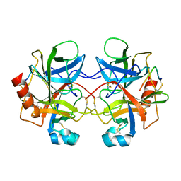
2CHA
DownloadVisualize
BU of 2cha by Molmil
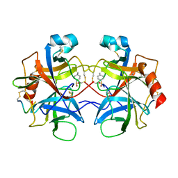
THE STRUCTURE OF CRYSTALLINE ALPHA-CHYMOTRYPSIN, $V.THE ATOMIC STRUCTURE OF TOSYL-ALPHA-CHYMOTRYPSIN AT 2 ANGSTROMS RESOLUTION
分子名称: ALPHA-CHYMOTRYPSIN A, PARA-TOLUENE SULFONATE
著者Birktoft, J.J, Blow, D.M.
登録日1975-01-01
公開日1977-01-18
最終更新日2023-09-27
実験手法X-RAY DIFFRACTION (2 Å)
主引用文献Structure of crystalline -chymotrypsin. V. The atomic structure of tosyl- -chymotrypsin at 2 A resolution.
J.Mol.Biol., 68, 1972
3FWE
DownloadVisualize
BU of 3fwe by Molmil
Crystal Structure of the Apo D138L CAP mutant
分子名称: Catabolite gene activator, PROLINE
著者Sharma, H, Wang, J, Kong, J, Yu, S, Steitz, T.
登録日2009-01-17
公開日2009-09-08
最終更新日2024-02-21
実験手法X-RAY DIFFRACTION (2.3 Å)
主引用文献Structure of apo-CAP reveals that large conformational changes are necessary for DNA binding
Proc.Natl.Acad.Sci.USA, 106, 2009
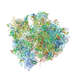
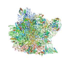
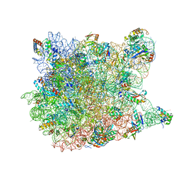
4V7M
DownloadVisualize
BU of 4v7m by Molmil
The structures of Capreomycin bound to the 70S ribosome.
分子名称: 16S ribosomal RNA, 23S ribosomal RNA, 30S ribosomal protein S10, ...
著者Stanley, R.E, Blaha, G.
登録日2009-11-12
公開日2014-07-09
最終更新日2017-11-22
実験手法X-RAY DIFFRACTION (3.45 Å)
主引用文献The structures of the anti-tuberculosis antibiotics viomycin and capreomycin bound to the 70S ribosome.
Nat.Struct.Mol.Biol., 17, 2010
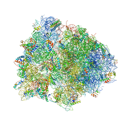
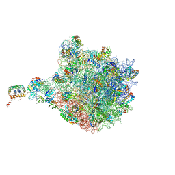
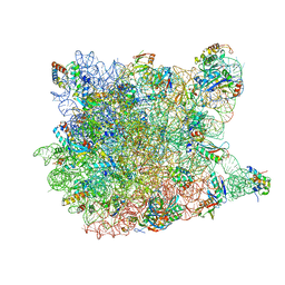
4V7L
DownloadVisualize
BU of 4v7l by Molmil
The structures of viomycin bound to the 70S ribosome.
分子名称: 16S ribosomal RNA, 23S ribosomal RNA, 30S ribosomal protein S10, ...
著者Stanley, R.E, Blaha, G.
登録日2009-11-12
公開日2014-07-09
最終更新日2018-07-11
実験手法X-RAY DIFFRACTION (3 Å)
主引用文献The structures of the anti-tuberculosis antibiotics viomycin and capreomycin bound to the 70S ribosome.
Nat.Struct.Mol.Biol., 17, 2010
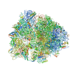
4V6A
DownloadVisualize
BU of 4v6a by Molmil
Structure of EF-P bound to the 70S ribosome.
分子名称: 16S ribosomal RNA, 23S ribosomal RNA, 30S ribosomal protein S10, ...
著者Stanley, R.E, Blaha, G.
登録日2009-06-15
公開日2014-07-09
最終更新日2023-09-20
実験手法X-RAY DIFFRACTION (3.1 Å)
主引用文献Formation of the first peptide bond: the structure of EF-P bound to the 70S ribosome.
Science, 325, 2009
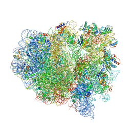
1K8A
DownloadVisualize
BU of 1k8a by Molmil
Co-crystal structure of Carbomycin A bound to the 50S ribosomal subunit of Haloarcula marismortui
分子名称: 23S RRNA, 5S RRNA, CADMIUM ION, ...
著者Hansen, J.L, Ippolito, J.A, Ban, N, Nissen, P, Moore, P.B, Steitz, T.
登録日2001-10-23
公開日2002-07-19
最終更新日2023-08-16
実験手法X-RAY DIFFRACTION (3 Å)
主引用文献The structures of four macrolide antibiotics bound to the large ribosomal subunit.
Mol.Cell, 10, 2002
4V9F
DownloadVisualize
BU of 4v9f by Molmil
The re-refined crystal structure of the Haloarcula marismortui large ribosomal subunit at 2.4 Angstrom resolution: more complete structure of the L7/L12 and L1 stalk, L5 and LX proteins
分子名称: 23S Ribosomal RNA, 50S ribosomal protein L10E, 50S ribosomal protein L10e, ...
著者Gabdulkhakov, A.
登録日2012-11-02
公開日2014-07-09
最終更新日2023-09-20
実験手法X-RAY DIFFRACTION (2.404 Å)
主引用文献Revisiting the Haloarcula marismortui 50S ribosomal subunit model.
Acta Crystallogr.,Sect.D, 69, 2013
4CHA
DownloadVisualize
BU of 4cha by Molmil
STRUCTURE OF ALPHA-*CHYMOTRYPSIN REFINED AT 1.68 ANGSTROMS RESOLUTION
分子名称: ALPHA-CHYMOTRYPSIN A
著者Tsukada, H, Blow, D.M.
登録日1984-11-26
公開日1985-04-01
最終更新日2024-06-05
実験手法X-RAY DIFFRACTION (1.68 Å)
主引用文献Structure of alpha-chymotrypsin refined at 1.68 A resolution.
J.Mol.Biol., 184, 1985
4TQF
DownloadVisualize
BU of 4tqf by Molmil
Crystal Structure of the C-terminal domain of IFRS bound with 2-(5-bromothienyl)-L-Ala and ATP
分子名称: 1,2-ETHANEDIOL, 3-(5-bromothiophen-2-yl)-L-alanine, ADENOSINE-5'-TRIPHOSPHATE, ...
著者Nakamura, A, O'Donoghue, P, Soll, D.
登録日2014-06-11
公開日2014-11-12
最終更新日2023-09-27
実験手法X-RAY DIFFRACTION (2.7143 Å)
主引用文献Polyspecific pyrrolysyl-tRNA synthetases from directed evolution.
Proc.Natl.Acad.Sci.USA, 111, 2014
4TQD
DownloadVisualize
BU of 4tqd by Molmil
Crystal Structure of the C-terminal domain of IFRS bound with 3-iodo-L-Phe and ATP
分子名称: 1,2-ETHANEDIOL, 3-iodo-L-phenylalanine, ADENOSINE-5'-TRIPHOSPHATE, ...
著者Nakamura, A, O'Donoghue, P, Soll, D.
登録日2014-06-11
公開日2014-11-12
最終更新日2023-11-15
実験手法X-RAY DIFFRACTION (2.1429 Å)
主引用文献Polyspecific pyrrolysyl-tRNA synthetases from directed evolution.
Proc.Natl.Acad.Sci.USA, 111, 2014
2BM1
DownloadVisualize
BU of 2bm1 by Molmil
Ribosomal elongation factor G (EF-G) Fusidic acid resistant mutant G16V
分子名称: ELONGATION FACTOR G, GUANOSINE-5'-DIPHOSPHATE, MAGNESIUM ION
著者Hansson, S, Singh, R, Gudkov, A.T, Liljas, A, Logan, D.T.
登録日2005-03-09
公開日2005-05-04
最終更新日2023-12-13
実験手法X-RAY DIFFRACTION (2.6 Å)
主引用文献Structural Insights Into Fusidic Acid Resistance and Sensitivity in EF-G
J.Mol.Biol., 348, 2005
2BM0
DownloadVisualize
BU of 2bm0 by Molmil
Ribosomal elongation factor G (EF-G) Fusidic acid resistant mutant T84A
分子名称: ELONGATION FACTOR G, GUANOSINE-5'-DIPHOSPHATE, MAGNESIUM ION
著者Hansson, S, Singh, R, Gudkov, A.T, Liljas, A, Logan, D.T.
登録日2005-03-09
公開日2005-05-04
最終更新日2023-12-13
実験手法X-RAY DIFFRACTION (2.4 Å)
主引用文献Structural Insights Into Fusidic Acid Resistance and Sensitivity in EF-G
J.Mol.Biol., 348, 2005
2BV3
DownloadVisualize
BU of 2bv3 by Molmil
Crystal structure of a mutant elongation factor G trapped with a GTP analogue
分子名称: ELONGATION FACTOR G, MAGNESIUM ION, PHOSPHOAMINOPHOSPHONIC ACID-GUANYLATE ESTER
著者Hansson, S, Singh, R, Gudkov, A.T, Liljas, A, Logan, D.T.
登録日2005-06-22
公開日2005-08-10
最終更新日2023-12-13
実験手法X-RAY DIFFRACTION (2.5 Å)
主引用文献Crystal Structure of a Mutant Elongation Factor G Trapped with a GTP Analogue.
FEBS Lett., 579, 2005
3CC2
DownloadVisualize
BU of 3cc2 by Molmil
The Refined Crystal Structure of the Haloarcula Marismortui Large Ribosomal Subunit at 2.4 Angstrom Resolution with rrnA Sequence for the 23S rRNA and Genome-derived Sequences for r-Proteins
分子名称: 23S RIBOSOMAL RNA, 50S ribosomal protein L10E, 50S ribosomal protein L10e, ...
著者Gurel, G, Blaha, G.
登録日2008-02-23
公開日2008-05-20
最終更新日2024-02-21
実験手法X-RAY DIFFRACTION (2.4 Å)
主引用文献Mutations outside the anisomycin-binding site can make ribosomes drug-resistant.
J.Mol.Biol., 379, 2008
3CC4
DownloadVisualize
BU of 3cc4 by Molmil
Co-crystal Structure of Anisomycin Bound to the 50S Ribosomal Subunit
分子名称: 23S ribosomal RNA, 50S ribosomal protein L10E, 50S ribosomal protein L10e, ...
著者Blaha, G, Gurel, G.
登録日2008-02-24
公開日2008-05-20
最終更新日2024-02-21
実験手法X-RAY DIFFRACTION (2.7 Å)
主引用文献Mutations outside the anisomycin-binding site can make ribosomes drug-resistant.
J.Mol.Biol., 379, 2008
1HW5
DownloadVisualize
BU of 1hw5 by Molmil
THE CAP/CRP VARIANT T127L/S128A
分子名称: ADENOSINE-3',5'-CYCLIC-MONOPHOSPHATE, CATABOLITE GENE ACTIVATOR
著者Chu, S.Y, Tordova, M, Gilliland, G.L, Gorshkova, I, Shi, Y.
登録日2001-01-09
公開日2001-01-17
最終更新日2023-08-09
実験手法X-RAY DIFFRACTION (1.82 Å)
主引用文献The structure of the T127L/S128A mutant of cAMP receptor protein facilitates promoter site binding
J.Biol.Chem., 276, 2001
4V4B
DownloadVisualize
BU of 4v4b by Molmil
Structure of the ribosomal 80S-eEF2-sordarin complex from yeast obtained by docking atomic models for RNA and protein components into a 11.7 A cryo-EM map.
分子名称: 18S ribosomal RNA, 40S ribosomal protein S0-A, 40S ribosomal protein S11, ...
著者Spahn, C.M, Gomez-Lorenzo, M.G, Grassucci, R.A, Jorgensen, R, Andersen, G.R, Beckmann, R, Penczek, P.A, Ballesta, J.P.G, Frank, J.
登録日2004-01-06
公開日2014-07-09
最終更新日2024-02-28
実験手法ELECTRON MICROSCOPY (11.7 Å)
主引用文献Domain movements of elongation factor eEF2 and the eukaryotic 80S ribosome facilitate tRNA translocation.
Embo J., 23, 2004
5CPA
DownloadVisualize
BU of 5cpa by Molmil
REFINED CRYSTAL STRUCTURE OF CARBOXYPEPTIDASE A AT 1.54 ANGSTROMS RESOLUTION.
分子名称: CARBOXYPEPTIDASE A, ZINC ION
著者Lipscomb, W.N.
登録日1982-05-06
公開日1982-07-29
最終更新日2017-11-29
実験手法X-RAY DIFFRACTION (1.54 Å)
主引用文献Refined crystal structure of carboxypeptidase A at 1.54 A resolution.
J.Mol.Biol., 168, 1983
5CRO
DownloadVisualize
BU of 5cro by Molmil
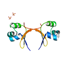
REFINED STRUCTURE OF CRO REPRESSOR PROTEIN FROM BACTERIOPHAGE LAMBDA
分子名称: CRO REPRESSOR PROTEIN, PHOSPHATE ION
著者Ohlendorf, D.H, Tronrud, D.E, Matthews, B.W.
登録日1998-04-17
公開日1998-06-17
最終更新日2024-03-06
実験手法X-RAY DIFFRACTION (2.3 Å)
主引用文献Refined structure of Cro repressor protein from bacteriophage lambda suggests both flexibility and plasticity.
J.Mol.Biol., 280, 1998
483D
DownloadVisualize
BU of 483d by Molmil
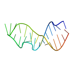
CRYSTAL STRUCTURE OF THE SARCIN/RICIN DOMAIN FROM E. COLI 23 S RRNA
分子名称: SARCIN/RICIN RRNA DOMAIN
著者Correll, C.C, Wool, I.G, Munishkin, A.
登録日1999-07-23
公開日1999-10-08
最終更新日2023-09-13
実験手法X-RAY DIFFRACTION (1.11 Å)
主引用文献The two faces of the Escherichia coli 23 S rRNA sarcin/ricin domain: the structure at 1.11 A resolution.
J.Mol.Biol., 292, 1999
480D
DownloadVisualize
BU of 480d by Molmil
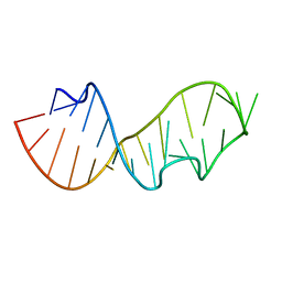
CRYSTAL STRUCTURE OF THE SARCIN/RICIN DOMAIN FROM E. COLI 23 S RRNA
分子名称: SARCIN/RICIN DOMAIN FROM 23 S RRNA
著者Correll, C.C, Wool, I.G, Munishkin, A.
登録日1999-07-20
公開日1999-10-08
最終更新日2023-09-13
実験手法X-RAY DIFFRACTION (1.5 Å)
主引用文献The two faces of the Escherichia coli 23 S rRNA sarcin/ricin domain: the structure at 1.11 A resolution.
J.Mol.Biol., 292, 1999

226262

件を2024-10-16に公開中

PDB statisticsPDBj update infoContact PDBjnumon