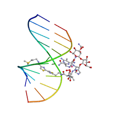
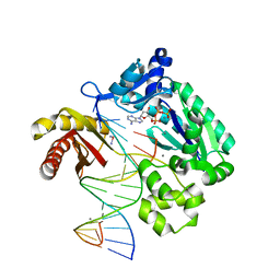
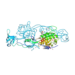
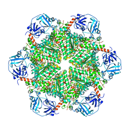
6FGC
 
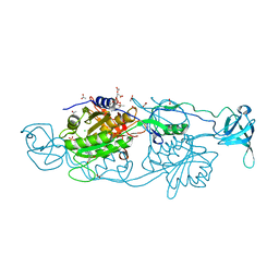 | | Crystal structure of Gephyrin E domain in complex with Artesunate | | 分子名称: | (4R)-2-METHYLPENTANE-2,4-DIOL, (4S)-2-METHYL-2,4-PENTANEDIOL, ACETATE ION, ... | | 著者 | Kasaragod, V.B, Schindelin, H. | | 登録日 | 2018-01-10 | | 公開日 | 2019-01-16 | | 最終更新日 | 2024-01-17 | | 実験手法 | X-RAY DIFFRACTION (1.5 Å) | | 主引用文献 | Elucidating the Molecular Basis for Inhibitory Neurotransmission Regulation by Artemisinins.
Neuron, 101, 2019
|
|
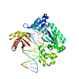
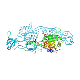
6FGD
 | |
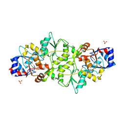
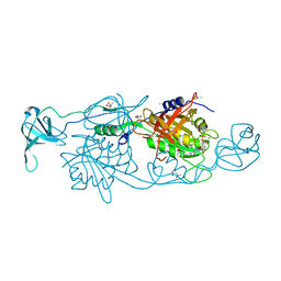
5YPP
 | | Crystal structure of IlvN.Val-1a | | 分子名称: | ACETATE ION, Acetolactate synthase isozyme 1 small subunit, DI(HYDROXYETHYL)ETHER, ... | | 著者 | Sarma, S.P, Bansal, A, Schindelin, H, Demeler, B. | | 登録日 | 2017-11-02 | | 公開日 | 2018-09-19 | | 最終更新日 | 2023-11-22 | | 実験手法 | X-RAY DIFFRACTION (1.9 Å) | | 主引用文献 | Crystallographic Structures of IlvN·Val/Ile Complexes: Conformational Selectivity for Feedback Inhibition of Aceto Hydroxy Acid Synthases.
Biochemistry, 58, 2019
|
|
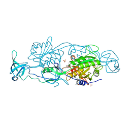
5YPW
 | | Crystal structure of IlvN.Val-1b | | 分子名称: | Acetolactate synthase isozyme 1 small subunit, VALINE | | 著者 | Sarma, S.P, Bansal, A, Schindelin, H, Demeler, B. | | 登録日 | 2017-11-03 | | 公開日 | 2018-09-19 | | 最終更新日 | 2023-11-22 | | 実験手法 | X-RAY DIFFRACTION (2.3 Å) | | 主引用文献 | Crystallographic Structures of IlvN·Val/Ile Complexes: Conformational Selectivity for Feedback Inhibition of Aceto Hydroxy Acid Synthases.
Biochemistry, 58, 2019
|
|
5W2A
 | |
1MXK
 | | NMR Structure of HO2-Co(III)bleomycin A(2) Bound to d(GGAAGCTTCC)(2) | | 分子名称: | 5'-D(*GP*GP*AP*AP*GP*CP*TP*TP*CP*C)-3', BLEOMYCIN A2, COBALT (III) ION, ... | | 著者 | Zhao, C, Xia, C, Mao, Q, Forsterling, H, DeRose, E, Antholine, W.E, Subczynski, W.K, Petering, D.H. | | 登録日 | 2002-10-02 | | 公開日 | 2002-10-16 | | 最終更新日 | 2024-05-22 | | 実験手法 | SOLUTION NMR | | 主引用文献 | Structures of HO(2)-Co(III)bleomycin A(2) Bound to d(GAGCTC)(2) and d(GGAAGCTTCC)(2): Structure-Reactivity Relationships of Co and Fe Bleomycins
J.Inorg.Biochem., 91, 2002
|
|
1MTG
 | | NMR Structure of HO2-Co(III)bleomycin A(2) bound to d(GAGCTC)(2) | | 分子名称: | 5'-D(*GP*AP*GP*CP*TP*C)-3', BLEOMYCIN A2, COBALT (III) ION, ... | | 著者 | Zhao, C, Xia, C, Mao, Q, Forsterling, H, DeRose, E, Antholine, W.E, Subczynski, W.K, Petering, D.H. | | 登録日 | 2002-09-20 | | 公開日 | 2002-10-16 | | 最終更新日 | 2024-05-01 | | 実験手法 | SOLUTION NMR | | 主引用文献 | Structures of HO(2)-Co(III)bleomycin A(2) Bound to d(GAGCTC)(2) and d(GGAAGCTTCC)(2): Structure-Reactivity Relationships of Co and Fe Bleomycins
J.Inorg.Biochem., 91, 2002
|
|
5W2C
 | |
3M9M
 | |
3M9O
 | |
3M9N
 | |
7PUX
 | |
7PO7
 | | Phosphoglycolate phosphatase from Mus musculus | | 分子名称: | ACETATE ION, CALCIUM ION, GLYCEROL, ... | | 著者 | Schloetzer, J, Schindelin, H, Fratz, S. | | 登録日 | 2021-09-08 | | 公開日 | 2022-12-21 | | 最終更新日 | 2024-07-03 | | 実験手法 | X-RAY DIFFRACTION (2.31 Å) | | 主引用文献 | Glycolytic flux control by drugging phosphoglycolate phosphatase.
Nat Commun, 13, 2022
|
|
5ERV
 | |
5ERT
 | | GephE in complex with Mn(2+) - ADP | | 分子名称: | (4S)-2-METHYL-2,4-PENTANEDIOL, ACETATE ION, ADENOSINE-5'-DIPHOSPHATE, ... | | 著者 | Kasaragod, V.B, Schindelin, H. | | 登録日 | 2015-11-15 | | 公開日 | 2016-05-04 | | 最終更新日 | 2024-01-10 | | 実験手法 | X-RAY DIFFRACTION (2 Å) | | 主引用文献 | Structural Framework for Metal Incorporation during Molybdenum Cofactor Biosynthesis.
Structure, 24, 2016
|
|
5ERU
 | |
5ERS
 | | GephE in complex with Mg(2+) - AMP | | 分子名称: | (4S)-2-METHYL-2,4-PENTANEDIOL, ACETATE ION, ADENOSINE MONOPHOSPHATE, ... | | 著者 | Kasaragod, V.B, Schindelin, H. | | 登録日 | 2015-11-15 | | 公開日 | 2016-05-04 | | 最終更新日 | 2024-01-10 | | 実験手法 | X-RAY DIFFRACTION (1.7 Å) | | 主引用文献 | Structural Framework for Metal Incorporation during Molybdenum Cofactor Biosynthesis.
Structure, 24, 2016
|
|
5ERR
 | | GephE in complex with Mg(2+) - ADP | | 分子名称: | (4S)-2-METHYL-2,4-PENTANEDIOL, ACETATE ION, ADENOSINE-5'-DIPHOSPHATE, ... | | 著者 | Kasaragod, V.B, Schindelin, H. | | 登録日 | 2015-11-15 | | 公開日 | 2016-05-04 | | 最終更新日 | 2024-01-10 | | 実験手法 | X-RAY DIFFRACTION (1.65 Å) | | 主引用文献 | Structural Framework for Metal Incorporation during Molybdenum Cofactor Biosynthesis.
Structure, 24, 2016
|
|
6ENS
 | | Structure of mouse wild-type RKIP | | 分子名称: | ACETATE ION, GLYCEROL, Phosphatidylethanolamine-binding protein 1 | | 著者 | Hirschbeck, M, Koelmel, W, Schindelin, H, Lorenz, K, Kisker, C. | | 登録日 | 2017-10-06 | | 公開日 | 2017-12-13 | | 最終更新日 | 2024-01-17 | | 実験手法 | X-RAY DIFFRACTION (1.3 Å) | | 主引用文献 | Conserved salt-bridge competition triggered by phosphorylation regulates the protein interactome.
Proc. Natl. Acad. Sci. U.S.A., 114, 2017
|
|
5ERQ
 | |
6ENT
 | | Structure of the rat RKIP variant delta143-146 | | 分子名称: | CHLORIDE ION, Phosphatidylethanolamine-binding protein 1, ZINC ION | | 著者 | Koelmel, W, Hirschbeck, M, Schindelin, H, Lorenz, K, Kisker, C. | | 登録日 | 2017-10-06 | | 公開日 | 2017-12-13 | | 最終更新日 | 2024-01-17 | | 実験手法 | X-RAY DIFFRACTION (2.66 Å) | | 主引用文献 | Conserved salt-bridge competition triggered by phosphorylation regulates the protein interactome.
Proc. Natl. Acad. Sci. U.S.A., 114, 2017
|
|
8S8A
 | | Human pyridoxal phosphatase in complex with 7,8-dihydroxyflavone without phosphate | | 分子名称: | 7,8-bis(oxidanyl)-2-phenyl-chromen-4-one, CHLORIDE ION, Chronophin, ... | | 著者 | Brenner, M, Gohla, A, Schindelin, H. | | 登録日 | 2024-03-06 | | 公開日 | 2024-06-12 | | 最終更新日 | 2024-06-26 | | 実験手法 | X-RAY DIFFRACTION (1.5 Å) | | 主引用文献 | 7,8-Dihydroxyflavone is a direct inhibitor of human and murine pyridoxal phosphatase.
Elife, 13, 2024
|
|
1MVG
 | | NMR solution structure of chicken Liver basic Fatty Acid Binding Protein (Lb-FABP) | | 分子名称: | Liver basic Fatty Acid Binding Protein | | 著者 | Vasile, F, Ragona, L, Catalano, M, Zetta, L, Perduca, M, Monaco, H, Molinari, H. | | 登録日 | 2002-09-25 | | 公開日 | 2003-03-04 | | 最終更新日 | 2024-05-22 | | 実験手法 | SOLUTION NMR | | 主引用文献 | Solution Structure of chicken Liver basic type Fatty Acid Binding Protein
J.BIOMOL.NMR, 25, 2003
|
|
5C1A
 | |
7PVN
 | | Crystal Structure of Human UBA6 in Complex with ATP | | 分子名称: | ADENOSINE-5'-TRIPHOSPHATE, CALCIUM ION, CHLORIDE ION, ... | | 著者 | Truongvan, N, Li, S, Schindelin, H. | | 登録日 | 2021-10-05 | | 公開日 | 2022-08-31 | | 最終更新日 | 2024-01-31 | | 実験手法 | X-RAY DIFFRACTION (2.71 Å) | | 主引用文献 | Structures of UBA6 explain its dual specificity for ubiquitin and FAT10.
Nat Commun, 13, 2022
|
|


