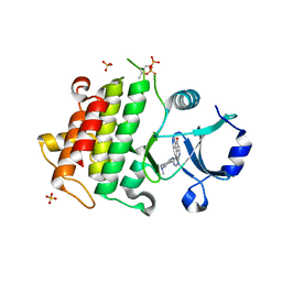
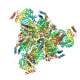
5K75
 
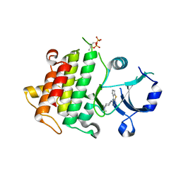 | | IRAK4 in complex with Compound 1 | | 分子名称: | Interleukin-1 receptor-associated kinase 4, SULFATE ION, ~{N}1-(7,8-dihydro-6~{H}-cyclopenta[2,3]thieno[2,4-~{c}]pyrimidin-1-yl)-~{N}4,~{N}4-dimethyl-cyclohexane-1,4-diamine | | 著者 | Ferguson, A.D. | | 登録日 | 2016-05-25 | | 公開日 | 2017-12-06 | | 最終更新日 | 2024-10-09 | | 実験手法 | X-RAY DIFFRACTION (2.03 Å) | | 主引用文献 | Discovery and Optimization of Pyrrolopyrimidine Inhibitors of Interleukin-1 Receptor Associated Kinase 4 (IRAK4) for the Treatment of Mutant MYD88L265P Diffuse Large B-Cell Lymphoma.
J. Med. Chem., 60, 2017
|
|
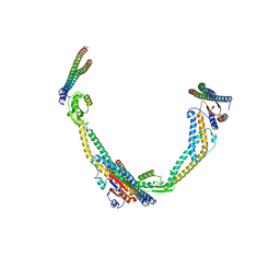
8CR1
 | | Homo sapiens Get1/Get2 heterotetramer in complex with a Get3 dimer | | 分子名称: | ATPase ASNA1, Guided entry of tail-anchored proteins factor CAMLG,Guided entry of tail-anchored proteins factor 1,GET2-GET1, ZINC ION | | 著者 | McDowell, M.A, Heimes, M, Wild, K, Sinning, I. | | 登録日 | 2023-03-07 | | 公開日 | 2023-11-29 | | 実験手法 | ELECTRON MICROSCOPY (3.2 Å) | | 主引用文献 | The GET insertase exhibits conformational plasticity and induces membrane thinning.
Nat Commun, 14, 2023
|
|
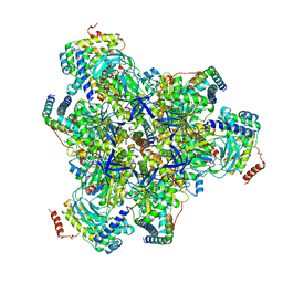
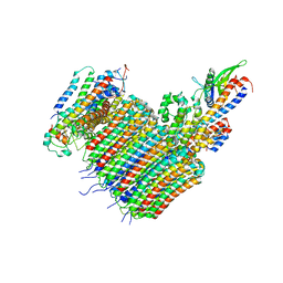
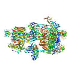
8EPL
 | | Human R-type voltage-gated calcium channel Cav2.3 at 3.1 Angstrom resolution | | 分子名称: | 1,2-Distearoyl-sn-glycerophosphoethanolamine, 2-acetamido-2-deoxy-beta-D-glucopyranose, 2-acetamido-2-deoxy-beta-D-glucopyranose-(1-4)-2-acetamido-2-deoxy-beta-D-glucopyranose, ... | | 著者 | Gao, S, Yao, X, Yan, N. | | 登録日 | 2022-10-06 | | 公開日 | 2022-12-14 | | 最終更新日 | 2023-07-12 | | 実験手法 | ELECTRON MICROSCOPY (3.1 Å) | | 主引用文献 | Structures of the R-type human Ca v 2.3 channel reveal conformational crosstalk of the intracellular segments.
Nat Commun, 13, 2022
|
|
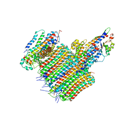
8EPM
 | | Human R-type voltage-gated calcium channel Cav2.3 CH2II-deleted mutant at 3.1 Angstrom resolution | | 分子名称: | 2-acetamido-2-deoxy-beta-D-glucopyranose, 2-acetamido-2-deoxy-beta-D-glucopyranose-(1-4)-2-acetamido-2-deoxy-beta-D-glucopyranose, 2-acetamido-2-deoxy-beta-D-glucopyranose-(1-4)-2-acetamido-2-deoxy-beta-D-glucopyranose-(1-4)-2-acetamido-2-deoxy-beta-D-glucopyranose-(1-4)-2-acetamido-2-deoxy-beta-D-glucopyranose, ... | | 著者 | Gao, S, Yao, X, Yan, N. | | 登録日 | 2022-10-06 | | 公開日 | 2022-12-14 | | 最終更新日 | 2023-07-12 | | 実験手法 | ELECTRON MICROSCOPY (3.1 Å) | | 主引用文献 | Structures of the R-type human Ca v 2.3 channel reveal conformational crosstalk of the intracellular segments.
Nat Commun, 13, 2022
|
|
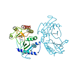
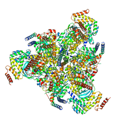
5K72
 | | IRAK4 in complex with Compound 21 | | 分子名称: | Interleukin-1 receptor-associated kinase 4, SULFATE ION, ~{N}4,~{N}4-dimethyl-~{N}1-[5-(oxan-4-yl)-7~{H}-pyrrolo[2,3-d]pyrimidin-4-yl]cyclohexane-1,4-diamine | | 著者 | Ferguson, A.D. | | 登録日 | 2016-05-25 | | 公開日 | 2017-12-06 | | 最終更新日 | 2024-10-16 | | 実験手法 | X-RAY DIFFRACTION (2.22 Å) | | 主引用文献 | Discovery and Optimization of Pyrrolopyrimidine Inhibitors of Interleukin-1 Receptor Associated Kinase 4 (IRAK4) for the Treatment of Mutant MYD88L265P Diffuse Large B-Cell Lymphoma.
J. Med. Chem., 60, 2017
|
|
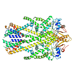
5E3T
 | | Crystal structure of phosphatidylinositol-4-phosphate 5-kinase | | 分子名称: | MANGANESE (II) ION, PHOSPHOAMINOPHOSPHONIC ACID-ADENYLATE ESTER, Phosphatidylinositol-4-phosphate 5-kinase, ... | | 著者 | Muftuoglu, Y. | | 登録日 | 2015-10-04 | | 公開日 | 2016-07-20 | | 最終更新日 | 2024-03-06 | | 実験手法 | X-RAY DIFFRACTION (3.3 Å) | | 主引用文献 | Mechanism of substrate specificity of phosphatidylinositol phosphate kinases.
Proc.Natl.Acad.Sci.USA, 113, 2016
|
|
6V9B
 | |
6VQJ
 | |
6VQI
 | |
6VQ7
 | |
6VQA
 | |
6VQH
 | |
6VQ9
 | |
6VQB
 | |
6VQ6
 | |
6VWK
 | |
6V9D
 | |
6VQC
 | |
6VLS
 | | Structure of C-terminal fragment of Vip3A toxin | | 分子名称: | DI(HYDROXYETHYL)ETHER, Maltose/maltodextrin-binding periplasmic protein,Vip3Aa | | 著者 | Jiang, K, Zhang, Y, Chen, Z, Gao, X. | | 登録日 | 2020-01-25 | | 公開日 | 2020-07-22 | | 最終更新日 | 2023-10-11 | | 実験手法 | X-RAY DIFFRACTION (3.2 Å) | | 主引用文献 | Structural and Functional Insights into the C-terminal Fragment of Insecticidal Vip3A Toxin ofBacillus thuringiensis.
Toxins, 12, 2020
|
|
6VQG
 | |
6VQK
 | |
6VQ8
 | |
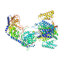
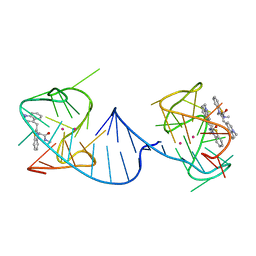
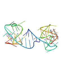
3KDJ
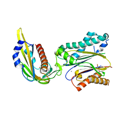 | | Complex structure of (+)-ABA-bound PYL1 and ABI1 | | 分子名称: | (2Z,4E)-5-[(1S)-1-hydroxy-2,6,6-trimethyl-4-oxocyclohex-2-en-1-yl]-3-methylpenta-2,4-dienoic acid, MANGANESE (II) ION, Protein phosphatase 2C 56, ... | | 著者 | Yin, P, Fan, H, Hao, Q, Yuan, X, Yan, N. | | 登録日 | 2009-10-23 | | 公開日 | 2009-11-10 | | 最終更新日 | 2023-11-01 | | 実験手法 | X-RAY DIFFRACTION (1.878 Å) | | 主引用文献 | Structural insights into the mechanism of abscisic acid signaling by PYL proteins
Nat.Struct.Mol.Biol., 16, 2009
|
|
3KDI
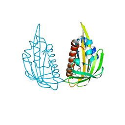 | | Structure of (+)-ABA bound PYL2 | | 分子名称: | (2Z,4E)-5-[(1S)-1-hydroxy-2,6,6-trimethyl-4-oxocyclohex-2-en-1-yl]-3-methylpenta-2,4-dienoic acid, Putative uncharacterized protein At2g26040 | | 著者 | Yin, P, Fan, H, Hao, Q, Yuan, X, Yan, N. | | 登録日 | 2009-10-22 | | 公開日 | 2009-11-10 | | 最終更新日 | 2023-11-01 | | 実験手法 | X-RAY DIFFRACTION (2.379 Å) | | 主引用文献 | Structural insights into the mechanism of abscisic acid signaling by PYL proteins
Nat.Struct.Mol.Biol., 16, 2009
|
|
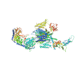
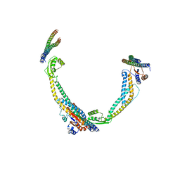
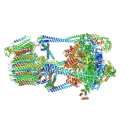
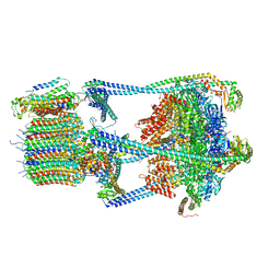
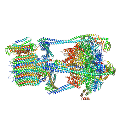
7U4T
 | | Human V-ATPase in state 2 with SidK and mEAK-7 | | 分子名称: | 2-acetamido-2-deoxy-beta-D-glucopyranose, ADENOSINE-5'-DIPHOSPHATE, MTOR-associated protein MEAK7, ... | | 著者 | Wang, L, Fu, T.M. | | 登録日 | 2022-03-01 | | 公開日 | 2022-08-24 | | 最終更新日 | 2023-09-06 | | 実験手法 | ELECTRON MICROSCOPY (3.6 Å) | | 主引用文献 | Identification of mEAK-7 as a human V-ATPase regulator via cryo-EM data mining.
Proc.Natl.Acad.Sci.USA, 119, 2022
|
|


