Loading
PDBj
MenuPDBj@FacebookPDBj@TwitterPDBj@YouTubewwPDB FoundationwwPDB
RCSB PDBPDBeBMRBAdv. SearchSearch help
Search by PDB author
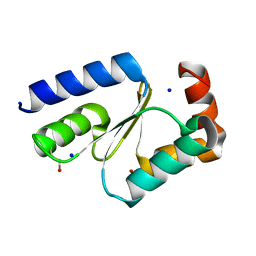
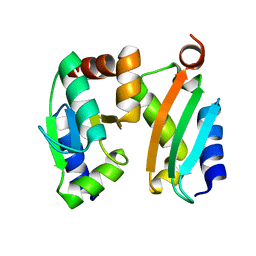
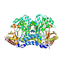
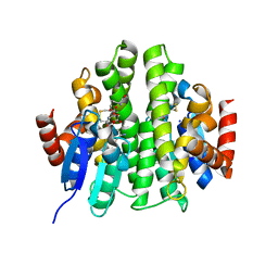
4Q75
DownloadVisualize
BU of 4q75 by Molmil
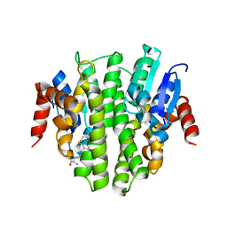
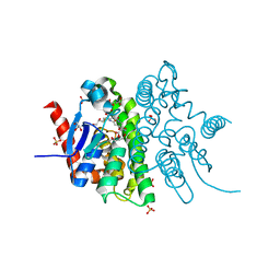
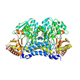
Crystal structure of Nfs2, the plastidial cysteine desulfurase from Arabidopsis thaliana
Descriptor: Cysteine desulfurase 2, chloroplastic
Authors:Roret, T, Didierjean, C.
Deposit date:2014-04-24
Release date:2014-09-10
Last modified:2023-12-06
Method:X-RAY DIFFRACTION (1.709 Å)
Cite:X-ray structures of Nfs2, the plastidial cysteine desulfurase from Arabidopsis thaliana.
Acta Crystallogr F Struct Biol Commun, 70, 2014
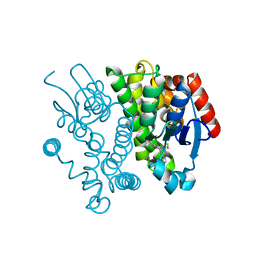
7NCW
DownloadVisualize
BU of 7ncw by Molmil
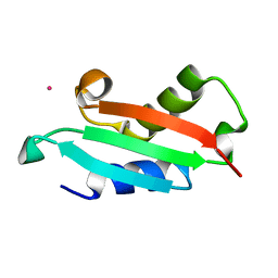
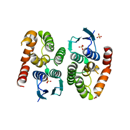
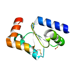
Crystal structure of oxidized glutaredoxin 2 from Chlamydomonas reinhardtii
Descriptor: ACETATE ION, Glutaredoxin, CPYC type
Authors:Roret, T, Didierjean, C.
Deposit date:2021-01-29
Release date:2021-05-26
Last modified:2024-10-23
Method:X-RAY DIFFRACTION (2.4 Å)
Cite:Atypical Iron-Sulfur Cluster Binding, Redox Activity and Structural Properties of Chlamydomonas reinhardtii Glutaredoxin 2.
Antioxidants (Basel), 10, 2021
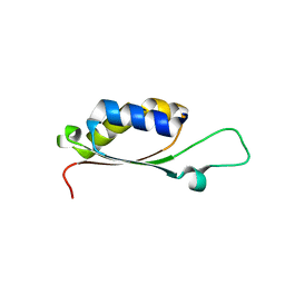
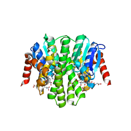
7NCV
DownloadVisualize
BU of 7ncv by Molmil
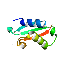
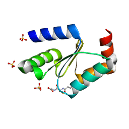
Crystal structure of reduced glutaredoxin 2 from Chlamydomonas reinhardtii
Descriptor: ACETATE ION, Glutaredoxin, CPYC type, ...
Authors:Roret, T, Didierjean, C.
Deposit date:2021-01-29
Release date:2021-05-26
Last modified:2024-10-23
Method:X-RAY DIFFRACTION (1.5 Å)
Cite:Atypical Iron-Sulfur Cluster Binding, Redox Activity and Structural Properties of Chlamydomonas reinhardtii Glutaredoxin 2.
Antioxidants (Basel), 10, 2021
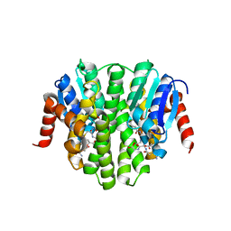
5NFL
DownloadVisualize
BU of 5nfl by Molmil
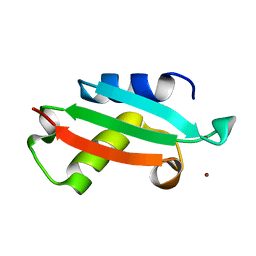
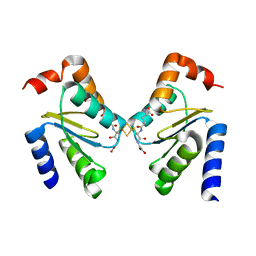
Crystal structure of YrbA from Sinorhizobium meliloti in complex with cobalt.
Descriptor: COBALT (II) ION, YrbA
Authors:Roret, T, Didierjean, C.
Deposit date:2017-03-14
Release date:2018-10-10
Last modified:2024-01-17
Method:X-RAY DIFFRACTION (1.903 Å)
Cite:Sinorhizobium meliloti YrbA binds divalent metal cations using two conserved histidines.
Biosci.Rep., 40, 2020
5NFK
DownloadVisualize
BU of 5nfk by Molmil
Crystal structure of YrbA from Sinorhizobium meliloti in complex with nickel.
Descriptor: LITHIUM ION, NICKEL (II) ION, YrbA
Authors:Roret, T, Didierjean, C.
Deposit date:2017-03-14
Release date:2018-10-10
Last modified:2024-05-08
Method:X-RAY DIFFRACTION (0.98 Å)
Cite:Sinorhizobium meliloti YrbA binds divalent metal cations using two conserved histidines.
Biosci.Rep., 40, 2020
5NFM
DownloadVisualize
BU of 5nfm by Molmil
Crystal structure of YrbA from Sinorhizobium meliloti in complex with copper.
Descriptor: COPPER (II) ION, LITHIUM ION, YrbA
Authors:Roret, T, Didierjean, C.
Deposit date:2017-03-14
Release date:2018-10-10
Last modified:2024-01-17
Method:X-RAY DIFFRACTION (0.8 Å)
Cite:Sinorhizobium meliloti YrbA binds divalent metal cations using two conserved histidines.
Biosci.Rep., 40, 2020
2MMA
DownloadVisualize
BU of 2mma by Molmil
NMR-based docking model of GrxS14-BolA2 apo-heterodimer from Arabidopsis thaliana
Descriptor: BolA2, Monothiol glutaredoxin-S14, chloroplastic
Authors:Roret, T, Tsan, P, Couturier, J, Rouhier, N, Didierjean, C.
Deposit date:2014-03-13
Release date:2014-07-23
Last modified:2024-05-01
Method:SOLUTION NMR
Cite:Structural and Spectroscopic Insights into BolA-Glutaredoxin Complexes.
J.Biol.Chem., 289, 2014
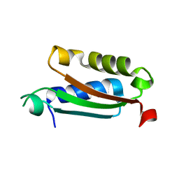
4PUG
DownloadVisualize
BU of 4pug by Molmil
BolA1 from Arabidopsis thaliana
Descriptor: BolA like protein
Authors:Roret, T, Didierjean, C.
Deposit date:2014-03-13
Release date:2014-07-23
Last modified:2023-09-20
Method:X-RAY DIFFRACTION (1.998 Å)
Cite:Structural and Spectroscopic Insights into BolA-Glutaredoxin Complexes.
J.Biol.Chem., 289, 2014
4PUI
DownloadVisualize
BU of 4pui by Molmil
BolA domain of SufE1 from Arabidopsis thaliana
Descriptor: SufE-like protein, chloroplastic
Authors:Roret, T, Didierjean, C.
Deposit date:2014-03-13
Release date:2014-07-23
Last modified:2023-09-20
Method:X-RAY DIFFRACTION (1.698 Å)
Cite:Structural and Spectroscopic Insights into BolA-Glutaredoxin Complexes.
J.Biol.Chem., 289, 2014
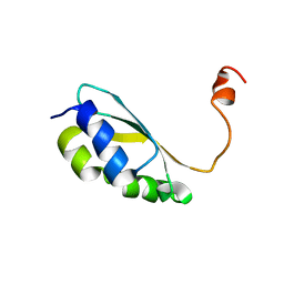
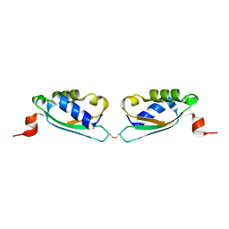
2MM9
DownloadVisualize
BU of 2mm9 by Molmil
Solution structure of reduced BolA2 from Arabidopsis thaliana
Descriptor: BolA2
Authors:Roret, T, Tsan, P, Couturier, J, Rouhier, N, Didierjean, C.
Deposit date:2014-03-13
Release date:2014-07-23
Last modified:2024-05-01
Method:SOLUTION NMR
Cite:Structural and Spectroscopic Insights into BolA-Glutaredoxin Complexes.
J.Biol.Chem., 289, 2014
4PUH
DownloadVisualize
BU of 4puh by Molmil
Oxidized BolA2 from Arabidopsis thaliana
Descriptor: At5g09830
Authors:Roret, T, Didierjean, C.
Deposit date:2014-03-13
Release date:2015-04-15
Last modified:2024-10-30
Method:X-RAY DIFFRACTION (1.898 Å)
Cite:Structural and spectroscopic insights into BolA-glutaredoxin complexes.
J.Biol.Chem., 289, 2014
4Q76
DownloadVisualize
BU of 4q76 by Molmil
Crystal structure of Nfs2 C384S mutant, the plastidial cysteine desulfurase from Arabidopsis thaliana
Descriptor: Cysteine desulfurase 2, chloroplastic
Authors:Roret, T, Didierjean, C.
Deposit date:2014-04-24
Release date:2014-09-10
Last modified:2023-12-06
Method:X-RAY DIFFRACTION (1.9 Å)
Cite:X-ray structures of Nfs2, the plastidial cysteine desulfurase from Arabidopsis thaliana.
Acta Crystallogr F Struct Biol Commun, 70, 2014
5O00
DownloadVisualize
BU of 5o00 by Molmil
Ure2p5 from Phanerochaete chrysosporium cocrystallized with 1-(S-glutathionyl)-2,4-dinitrobenzene.
Descriptor: DINITROPHENYLENE, GLYCEROL, Glutathione transferase Ure2p5, ...
Authors:Roret, T, Didierjean, C.
Deposit date:2017-05-15
Release date:2017-05-24
Last modified:2024-01-17
Method:X-RAY DIFFRACTION (2.03 Å)
Cite:Ure2p5 from Phanerochaete chrysosporium cocrystallized with 1-(S-glutathionyl)-2,4-dinitrobenzene.
To Be Published
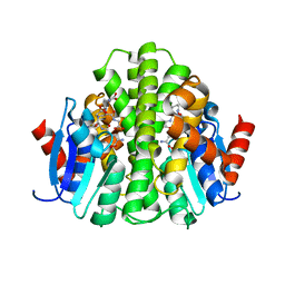
4ZBD
DownloadVisualize
BU of 4zbd by Molmil
Crystal structure of the glutathione transferase URE2P6 from Phanerochaete chrysosporium in complex with glutathione reduced by X-ray irradiation at 100K
Descriptor: GLUTATHIONE, PcUre2p6
Authors:Roret, T, Didierjean, C.
Deposit date:2015-04-14
Release date:2015-09-30
Last modified:2024-01-10
Method:X-RAY DIFFRACTION (1.12 Å)
Cite:Evolutionary divergence of Ure2pA glutathione transferases in wood degrading fungi.
Fungal Genet. Biol., 83, 2015
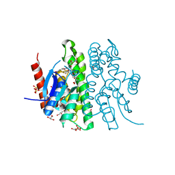
4ZB6
DownloadVisualize
BU of 4zb6 by Molmil
Crystal structure of glutathione transferase URE2P4 from Phanerochaete chrysosporium in complex with oxidized glutathione.
Descriptor: OXIDIZED GLUTATHIONE DISULFIDE, PcUre2p4, SODIUM ION
Authors:Roret, T, Didierjean, C.
Deposit date:2015-04-14
Release date:2015-09-30
Last modified:2024-01-10
Method:X-RAY DIFFRACTION (1.801 Å)
Cite:Evolutionary divergence of Ure2pA glutathione transferases in wood degrading fungi.
Fungal Genet. Biol., 83, 2015
4ZB8
DownloadVisualize
BU of 4zb8 by Molmil
Crystal structure of the glutathione transferase URE2P6 from Phanerochaete chrysosporium in complex with oxidized glutathione.
Descriptor: OXIDIZED GLUTATHIONE DISULFIDE, PcUre2p6
Authors:Roret, T, Didierjean, C.
Deposit date:2015-04-14
Release date:2015-09-30
Last modified:2024-01-10
Method:X-RAY DIFFRACTION (2 Å)
Cite:Evolutionary divergence of Ure2pA glutathione transferases in wood degrading fungi.
Fungal Genet. Biol., 83, 2015
4ZBB
DownloadVisualize
BU of 4zbb by Molmil
Crystal structure of the glutathione transferase URE2P8 from Phanerochaete chrysosporium complexed with glutathionyl-S-dinitrobenzene.
Descriptor: ACETATE ION, CHLORIDE ION, GLUTATHIONE S-(2,4 DINITROBENZENE), ...
Authors:Roret, T, Didierjean, C.
Deposit date:2015-04-14
Release date:2015-09-30
Last modified:2024-01-10
Method:X-RAY DIFFRACTION (1.8 Å)
Cite:Evolutionary divergence of Ure2pA glutathione transferases in wood degrading fungi.
Fungal Genet. Biol., 83, 2015
4ZBA
DownloadVisualize
BU of 4zba by Molmil
Crystal structure of the glutathione transferase URE2P8 from Phanerochaete chrysosporium with oxidized glutathione.
Descriptor: ACETATE ION, CHLORIDE ION, OXIDIZED GLUTATHIONE DISULFIDE, ...
Authors:Roret, T, Didierjean, C.
Deposit date:2015-04-14
Release date:2015-09-30
Last modified:2024-01-10
Method:X-RAY DIFFRACTION (1.501 Å)
Cite:Evolutionary divergence of Ure2pA glutathione transferases in wood degrading fungi.
Fungal Genet. Biol., 83, 2015
4ZB9
DownloadVisualize
BU of 4zb9 by Molmil
Crystal structure of the glutathione transferase URE2P8 from Phanerochaete chrysosporium, with one glutathione disulfide bound per dimer.
Descriptor: CHLORIDE ION, OXIDIZED GLUTATHIONE DISULFIDE, PcUre2p8
Authors:Roret, T, Didierjean, C.
Deposit date:2015-04-14
Release date:2015-09-30
Last modified:2024-01-10
Method:X-RAY DIFFRACTION (2.403 Å)
Cite:Evolutionary divergence of Ure2pA glutathione transferases in wood degrading fungi.
Fungal Genet. Biol., 83, 2015
4ZB7
DownloadVisualize
BU of 4zb7 by Molmil
Phanerochaete chrysosporium URE2P6 in apo form.
Descriptor: GLYCEROL, PcUre2p6, SULFATE ION
Authors:Roret, T, Didierjean, C.
Deposit date:2015-04-14
Release date:2015-09-30
Last modified:2024-01-10
Method:X-RAY DIFFRACTION (2.4 Å)
Cite:Evolutionary divergence of Ure2pA glutathione transferases in wood degrading fungi.
Fungal Genet. Biol., 83, 2015
3RHB
DownloadVisualize
BU of 3rhb by Molmil
Crystal structure of the apo form of glutaredoxin C5 from Arabidopsis thaliana
Descriptor: GLUTATHIONE, Glutaredoxin-C5, chloroplastic, ...
Authors:Roret, T, Couturier, J, Tsan, P, Jacquot, J.P, Rouhier, N, Didierjean, C.
Deposit date:2011-04-11
Release date:2011-06-01
Last modified:2023-09-13
Method:X-RAY DIFFRACTION (1.2 Å)
Cite:Arabidopsis chloroplastic glutaredoxin c5 as a model to explore molecular determinants for iron-sulfur cluster binding into glutaredoxins.
J.Biol.Chem., 286, 2011
3RHC
DownloadVisualize
BU of 3rhc by Molmil
Crystal structure of the holo form of glutaredoxin C5 from Arabidopsis thaliana
Descriptor: FE2/S2 (INORGANIC) CLUSTER, GLUTATHIONE, Glutaredoxin-C5, ...
Authors:Roret, T, Couturier, J, Tsan, P, Jacquot, J.P, Rouhier, N, Didierjean, C.
Deposit date:2011-04-11
Release date:2011-06-01
Last modified:2024-02-21
Method:X-RAY DIFFRACTION (2.4 Å)
Cite:Arabidopsis chloroplastic glutaredoxin c5 as a model to explore molecular determinants for iron-sulfur cluster binding into glutaredoxins.
J.Biol.Chem., 286, 2011
4F0C
DownloadVisualize
BU of 4f0c by Molmil
Crystal structure of the glutathione transferase URE2P5 from Phanerochaete chrysosporium
Descriptor: GLYCEROL, Glutathione transferase, OXIDIZED GLUTATHIONE DISULFIDE, ...
Authors:Didierjean, C, Favier, F, Roret, T.
Deposit date:2012-05-04
Release date:2013-06-12
Last modified:2023-09-13
Method:X-RAY DIFFRACTION (1.901 Å)
Cite:Evolutionary divergence of Ure2pA glutathione transferases in wood degrading fungi.
Fungal Genet Biol, 83, 2015
7AJ0
DownloadVisualize
BU of 7aj0 by Molmil
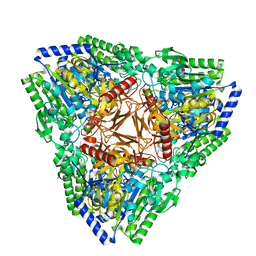
Crystal structure of PsFucS1 sulfatase from Pseudoalteromonas sp.
Descriptor: Arylsulfatase, CALCIUM ION, CHLORIDE ION
Authors:Roret, T, Mikkelsen, M.D, Czjzek, M, Meyer, A.S.
Deposit date:2020-09-28
Release date:2021-09-08
Last modified:2024-01-31
Method:X-RAY DIFFRACTION (2.5 Å)
Cite:A novel thermostable prokaryotic fucoidan active sulfatase PsFucS1 with an unusual quaternary hexameric structure.
Sci Rep, 11, 2021
6HHN
DownloadVisualize
BU of 6hhn by Molmil
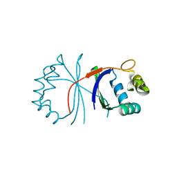
Crystal structure of L-rhamnose mutarotase FA22100 from Formosa agariphila
Descriptor: L-rhamnose mutarotase
Authors:Roret, T, Prechoux, A, Michel, G, Czjzek, M.
Deposit date:2018-08-28
Release date:2019-06-26
Last modified:2024-01-17
Method:X-RAY DIFFRACTION (1.47 Å)
Cite:A marine bacterial enzymatic cascade degrades the algal polysaccharide ulvan.
Nat.Chem.Biol., 15, 2019


123>

227111

PDB entries from 2024-11-06

PDB statisticsPDBj update infoContact PDBjnumon