Loading
PDBj
MenuPDBj@FacebookPDBj@TwitterPDBj@YouTubewwPDB FoundationwwPDB
RCSB PDBPDBeBMRBAdv. SearchSearch help
Search by PDB author
7UE2
DownloadVisualize
BU of 7ue2 by Molmil
Crystal structure of designed helical repeat protein RPB_PLP3_R6 bound to PLPx6 peptide
Descriptor: PLPx6 peptide, RPB_PLP3_R6
Authors:Chang, Y, Ekiert, D.C, Bhabha, G.
Deposit date:2022-03-21
Release date:2023-03-29
Last modified:2024-04-03
Method:X-RAY DIFFRACTION (2.68 Å)
Cite:De novo design of modular peptide-binding proteins by superhelical matching.
Nature, 616, 2023
3LR3
DownloadVisualize
BU of 3lr3 by Molmil
Periplasmic domain of the risS sensor protein from Burkholderia pesuromallei, low pH native structure
Descriptor: Sensor protein
Authors:Seattle Structural Genomics Center for Infectious Disease (SSGCID)
Deposit date:2010-02-10
Release date:2010-06-16
Last modified:2023-09-06
Method:X-RAY DIFFRACTION (2.1 Å)
Cite:Structural basis for pH sensing by the periplasmic domain of the risS histidine kinase from Burkholderia pseudomallei
To be Published
3LR0
DownloadVisualize
BU of 3lr0 by Molmil
Periplasmic domain of the risS sensor protein from Burkholderia pseudomallei, iodide phased at low pH
Descriptor: IODIDE ION, Sensor protein
Authors:Seattle Structural Genomics Center for Infectious Disease (SSGCID)
Deposit date:2010-02-10
Release date:2010-06-16
Last modified:2024-02-21
Method:X-RAY DIFFRACTION (1.9 Å)
Cite:Structural basis for pH sensing by the periplasmic domain of the risS histidine kinase from Bukholderia pseudomallei
To be Published
3LR5
DownloadVisualize
BU of 3lr5 by Molmil
Periplasmic domain of the risS sensor protein from Burkholderia pseudomallei, iodide phased at neutral pH
Descriptor: IODIDE ION, Sensor protein
Authors:Seattle Structural Genomics Center for Infectious Disease (SSGCID)
Deposit date:2010-02-10
Release date:2010-06-16
Last modified:2024-02-21
Method:X-RAY DIFFRACTION (2.3 Å)
Cite:Structural basis for pH sensing by the periplsamic domain of the risS histidine kinase from Burkholderia pseudomallei
To be Published
3LR4
DownloadVisualize
BU of 3lr4 by Molmil
Periplasmic domain of the risS sensor protein from Burkholderia pseudomallei, barium phased at low pH
Descriptor: BARIUM ION, CHLORIDE ION, Sensor protein
Authors:Seattle Structural Genomics Center for Infectious Disease (SSGCID)
Deposit date:2010-02-10
Release date:2010-06-16
Last modified:2024-02-21
Method:X-RAY DIFFRACTION (1.9 Å)
Cite:Structural basis for pH sensing by the periplasmic domain of the risS histidine kinase from Burkholderia pseudomallei
To be Published
3MBD
DownloadVisualize
BU of 3mbd by Molmil
Crystal structure of fructose bisphosphate aldolase from Encephalitozoon cuniculi, bound to phosphate
Descriptor: CHLORIDE ION, Fructose-bisphosphate aldolase, PHOSPHATE ION
Authors:Seattle Structural Genomics Center for Infectious Disease (SSGCID)
Deposit date:2010-03-25
Release date:2010-04-14
Last modified:2023-09-06
Method:X-RAY DIFFRACTION (2 Å)
Cite:Structure of fructose bisphosphate aldolase from Encephalitozoon cuniculi.
Acta Crystallogr.,Sect.F, 67, 2011
3MBF
DownloadVisualize
BU of 3mbf by Molmil
Crystal structure of fructose bisphosphate aldolase from Encephalitozoon cuniculi, bound to fructose 1,6-bisphosphate
Descriptor: 1,6-FRUCTOSE DIPHOSPHATE (LINEAR FORM), Fructose-bisphosphate aldolase
Authors:Seattle Structural Genomics Center for Infectious Disease (SSGCID)
Deposit date:2010-03-25
Release date:2010-04-07
Last modified:2023-09-06
Method:X-RAY DIFFRACTION (2.37 Å)
Cite:Structure of fructose bisphosphate aldolase from Encephalitozoon cuniculi.
Acta Crystallogr.,Sect.F, 67, 2011
3KRS
DownloadVisualize
BU of 3krs by Molmil
Structure of Triosephosphate Isomerase from Cryptosporidium Parvum at 1.55A Resolution
Descriptor: SODIUM ION, Triosephosphate isomerase, UNKNOWN ATOM OR ION
Authors:Seattle Structural Genomics Center for Infectious Disease (SSGCID)
Deposit date:2009-11-19
Release date:2009-12-01
Last modified:2023-09-06
Method:X-RAY DIFFRACTION (1.55 Å)
Cite:Structure of triosephosphate isomerase from Cryptosporidium parvum.
Acta Crystallogr.,Sect.F, 67, 2011
3LD9
DownloadVisualize
BU of 3ld9 by Molmil
Crystal structure of thymidylate kinase from Ehrlichia chaffeensis at 2.15A resolution
Descriptor: 1,2-ETHANEDIOL, SULFATE ION, Thymidylate kinase
Authors:Seattle Structural Genomics Center for Infectious Disease (SSGCID)
Deposit date:2010-01-12
Release date:2010-02-16
Last modified:2023-09-06
Method:X-RAY DIFFRACTION (2.15 Å)
Cite:Structure of thymidylate kinase from Ehrlichia chaffeensis.
Acta Crystallogr.,Sect.F, 67, 2011
3GMT
DownloadVisualize
BU of 3gmt by Molmil
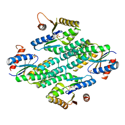
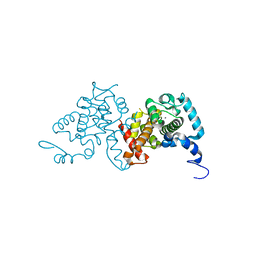
Crystal structure of adenylate kinase from burkholderia pseudomallei
Descriptor: Adenylate kinase, SULFATE ION
Authors:Abendroth, J, Staker, B.L, Robinson, H, Buchko, G.W, Hewitt, S.N, Napuli, A.J, Van Voorhis, W, Stacy, R, Myler, P.J, Stewart, L, Seattle Structural Genomics Center for Infectious Disease (SSGCID)
Deposit date:2009-03-15
Release date:2009-06-02
Last modified:2013-10-30
Method:X-RAY DIFFRACTION (2.1 Å)
Cite:Structural characterization of Burkholderia pseudomallei adenylate kinase (Adk): profound asymmetry in the crystal structure of the 'open' state.
Biochem.Biophys.Res.Commun., 394, 2010
3TMG
DownloadVisualize
BU of 3tmg by Molmil
Crystal structure of Glycine betaine, L-proline ABC transporter, glycine/betaine/L-proline-binding protein (ProX) from Borrelia burgdorferi
Descriptor: 1,2-ETHANEDIOL, Glycine betaine, L-proline ABC transporter, ...
Authors:Seattle Structural Genomics Center for Infectious Disease (SSGCID)
Deposit date:2011-08-31
Release date:2011-09-14
Last modified:2023-11-15
Method:X-RAY DIFFRACTION (1.9 Å)
Cite:Crystal structure of Glycine betaine, L-proline ABC transporter, glycine/betaine/L-proline-binding protein (ProX) from Borrelia burgdorferi
To be Published
3UAM
DownloadVisualize
BU of 3uam by Molmil
Crystal structure of a chitin binding domain from Burkholderia pseudomallei
Descriptor: Chitin binding domain, GLYCEROL, NITRATE ION
Authors:Seattle Structural Genomics Center for Infectious Disease (SSGCID)
Deposit date:2011-10-21
Release date:2011-11-02
Last modified:2023-09-13
Method:X-RAY DIFFRACTION (2 Å)
Cite:Crystal structure of a chitin binding domain from Burkholderia pseudomallei
to be published
3TSM
DownloadVisualize
BU of 3tsm by Molmil
Crystal structure of Indole-3-glycerol phosphate synthase from Brucella melitensis
Descriptor: GLYCEROL, Indole-3-glycerol phosphate synthase, SULFATE ION, ...
Authors:Seattle Structural Genomics Center for Infectious Disease (SSGCID)
Deposit date:2011-09-13
Release date:2011-10-05
Last modified:2023-09-13
Method:X-RAY DIFFRACTION (2.15 Å)
Cite:Crystal structure of Indole-3-glycerol phosphate synthase from Brucella melitensis
To be Published
3U5W
DownloadVisualize
BU of 3u5w by Molmil
Crystal Structure of a probable FAD-binding, putative uncharacterized protein from Brucella melitensis, apo form
Descriptor: 1,2-ETHANEDIOL, CHLORIDE ION, Putative uncharacterized protein
Authors:Seattle Structural Genomics Center for Infectious Disease (SSGCID)
Deposit date:2011-10-11
Release date:2011-10-19
Last modified:2023-09-13
Method:X-RAY DIFFRACTION (2.05 Å)
Cite:Crystal Structure of a probable FAD-binding, putative uncharacterized protein from Brucella melitensis, apo form
To be Published
3U0I
DownloadVisualize
BU of 3u0i by Molmil
Crystal Structure of a probable FAD-binding, putative uncharacterized protein from Brucella melitensis
Descriptor: 1,2-ETHANEDIOL, 2-[N-CYCLOHEXYLAMINO]ETHANE SULFONIC ACID, probable FAD-binding, ...
Authors:Seattle Structural Genomics Center for Infectious Disease (SSGCID)
Deposit date:2011-09-28
Release date:2011-10-12
Last modified:2023-09-13
Method:X-RAY DIFFRACTION (2.2 Å)
Cite:Crystal Structure of a probable FAD-binding, putative uncharacterized protein from Brucella melitensis
To be Published
3SC4
DownloadVisualize
BU of 3sc4 by Molmil
Crystal structure of a Short chain dehydrogenase (A0QTM2 homolog) Mycobacterium thermoresistibile
Descriptor: 1,2-ETHANEDIOL, CHLORIDE ION, GLYCEROL, ...
Authors:Seattle Structural Genomics Center for Infectious Disease (SSGCID)
Deposit date:2011-06-06
Release date:2011-06-29
Last modified:2023-09-13
Method:X-RAY DIFFRACTION (2.5 Å)
Cite:Crystal structure of a Short chain dehydrogenase (A0QTM2 homolog) Mycobacterium thermoresistibile
To be Published
3SIB
DownloadVisualize
BU of 3sib by Molmil
Crystal structure of URE3-binding protein, wild-type
Descriptor: CALCIUM ION, SODIUM ION, URE3-BP sequence specific DNA binding protein
Authors:Seattle Structural Genomics Center for Infectious Disease (SSGCID)
Deposit date:2011-06-17
Release date:2011-06-29
Last modified:2023-09-13
Method:X-RAY DIFFRACTION (1.9 Å)
Cite:Crystal structure of URE3-binding protein
To be Published
3SJS
DownloadVisualize
BU of 3sjs by Molmil
Crystal structure of URE3-binding protein from Entamoeba histolytica, (D127A,N129A) mutant, native form
Descriptor: CALCIUM ION, URE3-BP sequence specific DNA binding protein
Authors:Seattle Structural Genomics Center for Infectious Disease (SSGCID)
Deposit date:2011-06-21
Release date:2011-07-06
Last modified:2023-09-13
Method:X-RAY DIFFRACTION (1.9 Å)
Cite:Crystal structure of URE3-binding protein from Entamoeba histolytica
to be published
3SIA
DownloadVisualize
BU of 3sia by Molmil
Crystal structure of URE3-binding protein, (D127A,N129A) mutant, iodide phased
Descriptor: CALCIUM ION, CHLORIDE ION, IODIDE ION, ...
Authors:Seattle Structural Genomics Center for Infectious Disease (SSGCID), Skubak, P.
Deposit date:2011-06-17
Release date:2011-06-29
Last modified:2024-02-28
Method:X-RAY DIFFRACTION (2.05 Å)
Cite:Crystal structure of URE3-binding protein
To be Published
3SW5
DownloadVisualize
BU of 3sw5 by Molmil
Crystal structure of inorganic pyrophosphatase from Bartonella henselae
Descriptor: (2S)-2-hydroxybutanedioic acid, Inorganic pyrophosphatase
Authors:Seattle Structural Genomics Center for Infectious Disease, Seattle Structural Genomics Center for Infectious Disease (SSGCID)
Deposit date:2011-07-13
Release date:2011-08-10
Last modified:2023-09-13
Method:X-RAY DIFFRACTION (2 Å)
Cite:Crystal structure of inorganic pyrophosphatase from Bartonella henselae
To be Published
3S6O
DownloadVisualize
BU of 3s6o by Molmil
Crystal structure of a Polysaccharide deacetylase family protein from Burkholderia pseudomallei
Descriptor: 1,2-ETHANEDIOL, Polysaccharide deacetylase family protein
Authors:Seattle Structural Genomics Center for Infectious Disease (SSGCID)
Deposit date:2011-05-25
Release date:2011-06-15
Last modified:2023-09-13
Method:X-RAY DIFFRACTION (1.85 Å)
Cite:Crystal structure of a Polysaccharide deacetylase family protein from Burkholderia pseudomallei
To be Published
3UJH
DownloadVisualize
BU of 3ujh by Molmil
Crystal structure of substrate-bound Glucose-6-phosphate isomerase from Toxoplasma gondii
Descriptor: 1,2-ETHANEDIOL, CHLORIDE ION, GLUCOSE-6-PHOSPHATE, ...
Authors:Seattle Structural Genomics Center for Infectious Disease (SSGCID)
Deposit date:2011-11-07
Release date:2011-11-16
Last modified:2023-09-13
Method:X-RAY DIFFRACTION (2.1 Å)
Cite:Crystal structure of substrate-bound Glucose-6-phosphate isomerase from Toxoplasma gondii
To be Published
4G6C
DownloadVisualize
BU of 4g6c by Molmil
Crystal structure of beta-hexosaminidase 1 from Burkholderia cenocepacia J2315
Descriptor: Beta-hexosaminidase 1
Authors:Seattle Structural Genomics Center for Infectious Disease (SSGCID)
Deposit date:2012-07-18
Release date:2012-08-01
Last modified:2024-02-28
Method:X-RAY DIFFRACTION (1.38 Å)
Cite:Crystal structure of beta-hexosaminidase 1 from Burkholderia cenocepacia J2315
To be Published
4FUR
DownloadVisualize
BU of 4fur by Molmil
Crystal Structure of Urease subunit gamma 2 from Brucella melitensis biovar Abortus 2308
Descriptor: 1,2-ETHANEDIOL, CHLORIDE ION, Urease subunit gamma 2
Authors:Seattle Structural Genomics Center for Infectious Disease (SSGCID)
Deposit date:2012-06-28
Release date:2012-08-15
Last modified:2023-09-13
Method:X-RAY DIFFRACTION (2.1 Å)
Cite:Crystal Structure of Urease subunit gamma 2 from Brucella melitensis biovar Abortus 2308
TO BE PUBLISHED
4DS3
DownloadVisualize
BU of 4ds3 by Molmil
Crystal Structure of Phosphoribosylglycinamide formyltransferase from Brucella melitensis
Descriptor: CHLORIDE ION, GLYCEROL, Phosphoribosylglycinamide formyltransferase, ...
Authors:Seattle Structural Genomics Center for Infectious Disease (SSGCID)
Deposit date:2012-02-17
Release date:2012-04-04
Last modified:2023-09-13
Method:X-RAY DIFFRACTION (1.85 Å)
Cite:Crystal Structure of Phosphoribosylglycinamide formyltransferase from Brucella melitensis
TO BE PUBLISHED

222926

PDB entries from 2024-07-24

PDB statisticsPDBj update infoContact PDBjnumon