Loading
PDBj
MenuPDBj@FacebookPDBj@TwitterPDBj@YouTubewwPDB FoundationwwPDB
RCSB PDBPDBeBMRBAdv. SearchSearch help
Search by PDB author
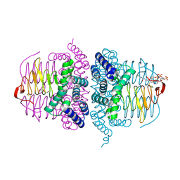
5XGG
DownloadVisualize
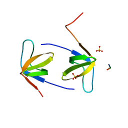
BU of 5xgg by Molmil
Crystal Structure C-terminal SH3 domain of Myosin IB from Entamoeba histolytica
Descriptor: SULFATE ION, Unconventional myosin IB
Authors:Gautam, G, Gourinath, S.
Deposit date:2017-04-13
Release date:2017-08-16
Last modified:2023-11-22
Method:X-RAY DIFFRACTION (1.72 Å)
Cite:Crystal structure of the PEG-bound SH3 domain of myosin IB from Entamoeba histolytica reveals its mode of ligand recognition
Acta Crystallogr D Struct Biol, 73, 2017
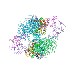
5ZKK
DownloadVisualize
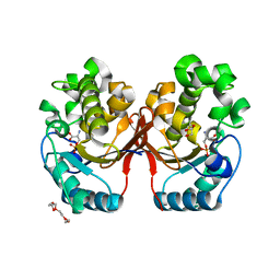
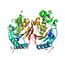
BU of 5zkk by Molmil
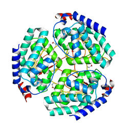
Crystal structure of Phosphoserine phosphatase from Entamoeba histolytica
Descriptor: BETA-MERCAPTOETHANOL, DI(HYDROXYETHYL)ETHER, GLYCEROL, ...
Authors:Kumari, P, Gourinath, S.
Deposit date:2018-03-24
Release date:2019-04-03
Last modified:2023-11-22
Method:X-RAY DIFFRACTION (2 Å)
Cite:Structural and functional characterisation of phosphoserine phosphatase, that plays critical role in the oxidative stress response in the parasite Entamoeba histolytica.
J.Struct.Biol., 206, 2019
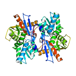
5Z51
DownloadVisualize
BU of 5z51 by Molmil
Helicase binding domain of primase from Mycobacterium tuberculosis
Descriptor: ACETATE ION, DI(HYDROXYETHYL)ETHER, DNA primase
Authors:Sharma, D.P, Gourinath, S.
Deposit date:2018-01-16
Release date:2018-11-28
Last modified:2024-10-30
Method:X-RAY DIFFRACTION (1.583 Å)
Cite:Structural insights into the interaction of helicase and primase inMycobacterium tuberculosis.
Biochem. J., 475, 2018
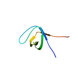
6A9C
DownloadVisualize
BU of 6a9c by Molmil
Crystal Structure c-terminal SH3 domain of Myosin IB from Entamoeba histolytica bound to EhFP10(GEF) peptide.
Descriptor: Peptide from Rho guanine nucleotide exchange factor, SULFATE ION, Unconventional myosin IB
Authors:Gautam, G, Gourinath, S.
Deposit date:2018-07-13
Release date:2019-06-12
Last modified:2023-11-22
Method:X-RAY DIFFRACTION (1.98 Å)
Cite:EhFP10: A FYVE family GEF interacts with myosin IB to regulate cytoskeletal dynamics during endocytosis in Entamoeba histolytica.
Plos Pathog., 15, 2019
5XG9
DownloadVisualize
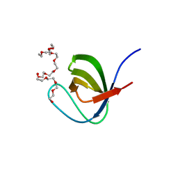
BU of 5xg9 by Molmil
Crystal Structure of PEG-bound SH3 domain of Myosin IB from Entamoeba histolytica
Descriptor: 1-(2-METHOXY-ETHOXY)-2-{2-[2-(2-METHOXY-ETHOXY]-ETHOXY}-ETHANE, 2,5,8,11,14,17,20,23,26,29,32,35,38,41,44,47,50,53,56,59,62,65,68,71,74,77,80-HEPTACOSAOXADOOCTACONTAN-82-OL, PENTAETHYLENE GLYCOL, ...
Authors:Gautam, G, Gourinath, S.
Deposit date:2017-04-13
Release date:2017-08-16
Last modified:2023-11-22
Method:X-RAY DIFFRACTION (1.78 Å)
Cite:Crystal structure of the PEG-bound SH3 domain of myosin IB from Entamoeba histolytica reveals its mode of ligand recognition
Acta Crystallogr D Struct Biol, 73, 2017
5ZR2
DownloadVisualize
BU of 5zr2 by Molmil
Crystal Structure of Phosphoserine Phosphatase Mutant (H9A) from Entamoeba histolytica in complex with Phosphoserine
Descriptor: DI(HYDROXYETHYL)ETHER, PHOSPHOSERINE, Phosphoglycerate mutase family protein, ...
Authors:Kumari, P, Gourinath, S.
Deposit date:2018-04-23
Release date:2019-04-24
Last modified:2023-11-22
Method:X-RAY DIFFRACTION (2.95 Å)
Cite:Structural and functional characterisation of phosphoserine phosphatase, that plays critical role in the oxidative stress response in the parasite Entamoeba histolytica.
J.Struct.Biol., 206, 2019
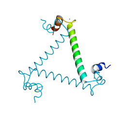
3LI6
DownloadVisualize
BU of 3li6 by Molmil
Crystal structure and trimer-monomer transition of N-terminal domain of EhCaBP1 from Entamoeba histolytica
Descriptor: CALCIUM ION, Calcium-binding protein
Authors:Kumar, S, Ahmad, E, Kumar, S, Mansuri, M.S, Khan, R.H, Samudrala, G.
Deposit date:2010-01-24
Release date:2010-02-02
Last modified:2023-11-01
Method:X-RAY DIFFRACTION (2.502 Å)
Cite:Crystal structure and trimer-monomer transition of N-terminal domain of EhCaBP1 from Entamoeba histolytica
Biophys.J., 98, 2010
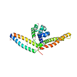
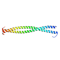
3BAS
DownloadVisualize
BU of 3bas by Molmil
Crystal structure of the N-terminal region of the scallop myosin rod, monoclinic (C2) form
Descriptor: IODIDE ION, Myosin heavy chain, striated muscle/General control protein GCN4 chimera
Authors:Brown, J.H, Cohen, C.
Deposit date:2007-11-08
Release date:2008-01-08
Last modified:2023-08-30
Method:X-RAY DIFFRACTION (2.3 Å)
Cite:An unstable head-rod junction may promote folding into the compact off-state conformation of regulated myosins.
J.Mol.Biol., 375, 2008
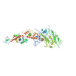
2EC6
DownloadVisualize
BU of 2ec6 by Molmil
Placopecten Striated Muscle Myosin II
Descriptor: CALCIUM ION, Myosin essential light chain, Myosin heavy chain, ...
Authors:Yang, Y, Brown, J, Samudrala, G, Reutzel, R, Szent-Gyorgyi, A.
Deposit date:2007-02-10
Release date:2008-02-26
Last modified:2024-05-29
Method:X-RAY DIFFRACTION (3.25 Å)
Cite:Rigor-like structures from muscle myosins reveal key mechanical elements in the transduction pathways of this allosteric motor.
Structure, 15, 2007
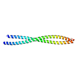
3BAT
DownloadVisualize
BU of 3bat by Molmil
Crystal structure of the N-terminal region of the scallop myosin rod, monoclinic (P21) form
Descriptor: Myosin heavy chain, striated muscle/General control protein GCN4
Authors:Brown, J.H, Cohen, C.
Deposit date:2007-11-08
Release date:2008-01-08
Last modified:2023-08-30
Method:X-RAY DIFFRACTION (2.3 Å)
Cite:An unstable head-rod junction may promote folding into the compact off-state conformation of regulated myosins.
J.Mol.Biol., 375, 2008
7BW9
DownloadVisualize
BU of 7bw9 by Molmil
Crystal structure of Serine acetyltransferase isoform 3 in complex with cysteine from Entamoeba histolytica
Descriptor: CYSTEINE, Serine acetyltransferase 1, putative
Authors:Dharavath, S, Kumar, S, Gourinath, S.
Deposit date:2020-04-14
Release date:2021-04-14
Last modified:2023-11-29
Method:X-RAY DIFFRACTION (2.131 Å)
Cite:Crystal structure of Serine acetyltransferase isoform 3 in complex with cysteine from Entamoeba histolytica
To Be Published
4TUN
DownloadVisualize
BU of 4tun by Molmil
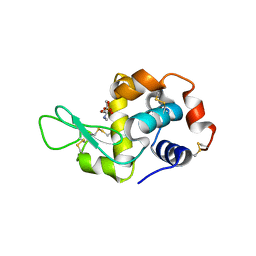
Crystal structure of Chicken egg white lysozyme adduct with Organophosphorus pesticide Monochrotophos
Descriptor: ACETATE ION, Lysozyme C, methyl [4-(methylamino)-4-oxidanylidene-but-2-en-2-yl] hydrogen phosphate
Authors:Amaraneni, S.R, Kumar, S, Samudrala, G.
Deposit date:2014-06-24
Release date:2014-07-09
Last modified:2023-12-27
Method:X-RAY DIFFRACTION (1.925 Å)
Cite:Biophysical aspects of lysozyme adduct with monocrotophos.
Anal Bioanal Chem, 406, 2014
2OSN
DownloadVisualize
BU of 2osn by Molmil
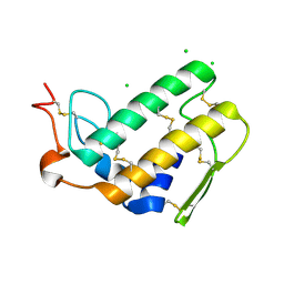
An alternate description of a crystal structure of phospholipase A2 from Bungarus caeruleus
Descriptor: CHLORIDE ION, Phospholipase A2 isoform 3
Authors:Stenkamp, R.E, Le Trong, I.
Deposit date:2007-02-06
Release date:2007-03-06
Last modified:2023-08-30
Method:X-RAY DIFFRACTION (2.5 Å)
Cite:An alternate description of two crystal structures of phospholipase A(2) from Bungarus caeruleus.
Acta Crystallogr.,Sect.D, 63, 2007
4HZC
DownloadVisualize
BU of 4hzc by Molmil
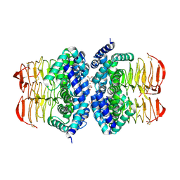
Crystal structure of Serine acetyltransferase from Brucella abortus strain S19
Descriptor: 2-AMINO-2-HYDROXYMETHYL-PROPANE-1,3-DIOL, CHLORIDE ION, CysE, ...
Authors:Kumar, S, Samudrala, G.
Deposit date:2012-11-15
Release date:2014-02-19
Last modified:2024-10-30
Method:X-RAY DIFFRACTION (1.97 Å)
Cite:Crystal structure of serine acetyl transferase from Brucella abortus and its complex with coenzyme A.
Biochim.Biophys.Acta, 1844, 2014
4HZD
DownloadVisualize
BU of 4hzd by Molmil
Crystal structure of Serine acetyltransferase in complex with Coenzyme A from Brucella abortus strain S19
Descriptor: COENZYME A, CysE, serine acetyltransferase, ...
Authors:Kumar, S, Samudrala, G.
Deposit date:2012-11-15
Release date:2014-02-19
Last modified:2014-08-13
Method:X-RAY DIFFRACTION (1.87 Å)
Cite:Crystal structure of serine acetyl transferase from Brucella abortus and its complex with coenzyme A.
Biochim.Biophys.Acta, 1844, 2014
1XJW
DownloadVisualize
BU of 1xjw by Molmil
The Structure of E. coli Aspartate Transcarbamoylase Q137A Mutant in The R-State
Descriptor: Aspartate carbamoyltransferase catalytic chain, Aspartate carbamoyltransferase regulatory chain, N-(PHOSPHONACETYL)-L-ASPARTIC ACID, ...
Authors:Stieglitz, K.A, Alam, N, Xia, J, Gourinath, S, Tsuruta, H, Kantrowitz, E.R.
Deposit date:2004-09-25
Release date:2005-05-24
Last modified:2023-08-23
Method:X-RAY DIFFRACTION (2.71 Å)
Cite:A Single Amino Acid Substitution in the Active Site of Escherichia coli Aspartate Transcarbamoylase Prevents the Allosteric Transition.
J.Mol.Biol., 349, 2005
6AHI
DownloadVisualize
BU of 6ahi by Molmil
Crystal structure of O-acetylserine dependent cystathionine beta-synthase from Helicobacter pylori.
Descriptor: Cysteine synthase, METHIONINE
Authors:Tarique, F.K, Devi, S, Rehman, S.A.A.
Deposit date:2018-08-19
Release date:2018-09-05
Last modified:2023-11-22
Method:X-RAY DIFFRACTION (1.9 Å)
Cite:Identification and characterization of Helicobacter pylori O-acetylserine-dependent cystathionine beta-synthase, a distinct member of the PLP-II family.
Mol.Microbiol., 112, 2019

226707

PDB entries from 2024-10-30

PDB statisticsPDBj update infoContact PDBjnumon