Loading
PDBj
MenuPDBj@FacebookPDBj@TwitterPDBj@YouTubewwPDB FoundationwwPDB
RCSB PDBPDBeBMRBAdv. SearchSearch help
Search by PDB author
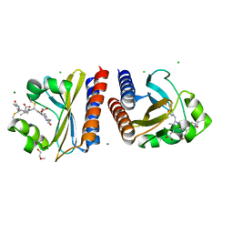
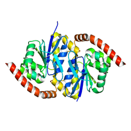
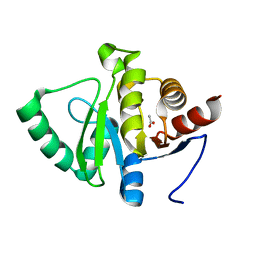
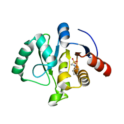
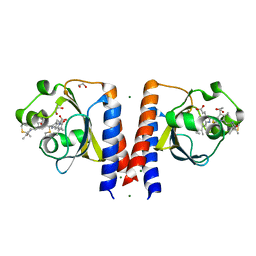
6P58
DownloadVisualize
BU of 6p58 by Molmil
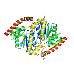
Dark and Steady State-Illuminated Crystal Structure of Cyanobacteriochrome Receptor PixJ at 150K
Descriptor: 1,2-ETHANEDIOL, MAGNESIUM ION, Methyl-accepting chemotaxis protein, ...
Authors:Clinger, J.A, Miller, M.D, Buirgie, E.S, Vierstra, R.D, Phillips Jr, G.N.
Deposit date:2019-05-29
Release date:2019-12-18
Last modified:2024-10-09
Method:X-RAY DIFFRACTION (1.499 Å)
Cite:Photoreversible interconversion of a phytochrome photosensory module in the crystalline state.
Proc.Natl.Acad.Sci.USA, 117, 2020
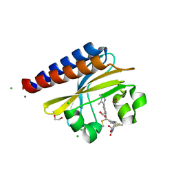
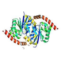
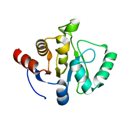
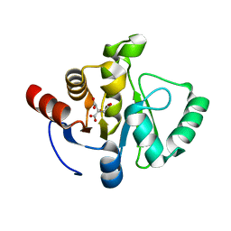
6PRY
DownloadVisualize
BU of 6pry by Molmil
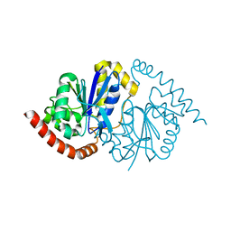
X-ray crystal structure of the blue-light absorbing state of PixJ from Thermosynechococcus elongatus by serial femtosecond crystallographic analysis
Descriptor: CHLORIDE ION, DI(HYDROXYETHYL)ETHER, MAGNESIUM ION, ...
Authors:Burgie, E.S, Clinger, J.A, Miller, M.D, Phillips Jr, G.N, Vierstra, R.D, Orville, A.M, Kern, J.F.
Deposit date:2019-07-12
Release date:2019-12-18
Last modified:2024-10-16
Method:X-RAY DIFFRACTION (1.55 Å)
Cite:Photoreversible interconversion of a phytochrome photosensory module in the crystalline state.
Proc.Natl.Acad.Sci.USA, 117, 2020
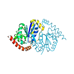
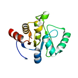
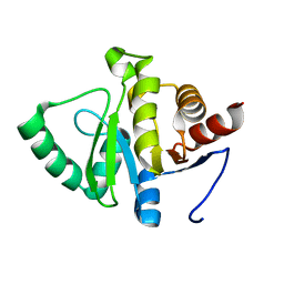
6PRU
DownloadVisualize
BU of 6pru by Molmil
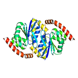
Photoconvertible crystals of PixJ from Thermosynechococcus elongatus
Descriptor: 1,2-ETHANEDIOL, CHLORIDE ION, DI(HYDROXYETHYL)ETHER, ...
Authors:Burgie, E.S, Clinger, J.A, Miller, M.D, Phillips Jr, G.N, Vierstra, R.D.
Deposit date:2019-07-11
Release date:2019-12-18
Last modified:2024-10-30
Method:X-RAY DIFFRACTION (1.539 Å)
Cite:Photoreversible interconversion of a phytochrome photosensory module in the crystalline state.
Proc.Natl.Acad.Sci.USA, 117, 2020
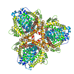
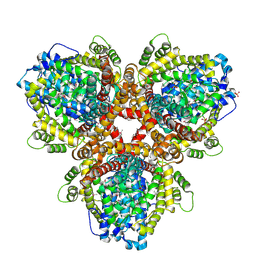
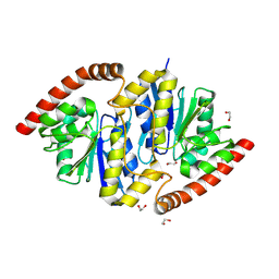
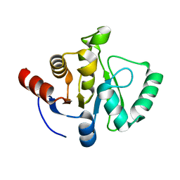
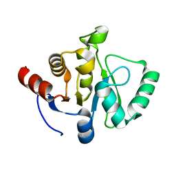
6OIW
DownloadVisualize
BU of 6oiw by Molmil
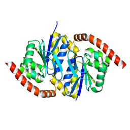
Structure of Escherichia coli dGTPase bound to dGTP-1-thiol
Descriptor: 2'-deoxyguanosine-5'-O-(1-thiotriphosphate), Deoxyguanosinetriphosphate triphosphohydrolase, MAGNESIUM ION, ...
Authors:Barnes, C.O, Wu, Y, Calero, G.
Deposit date:2019-04-09
Release date:2019-05-15
Last modified:2024-03-13
Method:X-RAY DIFFRACTION (3.35 Å)
Cite:The crystal structure of dGTPase reveals the molecular basis of dGTP selectivity.
Proc.Natl.Acad.Sci.USA, 116, 2019
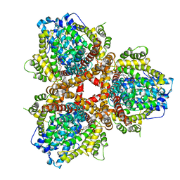
6OIY
DownloadVisualize
BU of 6oiy by Molmil
Structure of Escherichia coli bound to dGTP
Descriptor: 2'-DEOXYGUANOSINE-5'-TRIPHOSPHATE, Deoxyguanosinetriphosphate triphosphohydrolase, MANGANESE (II) ION
Authors:Barnes, C.O, Wu, Y, Calero, G.
Deposit date:2019-04-09
Release date:2019-05-15
Last modified:2024-03-13
Method:X-RAY DIFFRACTION (3.29 Å)
Cite:The crystal structure of dGTPase reveals the molecular basis of dGTP selectivity.
Proc.Natl.Acad.Sci.USA, 116, 2019
6OI7
DownloadVisualize
BU of 6oi7 by Molmil
Se-Met structure of apo- Escherichia coli dGTPase
Descriptor: Deoxyguanosinetriphosphate triphosphohydrolase, MANGANESE (II) ION, SULFATE ION
Authors:Calero, G, Barnes, C.O, Wu, Y.
Deposit date:2019-04-08
Release date:2019-05-29
Last modified:2020-01-01
Method:X-RAY DIFFRACTION (2.9 Å)
Cite:The crystal structure of dGTPase reveals the molecular basis of dGTP selectivity.
Proc.Natl.Acad.Sci.USA, 116, 2019
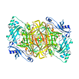
1N9E
DownloadVisualize
BU of 1n9e by Molmil
Crystal structure of Pichia pastoris Lysyl Oxidase PPLO
Descriptor: 2-acetamido-2-deoxy-beta-D-glucopyranose, 2-acetamido-2-deoxy-beta-D-glucopyranose-(1-4)-2-acetamido-2-deoxy-beta-D-glucopyranose, CALCIUM ION, ...
Authors:Guss, J.M, Duff, A.P.
Deposit date:2002-11-24
Release date:2004-01-13
Last modified:2023-10-25
Method:X-RAY DIFFRACTION (1.65 Å)
Cite:The Crystal Structure of Pichia pastoris Lysyl Oxidase
Biochemistry, 42, 2003
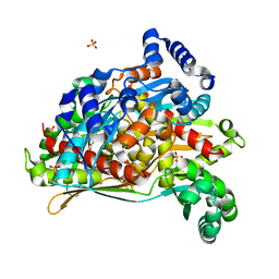
8SZ6
DownloadVisualize
BU of 8sz6 by Molmil
PmHMGR bound to mevaldehyde and CoA
Descriptor: (3R)-3,5,5-trihydroxy-3-methylpentanoic acid, 3-hydroxy-3-methylglutaryl-coenzyme A reductase, Mevaldyl-Coenzyme A, ...
Authors:Purohit, V, Stauffacher, C.V, Steussy, C.N.
Deposit date:2023-05-27
Release date:2024-03-20
Method:X-RAY DIFFRACTION (1.65 Å)
Cite:pH-dependent reaction triggering in PmHMGR crystals for time-resolved crystallography.
Biophys.J., 123, 2024
8TSY
DownloadVisualize
BU of 8tsy by Molmil
Pseudomonas fluorescens G150T-2 isocyanide hydratase at 274 K
Descriptor: CHLORIDE ION, Isonitrile hydratase InhA
Authors:Wilson, M.A, Smith, N, Dasgupta, M, Dolamore, C.
Deposit date:2023-08-12
Release date:2023-09-20
Method:X-RAY DIFFRACTION (1.2 Å)
Cite:Changes in an Enzyme Ensemble During Catalysis Observed by High Resolution XFEL Crystallography.
Biorxiv, 2023
8TT0
DownloadVisualize
BU of 8tt0 by Molmil
Pseudomonas fluorescens isocyanide hydratase pH=4.2
Descriptor: Isonitrile hydratase InhA
Authors:Wilson, M.A, Smith, N, Dasgupta, M, Dolamore, C.
Deposit date:2023-08-12
Release date:2023-09-20
Method:X-RAY DIFFRACTION (1.5 Å)
Cite:Changes in an Enzyme Ensemble During Catalysis Observed by High Resolution XFEL Crystallography.
Biorxiv, 2023
8TT2
DownloadVisualize
BU of 8tt2 by Molmil
Pseudomonas fluorescens isocyanide hydratase pH=5.4
Descriptor: 1,2-ETHANEDIOL, Isonitrile hydratase InhA
Authors:Wilson, M.A, Smith, N, Dasgupta, M, Dolamore, C.
Deposit date:2023-08-12
Release date:2023-09-20
Method:X-RAY DIFFRACTION (1.33 Å)
Cite:Changes in an Enzyme Ensemble During Catalysis Observed by High Resolution XFEL Crystallography.
Biorxiv, 2023
8TSZ
DownloadVisualize
BU of 8tsz by Molmil
Pseudomonas fluorescens G150T-3 isocyanide hydratase at 274 K
Descriptor: CHLORIDE ION, Isonitrile hydratase InhA
Authors:Wilson, M.A, Smith, N, Dasgupta, M, Dolamore, C.
Deposit date:2023-08-12
Release date:2023-09-20
Method:X-RAY DIFFRACTION (1.1 Å)
Cite:Changes in an Enzyme Ensemble During Catalysis Observed by High Resolution XFEL Crystallography.
Biorxiv, 2023
8TT4
DownloadVisualize
BU of 8tt4 by Molmil
Pseudomonas fluorescens isocyanide hydratase pH=6.0
Descriptor: 1,2-ETHANEDIOL, Isonitrile hydratase InhA
Authors:Wilson, M.A, Smith, N, Dasgupta, M, Dolamore, C.
Deposit date:2023-08-12
Release date:2023-09-20
Method:X-RAY DIFFRACTION (1.2 Å)
Cite:Changes in an Enzyme Ensemble During Catalysis Observed by High Resolution XFEL Crystallography.
Biorxiv, 2023
8TT5
DownloadVisualize
BU of 8tt5 by Molmil
Pseudomonas fluorescens isocyanide hydratase pH=8.3
Descriptor: 1,2-ETHANEDIOL, Isonitrile hydratase InhA
Authors:Wilson, M.A, Smith, N, Dasgupta, M, Dolamore, C.
Deposit date:2023-08-12
Release date:2023-09-20
Method:X-RAY DIFFRACTION (1.02 Å)
Cite:Changes in an Enzyme Ensemble During Catalysis Observed by High Resolution XFEL Crystallography.
Biorxiv, 2023
8TSU
DownloadVisualize
BU of 8tsu by Molmil
Pseudomonas fluorescens G150T-1 isocyanide hydratase at 274 K
Descriptor: CHLORIDE ION, Isonitrile hydratase InhA
Authors:Wilson, M.A, Smith, N, Dasgupta, M, Dolamore, C.
Deposit date:2023-08-11
Release date:2023-09-20
Method:X-RAY DIFFRACTION (1.15 Å)
Cite:Changes in an Enzyme Ensemble During Catalysis Observed by High Resolution XFEL Crystallography.
Biorxiv, 2023
8TSX
DownloadVisualize
BU of 8tsx by Molmil
Pseudomonas fluorescens G150T isocyanide hydratase at 100 K
Descriptor: CHLORIDE ION, Isonitrile hydratase InhA
Authors:Wilson, M.A, Smith, N, Dasgupta, M, Dolamore, C.
Deposit date:2023-08-12
Release date:2023-09-20
Method:X-RAY DIFFRACTION (1 Å)
Cite:Changes in an Enzyme Ensemble During Catalysis Observed by High Resolution XFEL Crystallography.
Biorxiv, 2023
8TT1
DownloadVisualize
BU of 8tt1 by Molmil
Pseudomonas fluorescens isocyanide hydratase pH=5.0
Descriptor: Isonitrile hydratase InhA
Authors:Wilson, M.A, Smith, N, Dasgupta, M, Dolamore, C.
Deposit date:2023-08-12
Release date:2023-09-20
Method:X-RAY DIFFRACTION (1.45 Å)
Cite:Changes in an Enzyme Ensemble During Catalysis Observed by High Resolution XFEL Crystallography.
Biorxiv, 2023
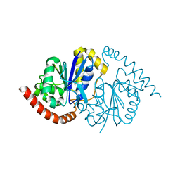
7TWR
DownloadVisualize
BU of 7twr by Molmil
Crystal structure of SARS-CoV-2 NSP3 macrodomain at pH 8 (P43 crystal form)
Descriptor: ACETATE ION, Non-structural protein 3
Authors:Correy, G.J, Fraser, J.S.
Deposit date:2022-02-07
Release date:2022-02-23
Last modified:2023-10-18
Method:X-RAY DIFFRACTION (0.9 Å)
Cite:The mechanisms of catalysis and ligand binding for the SARS-CoV-2 NSP3 macrodomain from neutron and x-ray diffraction at room temperature.
Sci Adv, 8, 2022
7TWF
DownloadVisualize
BU of 7twf by Molmil
Crystal structure of SARS-CoV-2 NSP3 macrodomain at 293 K (P43 crystal form, 73 kGy)
Descriptor: Non-structural protein 3
Authors:Correy, G.J, Fraser, J.S.
Deposit date:2022-02-07
Release date:2022-02-23
Last modified:2023-10-18
Method:X-RAY DIFFRACTION (1.1 Å)
Cite:The mechanisms of catalysis and ligand binding for the SARS-CoV-2 NSP3 macrodomain from neutron and x-ray diffraction at room temperature.
Sci Adv, 8, 2022
7TWP
DownloadVisualize
BU of 7twp by Molmil
Crystal structure of SARS-CoV-2 NSP3 macrodomain at pH 7 (P43 crystal form)
Descriptor: ACETATE ION, Non-structural protein 3
Authors:Correy, G.J, Fraser, J.S.
Deposit date:2022-02-07
Release date:2022-02-23
Last modified:2023-10-18
Method:X-RAY DIFFRACTION (0.9 Å)
Cite:The mechanisms of catalysis and ligand binding for the SARS-CoV-2 NSP3 macrodomain from neutron and x-ray diffraction at room temperature.
Sci Adv, 8, 2022
7TWH
DownloadVisualize
BU of 7twh by Molmil
Crystal structure of SARS-CoV-2 NSP3 macrodomain at 293 K (P43 crystal form, 290 kGy)
Descriptor: Non-structural protein 3
Authors:Correy, G.J, Fraser, J.S.
Deposit date:2022-02-07
Release date:2022-02-23
Last modified:2023-10-18
Method:X-RAY DIFFRACTION (1.1 Å)
Cite:The mechanisms of catalysis and ligand binding for the SARS-CoV-2 NSP3 macrodomain from neutron and x-ray diffraction at room temperature.
Sci Adv, 8, 2022
7TX3
DownloadVisualize
BU of 7tx3 by Molmil
Neutron crystal structure of SARS-CoV-2 NSP3 macrodomain at 293 K (P43 crystal form)
Descriptor: 2-[N-CYCLOHEXYLAMINO]ETHANE SULFONIC ACID, Non-structural protein 3
Authors:Correy, G.J, Fraser, J.S, Meilleur, F.
Deposit date:2022-02-07
Release date:2022-02-23
Last modified:2023-10-25
Method:NEUTRON DIFFRACTION (1.6 Å), X-RAY DIFFRACTION
Cite:The mechanisms of catalysis and ligand binding for the SARS-CoV-2 NSP3 macrodomain from neutron and x-ray diffraction at room temperature.
Sci Adv, 8, 2022
7TWN
DownloadVisualize
BU of 7twn by Molmil
Crystal structure of SARS-CoV-2 NSP3 macrodomain at pH 5 (P43 crystal form)
Descriptor: 2-[N-CYCLOHEXYLAMINO]ETHANE SULFONIC ACID, CITRIC ACID, Non-structural protein 3
Authors:Correy, G.J, Fraser, J.S.
Deposit date:2022-02-07
Release date:2022-02-23
Last modified:2023-10-18
Method:X-RAY DIFFRACTION (0.9 Å)
Cite:The mechanisms of catalysis and ligand binding for the SARS-CoV-2 NSP3 macrodomain from neutron and x-ray diffraction at room temperature.
Sci Adv, 8, 2022
7TWQ
DownloadVisualize
BU of 7twq by Molmil
Crystal structure of SARS-CoV-2 NSP3 macrodomain at pH 9 (P43 crystal form)
Descriptor: Non-structural protein 3
Authors:Correy, G.J, Fraser, J.S.
Deposit date:2022-02-07
Release date:2022-02-23
Last modified:2023-10-18
Method:X-RAY DIFFRACTION (0.9 Å)
Cite:The mechanisms of catalysis and ligand binding for the SARS-CoV-2 NSP3 macrodomain from neutron and x-ray diffraction at room temperature.
Sci Adv, 8, 2022
7TWI
DownloadVisualize
BU of 7twi by Molmil
Crystal structure of SARS-CoV-2 NSP3 macrodomain at 293 K (P43 crystal form, 539 kGy)
Descriptor: Non-structural protein 3
Authors:Correy, G.J, Fraser, J.S.
Deposit date:2022-02-07
Release date:2022-02-23
Last modified:2023-10-18
Method:X-RAY DIFFRACTION (1.1 Å)
Cite:The mechanisms of catalysis and ligand binding for the SARS-CoV-2 NSP3 macrodomain from neutron and x-ray diffraction at room temperature.
Sci Adv, 8, 2022

226707

PDB entries from 2024-10-30

PDB statisticsPDBj update infoContact PDBjnumon