Loading
PDBj
MenuPDBj@FacebookPDBj@TwitterPDBj@YouTubewwPDB FoundationwwPDB
RCSB PDBPDBeBMRBAdv. SearchSearch help
Search by PDB author
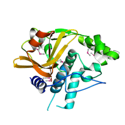
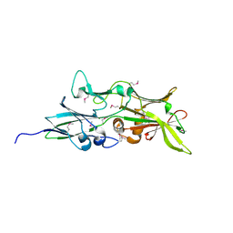
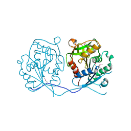
4FDY
DownloadVisualize
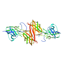
BU of 4fdy by Molmil
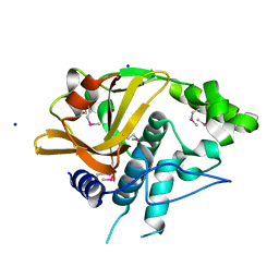
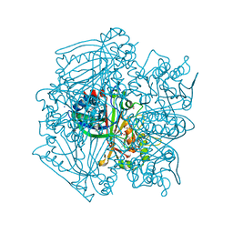
Crystal structure of a similar to lipoprotein, NLP/P60 family (SAV0400) from Staphylococcus aureus subsp. aureus Mu50 at 2.23 A resolution
Descriptor: Similar to lipoprotein, NLP/P60 family
Authors:Joint Center for Structural Genomics (JCSG)
Deposit date:2012-05-29
Release date:2012-07-25
Last modified:2023-02-01
Method:X-RAY DIFFRACTION (2.23 Å)
Cite:Structures of a Bifunctional Cell Wall Hydrolase CwlT Containing a Novel Bacterial Lysozyme and an NlpC/P60 dl-Endopeptidase.
J.Mol.Biol., 426, 2014
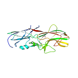
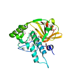
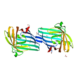
4H40
DownloadVisualize
BU of 4h40 by Molmil
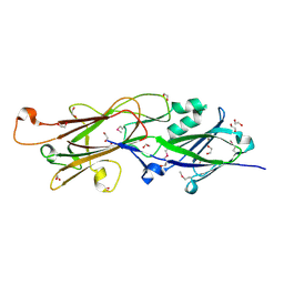
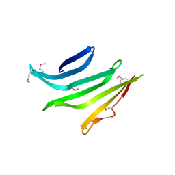
Crystal structure of a putative cell adhesion protein (BF2867) from Bacteroides fragilis NCTC 9343 at 2.57 A resolution
Descriptor: CHLORIDE ION, putative cell adhesion protein
Authors:Joint Center for Structural Genomics (JCSG)
Deposit date:2012-09-14
Release date:2012-10-03
Last modified:2023-02-01
Method:X-RAY DIFFRACTION (2.57 Å)
Cite:A Distinct Type of Pilus from the Human Microbiome.
Cell, 165, 2016
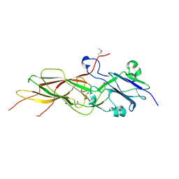
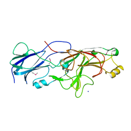
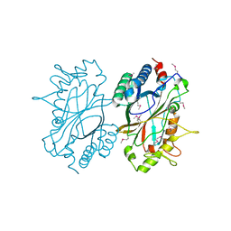
4H4J
DownloadVisualize
BU of 4h4j by Molmil
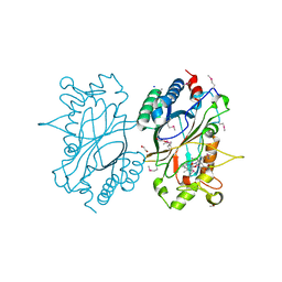
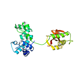
Crystal structure of a N-acetylmuramoyl-L-alanine amidase (BACUNI_02947) from Bacteroides uniformis ATCC 8492 at 1.15 A resolution
Descriptor: hypothetical protein
Authors:Joint Center for Structural Genomics (JCSG)
Deposit date:2012-09-17
Release date:2012-10-17
Last modified:2023-02-01
Method:X-RAY DIFFRACTION (1.15 Å)
Cite:Structure-guided functional characterization of DUF1460 reveals a highly specific NlpC/P60 amidase family.
Structure, 22, 2014
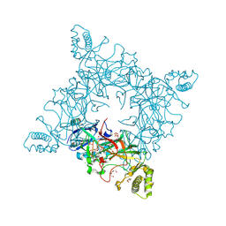
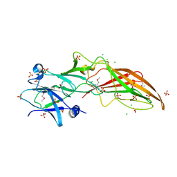
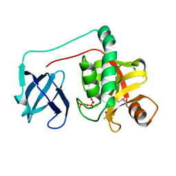
4GPV
DownloadVisualize
BU of 4gpv by Molmil
Crystal structure of a putative cell adhesion protein (BACEGG_00536) from Bacteroides eggerthii DSM 20697 at 1.67 A resolution
Descriptor: 2-AMINO-2-HYDROXYMETHYL-PROPANE-1,3-DIOL, putative cell adhesion protein
Authors:Joint Center for Structural Genomics (JCSG)
Deposit date:2012-08-21
Release date:2012-09-19
Last modified:2023-02-01
Method:X-RAY DIFFRACTION (1.67 Å)
Cite:A Distinct Type of Pilus from the Human Microbiome.
Cell, 165, 2016
4JG5
DownloadVisualize
BU of 4jg5 by Molmil
Crystal structure of a putative cell adhesion protein (BDI_3519) from Parabacteroides distasonis ATCC 8503 at 2.34 A resolution (PSI Community Target, Nakayama)
Descriptor: Putative cell adhesion protein
Authors:Joint Center for Structural Genomics (JCSG)
Deposit date:2013-02-28
Release date:2013-03-20
Last modified:2023-02-01
Method:X-RAY DIFFRACTION (2.34 Å)
Cite:A Distinct Type of Pilus from the Human Microbiome.
Cell, 165, 2016
4JRF
DownloadVisualize
BU of 4jrf by Molmil
Crystal structure of a putative cell adhesion protein (BACOVA_01548) from Bacteroides ovatus ATCC 8483 at 1.98 A resolution (PSI Community Target, Nakayama)
Descriptor: CALCIUM ION, CHLORIDE ION, SULFATE ION, ...
Authors:Joint Center for Structural Genomics (JCSG)
Deposit date:2013-03-21
Release date:2013-04-03
Last modified:2023-02-01
Method:X-RAY DIFFRACTION (1.98 Å)
Cite:A Distinct Type of Pilus from the Human Microbiome.
Cell, 165, 2016
4QDG
DownloadVisualize
BU of 4qdg by Molmil
Crystal structure of a putative adhesin (BT2657) from Bacteroides thetaiotaomicron VPI-5482 at 2.20 A resolution (PSI Community Target, Nakayama)
Descriptor: 1,2-ETHANEDIOL, MAGNESIUM ION, Putative adhesin
Authors:Joint Center for Structural Genomics (JCSG)
Deposit date:2014-05-13
Release date:2014-06-11
Last modified:2023-02-01
Method:X-RAY DIFFRACTION (2.2 Å)
Cite:A Distinct Type of Pilus from the Human Microbiome.
Cell, 165, 2016
4Q5K
DownloadVisualize
BU of 4q5k by Molmil
Crystal structure of a N-acetylmuramoyl-L-alanine amidase (BACUNI_02947) from Bacteroides uniformis ATCC 8492 at 1.30 A resolution
Descriptor: (2R)-2-[[(1R,2S,3R,4R,5R)-4-acetamido-2-[(2S,3R,4R,5S,6R)-3-acetamido-6-(hydroxymethyl)-4,5-bis(oxidanyl)oxan-2-yl]oxy-6,8-dioxabicyclo[3.2.1]octan-3-yl]oxy]propanoic acid, SODIUM ION, Uncharacterized protein
Authors:Joint Center for Structural Genomics (JCSG)
Deposit date:2014-04-17
Release date:2014-05-21
Last modified:2023-12-06
Method:X-RAY DIFFRACTION (1.3 Å)
Cite:Structure-guided functional characterization of DUF1460 reveals a highly specific NlpC/P60 amidase family.
Structure, 22, 2014
4Q98
DownloadVisualize
BU of 4q98 by Molmil
Crystal structure of a fimbrilin (fimA) from Porphyromonas gingivalis W83 at 1.30 A resolution (PSI Community Target, Nakayama)
Descriptor: Major fimbrial subunit protein, SODIUM ION
Authors:Joint Center for Structural Genomics (JCSG)
Deposit date:2014-04-29
Release date:2014-06-04
Last modified:2023-02-01
Method:X-RAY DIFFRACTION (1.3 Å)
Cite:A Distinct Type of Pilus from the Human Microbiome.
Cell, 165, 2016
4QB7
DownloadVisualize
BU of 4qb7 by Molmil
Crystal structure of a fimbrial protein (BVU_2522) from Bacteroides vulgatus ATCC 8482 at 2.55 A resolution
Descriptor: CHLORIDE ION, SULFATE ION, Uncharacterized protein
Authors:Joint Center for Structural Genomics (JCSG)
Deposit date:2014-05-06
Release date:2014-07-02
Last modified:2023-02-01
Method:X-RAY DIFFRACTION (2.55 Å)
Cite:A Distinct Type of Pilus from the Human Microbiome.
Cell, 165, 2016
4QK4
DownloadVisualize
BU of 4qk4 by Molmil
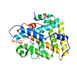
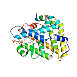
Crystal structure of human nuclear receptor sf-1 (nr5a1) bound to pip2 at 2.8 a resolution
Descriptor: (2S)-3-{[(R)-hydroxy{[(1R,2R,3S,4R,5R,6S)-2,3,6-trihydroxy-4,5-bis(phosphonooxy)cyclohexyl]oxy}phosphoryl]oxy}propane-1,2-diyl dihexadecanoate, 1,2-ETHANEDIOL, Peroxisome proliferator-activated receptor gamma coactivator 1-alpha, ...
Authors:Joint Center for Structural Genomics (JCSG), Partnership for Stem Cell Biology (STEMCELL)
Deposit date:2014-06-05
Release date:2014-07-30
Last modified:2024-03-13
Method:X-RAY DIFFRACTION (2.81 Å)
Cite:The signaling phospholipid PIP3 creates a new interaction surface on the nuclear receptor SF-1.
Proc.Natl.Acad.Sci.USA, 111, 2014
4R03
DownloadVisualize
BU of 4r03 by Molmil
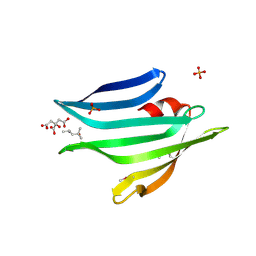
Crystal structure of a DUF3836 family protein (BDI_3222) from Parabacteroides distasonis ATCC 8503 at 1.50 A resolution
Descriptor: (4S)-2-METHYL-2,4-PENTANEDIOL, CITRIC ACID, PHOSPHATE ION, ...
Authors:Joint Center for Structural Genomics (JCSG)
Deposit date:2014-07-29
Release date:2014-08-27
Last modified:2023-02-01
Method:X-RAY DIFFRACTION (1.5 Å)
Cite:Structures of single-layer beta-sheet proteins evolved from beta-hairpin repeats.
Protein Sci., 28, 2019
4RDB
DownloadVisualize
BU of 4rdb by Molmil
Crystal structure of an immunoreactive 32 kDa antigen PG49 (PG_0181) from Porphyromonas gingivalis W83 at 1.45 A resolution (PSI Community Target, Nakayama)
Descriptor: 1,2-ETHANEDIOL, ACETONE, Immunoreactive 32 kDa antigen PG49
Authors:Joint Center for Structural Genomics (JCSG)
Deposit date:2014-09-18
Release date:2014-12-03
Last modified:2023-02-01
Method:X-RAY DIFFRACTION (1.45 Å)
Cite:A Distinct Type of Pilus from the Human Microbiome.
Cell, 165, 2016
4RBO
DownloadVisualize
BU of 4rbo by Molmil
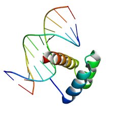
Crystal structure of a Nanog homeobox (NANOG) from Homo sapiens at 3.30 A resolution
Descriptor: 5'-D(*CP*TP*TP*GP*AP*AP*TP*GP*GP*GP*CP*C)-3', 5'-D(*GP*GP*CP*CP*CP*AP*TP*TP*CP*AP*AP*G)-3', Putative homeobox protein NANOGP8
Authors:Joint Center for Structural Genomics (JCSG), Partnership for Stem Cell Biology (STEMCELL)
Deposit date:2014-09-12
Release date:2014-10-01
Last modified:2023-02-01
Method:X-RAY DIFFRACTION (3.3 Å)
Cite:Structure-based discovery of NANOG variant with enhanced properties to promote self-renewal and reprogramming of pluripotent stem cells.
Proc.Natl.Acad.Sci.USA, 112, 2015
4R8O
DownloadVisualize
BU of 4r8o by Molmil
Crystal structure of a DUF3836 family protein (BVU_1206) from Bacteroides vulgatus ATCC 8482 at 2.50 A resolution
Descriptor: Uncharacterized protein
Authors:Joint Center for Structural Genomics (JCSG)
Deposit date:2014-09-02
Release date:2014-09-17
Last modified:2023-02-01
Method:X-RAY DIFFRACTION (2.5 Å)
Cite:Structures of single-layer beta-sheet proteins evolved from beta-hairpin repeats.
Protein Sci., 28, 2019
4Q68
DownloadVisualize
BU of 4q68 by Molmil
Crystal structure of a N-acetylmuramoyl-L-alanine amidase (BACUNI_02947) from Bacteroides uniformis ATCC 8492 at 1.07 A resolution
Descriptor: 2-acetamido-2-deoxy-beta-D-glucopyranose, SODIUM ION, Uncharacterized protein
Authors:Joint Center for Structural Genomics (JCSG)
Deposit date:2014-04-21
Release date:2014-05-07
Last modified:2023-12-06
Method:X-RAY DIFFRACTION (1.07 Å)
Cite:Structure-guided functional characterization of DUF1460 reveals a highly specific NlpC/P60 amidase family.
Structure, 22, 2014
4QJR
DownloadVisualize
BU of 4qjr by Molmil
Crystal structure of human nuclear receptor sf-1 (nr5a1) bound to its hormone pip3 at 2.4 a resolution
Descriptor: (2S)-3-{[(R)-{[(1S,2S,3R,4S,5S,6S)-2,6-dihydroxy-3,4,5-tris(phosphonooxy)cyclohexyl]oxy}(hydroxy)phosphoryl]oxy}propane -1,2-diyl dihexadecanoate, ACETATE ION, Peroxisome proliferator-activated receptor gamma coactivator 1-alpha, ...
Authors:Joint Center for Structural Genomics (JCSG), Partnership for Stem Cell Biology (STEMCELL)
Deposit date:2014-06-04
Release date:2014-07-30
Last modified:2024-03-13
Method:X-RAY DIFFRACTION (2.4 Å)
Cite:The signaling phospholipid PIP3 creates a new interaction surface on the nuclear receptor SF-1.
Proc.Natl.Acad.Sci.USA, 111, 2014
2IIZ
DownloadVisualize
BU of 2iiz by Molmil
Crystal structure of putative melanin biosynthesis protein TyrA with bound heme (NP_716371.1) from Shewanella Oneidensis at 2.30 A resolution
Descriptor: 1,2-ETHANEDIOL, ISOPROPYL ALCOHOL, Melanin biosynthesis protein TyrA, ...
Authors:Joint Center for Structural Genomics (JCSG)
Deposit date:2006-09-28
Release date:2006-11-14
Last modified:2023-11-15
Method:X-RAY DIFFRACTION (2.3 Å)
Cite:Identification and structural characterization of heme binding in a novel dye-decolorizing peroxidase, TyrA.
Proteins, 69, 2007
2GHR
DownloadVisualize
BU of 2ghr by Molmil
Crystal structure of homoserine o-succinyltransferase (NP_981826.1) from Bacillus cereus ATCC 10987 at 2.40 A resolution
Descriptor: Homoserine O-succinyltransferase, SULFATE ION
Authors:Joint Center for Structural Genomics (JCSG)
Deposit date:2006-03-27
Release date:2006-04-11
Last modified:2023-01-25
Method:X-RAY DIFFRACTION (2.4 Å)
Cite:Crystal structure of homoserine O-succinyltransferase from Bacillus cereus at 2.4 A resolution
Proteins, 68, 2007
2H1T
DownloadVisualize
BU of 2h1t by Molmil
Crystal structure of a duf1089 family protein (pa1994) from pseudomonas aeruginosa at 1.80 A resolution
Descriptor: (4S)-2-METHYL-2,4-PENTANEDIOL, 1,2-ETHANEDIOL, Hypothetical protein
Authors:Joint Center for Structural Genomics (JCSG)
Deposit date:2006-05-16
Release date:2006-05-30
Last modified:2023-01-25
Method:X-RAY DIFFRACTION (1.8 Å)
Cite:The structure of the first representative of Pfam family PF06475 reveals a new fold with possible involvement in glycolipid metabolism.
Acta Crystallogr.,Sect.F, 66, 2010
2HAG
DownloadVisualize
BU of 2hag by Molmil
Crystal structure of a putative dyp-type peroxidase protein (so_0740) from shewanella oneidensis at 2.75 A resolution
Descriptor: Melanin biosynthesis protein TyrA, putative
Authors:Joint Center for Structural Genomics (JCSG)
Deposit date:2006-06-12
Release date:2006-08-08
Last modified:2023-01-25
Method:X-RAY DIFFRACTION (2.75 Å)
Cite:Crystal structures of two novel dye-decolorizing peroxidases reveal a beta-barrel fold with a conserved heme-binding motif.
Proteins, 69, 2007
2HBW
DownloadVisualize
BU of 2hbw by Molmil
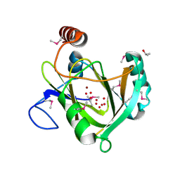
Crystal structure of a putative endopeptidase (ava_3396) from anabaena variabilis atcc 29413 at 1.05 A resolution
Descriptor: ACETATE ION, NLP/P60 protein, UNKNOWN LIGAND
Authors:Joint Center for Structural Genomics (JCSG)
Deposit date:2006-06-14
Release date:2006-08-08
Last modified:2023-01-25
Method:X-RAY DIFFRACTION (1.05 Å)
Cite:Structural Basis of Murein Peptide Specificity of a gamma-D-Glutamyl-L-Diamino Acid Endopeptidase.
Structure, 17, 2009
2GVK
DownloadVisualize
BU of 2gvk by Molmil
Crystal structure of a dye-decolorizing peroxidase (DyP) from Bacteroides thetaiotaomicron VPI-5482 at 1.6 A resolution
Descriptor: 1,2-ETHANEDIOL, ACETIC ACID, CHLORIDE ION, ...
Authors:Joint Center for Structural Genomics (JCSG)
Deposit date:2006-05-02
Release date:2006-05-16
Last modified:2023-01-25
Method:X-RAY DIFFRACTION (1.6 Å)
Cite:Crystal structures of two novel dye-decolorizing peroxidases reveal a beta-barrel fold with a conserved heme-binding motif.
Proteins, 69, 2007
2HUJ
DownloadVisualize
BU of 2huj by Molmil
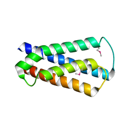
Crystal structure of a protein of uknown function (NP_471338.1) from Listeria innocua at 1.74 A resolution
Descriptor: Lin2004 protein
Authors:Joint Center for Structural Genomics (JCSG)
Deposit date:2006-07-26
Release date:2006-08-08
Last modified:2023-01-25
Method:X-RAY DIFFRACTION (1.74 Å)
Cite:Crystal structures of MW1337R and lin2004: representatives of a novel protein family that adopt a four-helical bundle fold.
Proteins, 71, 2008
1VR3
DownloadVisualize
BU of 1vr3 by Molmil
Crystal structure of Acireductone dioxygenase (13543033) from Mus musculus at 2.06 A resolution
Descriptor: Acireductone dioxygenase, ISOPROPYL ALCOHOL, NICKEL (II) ION, ...
Authors:Joint Center for Structural Genomics (JCSG)
Deposit date:2005-01-28
Release date:2005-02-15
Last modified:2023-01-25
Method:X-RAY DIFFRACTION (2.06 Å)
Cite:Crystal structure of acireductone dioxygenase (ARD) from Mus musculus at 2.06 angstrom resolution.
Proteins, 64, 2006

223790

PDB entries from 2024-08-14

PDB statisticsPDBj update infoContact PDBjnumon