Loading
PDBj
MenuPDBj@FacebookPDBj@X(formerly Twitter)PDBj@BlueSkyPDBj@YouTubewwPDB FoundationwwPDBDonate
RCSB PDBPDBeBMRBAdv. SearchSearch help
Search by PDB author
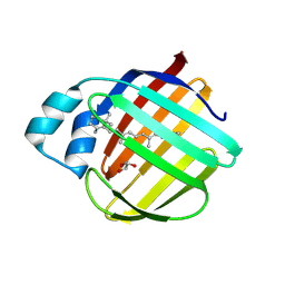
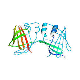
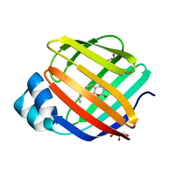
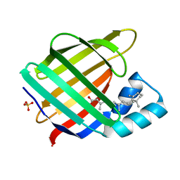
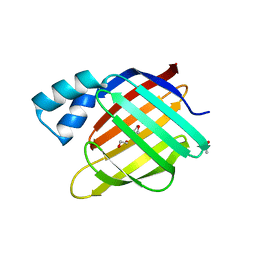
3FA7
DownloadVisualize
BU of 3fa7 by Molmil
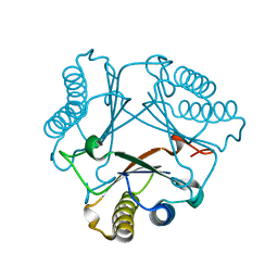
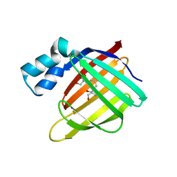
Crystal structure of the apo R132K:R111L:L121E:R59E mutant of cellular retinoic acid-binding protein II at 1.90 angstrom resolution
Descriptor: 2-[3-(2-HYDROXY-1,1-DIHYDROXYMETHYL-ETHYLAMINO)-PROPYLAMINO]-2-HYDROXYMETHYL-PROPANE-1,3-DIOL, Cellular retinoic acid-binding protein 2
Authors:Jia, X, Geiger, J.H.
Deposit date:2008-11-15
Release date:2009-10-27
Last modified:2023-12-27
Method:X-RAY DIFFRACTION (1.9 Å)
Cite:Crystal structures of apo cellular retinoic acid-binding protein II mutants: Structural integrity investigated through multiple site mutations
To be Published
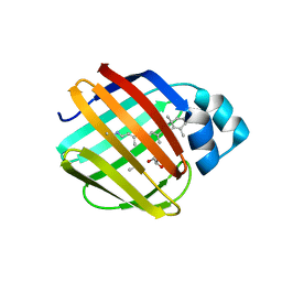
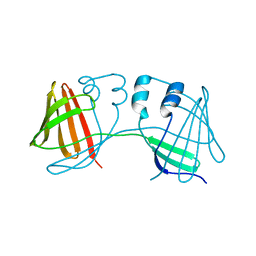
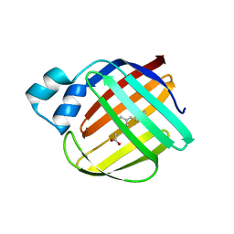
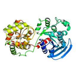
3FEK
DownloadVisualize
BU of 3fek by Molmil
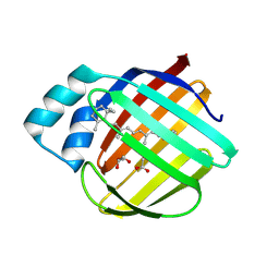
Crystal structure of the R132K:Y134F:R111L:L121D:T54V mutant of cellular retinoic acid-binding protein II at 1.51 angstrom resolution
Descriptor: ACETATE ION, Cellular retinoic acid-binding protein 2, DI(HYDROXYETHYL)ETHER
Authors:Jia, X, Geiger, J.H.
Deposit date:2008-11-30
Release date:2009-11-10
Last modified:2023-09-06
Method:X-RAY DIFFRACTION (1.51 Å)
Cite:Crystal structures of apo cellular retinoic acid-binding protein II mutants: Structural integrity investigated through multiple site mutations
To be Published
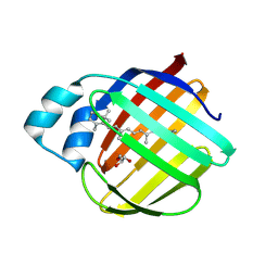
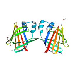
7LHN
DownloadVisualize
BU of 7lhn by Molmil
Crystal structure of Q108K:K40L:T51V:T53C:R58W:T29L:Y19W:Q4A mutant of cellular retinol binding protein II complex with all-trans-retinal after exposure to visible light
Descriptor: GLYCEROL, RETINAL, Retinol-binding protein 2
Authors:Ehyaei, N, Geiger, J.H, Borhan, B.
Deposit date:2021-01-25
Release date:2021-04-14
Last modified:2024-10-30
Method:X-RAY DIFFRACTION (2.11 Å)
Cite:Crystal structure of Q108K:K40L:T51V:T53C:R58W:T29L:Y19W:Q4A mutant of cellular retinol binding protein II complex with all-trans-retinal after exposure to visible light
To Be Published
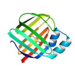
7LHM
DownloadVisualize
BU of 7lhm by Molmil
Crystal structure of Q108K:K40L:T51V:T53C:R58W:T29L:Y19W:Q4A:Q38L mutant of cellular retinol binding protein II complex with all-trans-retinal
Descriptor: ACETATE ION, GLYCEROL, RETINAL, ...
Authors:Ehyaei, N, Geiger, J.H, Borhan, B.
Deposit date:2021-01-25
Release date:2021-04-14
Last modified:2024-10-23
Method:X-RAY DIFFRACTION (1.5 Å)
Cite:Crystal structure of Q108K:K40L:T51V:T53C:R58W:T29L:Y19W:Q4A:Q38L mutant of cellular retinol binding protein II complex with all-trans-retinal
To Be Published
7LHO
DownloadVisualize
BU of 7lho by Molmil
Crystal structure of Q108K:K40L:T51V:T53C:R58W:T29L:Y19W:Q4A mutant of cellular retinol binding protein II complex with all-trans-retinal in the dark
Descriptor: GLYCEROL, RETINAL, Retinol-binding protein 2
Authors:Ehyaei, N, Geiger, J.H, Borhan, B.
Deposit date:2021-01-26
Release date:2021-04-14
Last modified:2024-10-16
Method:X-RAY DIFFRACTION (1.4 Å)
Cite:Crystal structure of Q108K:K40L:T51V:T53C:R58W:T29L:Y19W:Q4A mutant of cellular retinol binding protein II complex with all-trans-retinal in the dark
To Be Published
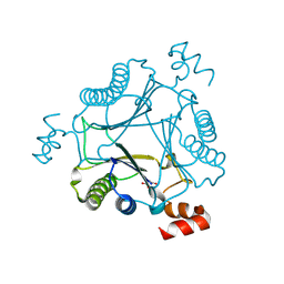
7MS1
DownloadVisualize
BU of 7ms1 by Molmil
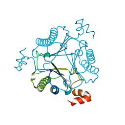
Crystal structure of H28A mutant of Cg10062 with a covalent intermediate of the hydration of acetylenecarboxylic acid
Descriptor: 3-HYDROXY-PROPANOIC ACID, 4-oxalocrotonate tautomerase
Authors:Nayebi, G.H, Geiger, J.H, Draths, K.
Deposit date:2021-05-10
Release date:2022-02-02
Last modified:2024-10-30
Method:X-RAY DIFFRACTION (2.95 Å)
Cite:Cg10062 Catalysis Forges a Link between Acetylenecarboxylic Acid and Bacterial Metabolism.
Biochemistry, 60, 2021
7MS0
DownloadVisualize
BU of 7ms0 by Molmil
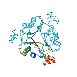
Crystal structure of native Cg10062
Descriptor: 4-oxalocrotonate tautomerase
Authors:Nayebi, G.H, Geiger, J.H, Draths, K.
Deposit date:2021-05-10
Release date:2022-02-02
Last modified:2023-10-18
Method:X-RAY DIFFRACTION (1.37 Å)
Cite:Cg10062 Catalysis Forges a Link between Acetylenecarboxylic Acid and Bacterial Metabolism.
Biochemistry, 60, 2021
7MS8
DownloadVisualize
BU of 7ms8 by Molmil
Crystal structure of Y103F mutant of Cg10062 with a covalent intermediate of the hydration of acetylenecarboxylic acid
Descriptor: 3-HYDROXY-PROPANOIC ACID, 4-oxalocrotonate tautomerase
Authors:Nayebi, G.H, Geiger, J.H, Draths, K.
Deposit date:2021-05-10
Release date:2022-02-02
Last modified:2024-10-30
Method:X-RAY DIFFRACTION (2.5 Å)
Cite:Cg10062 Catalysis Forges a Link between Acetylenecarboxylic Acid and Bacterial Metabolism.
Biochemistry, 60, 2021
7MS3
DownloadVisualize
BU of 7ms3 by Molmil
Crystal structure of R73A mutant of Cg10062 with a covalent intermediate of the hydration of acetylenecarboxylic acid
Descriptor: 3-HYDROXY-PROPANOIC ACID, 4-oxalocrotonate tautomerase
Authors:Nayebi, G.H, Geiger, J.H, Draths, K.
Deposit date:2021-05-10
Release date:2022-02-02
Last modified:2024-10-16
Method:X-RAY DIFFRACTION (3 Å)
Cite:Cg10062 Catalysis Forges a Link between Acetylenecarboxylic Acid and Bacterial Metabolism.
Biochemistry, 60, 2021
7LHJ
DownloadVisualize
BU of 7lhj by Molmil
Crystal structure of Q108K:K40L:T51V:T53C:R58W:T29L:Y19W:Q4A mutant of cellular retinol binding protein II complex with 15-cis-retinal
Descriptor: ACETATE ION, GLYCEROL, RETINAL, ...
Authors:Ehyaei, N, Geiger, J.H, Borhan, B.
Deposit date:2021-01-25
Release date:2021-02-10
Last modified:2024-10-30
Method:X-RAY DIFFRACTION (1.26 Å)
Cite:Crystal structure of Q108K:K40L:T51V:T53C:R58W:T29L:Y19W:Q4A mutant of cellular retinol binding protein II complex with 15cis- retinal
To Be Published
4ZH6
DownloadVisualize
BU of 4zh6 by Molmil
Crystal Structure of the Domain-Swapped Dimer Y60L mutant of Human Cellular Retinol Binding Protein II
Descriptor: ACETATE ION, Retinol-binding protein 2
Authors:Assar, Z, Nossoni, Z, Geiger, J.H.
Deposit date:2015-04-24
Release date:2016-06-08
Last modified:2023-09-27
Method:X-RAY DIFFRACTION (1.5497 Å)
Cite:Domain-Swapped Dimers of Intracellular Lipid-Binding Proteins: Evidence for Ordered Folding Intermediates.
Structure, 24, 2016
4ZH9
DownloadVisualize
BU of 4zh9 by Molmil
Crystal Structure of the Domain-Swapped Dimer Wild-Type of Human Cellular Retinol Binding Protein II
Descriptor: Retinol-binding protein 2
Authors:Assar, Z, Nossoni, Z, Geiger, J.H.
Deposit date:2015-04-24
Release date:2016-06-08
Last modified:2023-09-27
Method:X-RAY DIFFRACTION (2.66 Å)
Cite:Domain-Swapped Dimers of Intracellular Lipid-Binding Proteins: Evidence for Ordered Folding Intermediates.
Structure, 24, 2016
4ZR2
DownloadVisualize
BU of 4zr2 by Molmil
Crystal Structure of the Domain-Swapped Dimer K40L:Q108K:Y60W mutant of Human Cellular Retinol Binding Protein II
Descriptor: ACETATE ION, RETINAL, Retinol-binding protein 2
Authors:Assar, Z, Nossoni, Z, Geiger, J.H.
Deposit date:2015-05-11
Release date:2016-06-08
Last modified:2024-10-30
Method:X-RAY DIFFRACTION (1.8024 Å)
Cite:Domain-Swapped Dimers of Intracellular Lipid-Binding Proteins: Evidence for Ordered Folding Intermediates.
Structure, 24, 2016
8DB2
DownloadVisualize
BU of 8db2 by Molmil
Q108K:K40L:T51C:T53A:R58L:Q38F mutant of hCRBPII bound to synthetic fluorophore CM1V
Descriptor: (2E)-3-[7-(diethylamino)-2-oxo-2H-1-benzopyran-3-yl]prop-2-enal, bound form, Retinol-binding protein 2
Authors:Bingham, C.R, Geiger, J.H, Borhan, B, Staples, R.
Deposit date:2022-06-14
Release date:2023-02-01
Last modified:2024-11-13
Method:X-RAY DIFFRACTION (1.5 Å)
Cite:Light controlled reversible Michael addition of cysteine: a new tool for dynamic site-specific labeling of proteins.
Analyst, 148, 2023
8DN1
DownloadVisualize
BU of 8dn1 by Molmil
Q108K:K40L:T51C:T53A:R58L:Q38F:Q4F mutant of hCRBPII bound to synthetic fluorophore CM1V at pH 7.2
Descriptor: (2E)-3-[7-(diethylamino)-2-oxo-2H-1-benzopyran-3-yl]prop-2-enal, bound form, GLYCEROL, ...
Authors:Bingham, C.R, Borhan, B, Geiger, J.H.
Deposit date:2022-07-10
Release date:2023-02-01
Last modified:2023-10-25
Method:X-RAY DIFFRACTION (1.32 Å)
Cite:Light controlled reversible Michael addition of cysteine: a new tool for dynamic site-specific labeling of proteins.
Analyst, 148, 2023
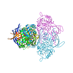
1JKI
DownloadVisualize
BU of 1jki by Molmil
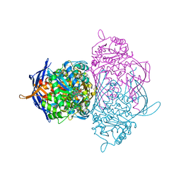
myo-Inositol-1-phosphate Synthase Complexed with an Inhibitor, 2-deoxy-glucitol-6-phosphate
Descriptor: 1,4-DIHYDRONICOTINAMIDE ADENINE DINUCLEOTIDE, 2-DEOXY-GLUCITOL-6-PHOSPHATE, AMMONIUM ION, ...
Authors:Stein, A.J, Geiger, J.H.
Deposit date:2001-07-12
Release date:2002-04-10
Last modified:2023-08-16
Method:X-RAY DIFFRACTION (2.2 Å)
Cite:The crystal structure and mechanism of 1-L-myo-inositol- 1-phosphate synthase
J.Biol.Chem., 277, 2002
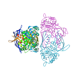
1JKF
DownloadVisualize
BU of 1jkf by Molmil
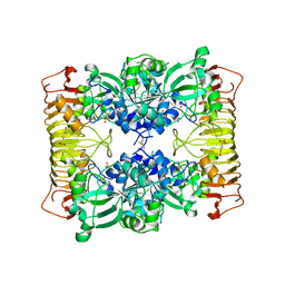
Holo 1L-myo-inositol-1-phosphate Synthase
Descriptor: NICOTINAMIDE-ADENINE-DINUCLEOTIDE, myo-inositol-1-phosphate synthase
Authors:Stein, A.J, Geiger, J.H.
Deposit date:2001-07-12
Release date:2002-04-10
Last modified:2024-12-25
Method:X-RAY DIFFRACTION (2.4 Å)
Cite:The crystal structure and mechanism of 1-L-myo-inositol- 1-phosphate synthase
J.Biol.Chem., 277, 2002
4ZJ0
DownloadVisualize
BU of 4zj0 by Molmil
The crystal structure of monomer Q108K:K40L:Y60W CRBPII bound to all-trans-retinal
Descriptor: ACETATE ION, RETINAL, Retinol-binding protein 2
Authors:Nossoni, Z, Assar, Z, Wang, W, Vasileiou, C, Borhan, B, Geiger, J.H.
Deposit date:2015-04-28
Release date:2016-05-11
Last modified:2024-11-20
Method:X-RAY DIFFRACTION (1.5 Å)
Cite:Domain-Swapped Dimers of Intracellular Lipid-Binding Proteins: Evidence for Ordered Folding Intermediates.
Structure, 24, 2016
3CWK
DownloadVisualize
BU of 3cwk by Molmil
Crystal Structure of the R132K:Y134F:R111L:T54V:L121E Mutant of Cellular Retinoic Acid Binding Protein Type II in Complex with All-trans-Retinoic Acid at 1.57 Angstroms Resolution
Descriptor: Cellular retinoic acid-binding protein 2, RETINOIC ACID, SULFATE ION
Authors:Vaezeslami, S, Geiger, J.H.
Deposit date:2008-04-22
Release date:2008-09-09
Last modified:2024-02-21
Method:X-RAY DIFFRACTION (1.6 Å)
Cite:Structural analysis of site-directed mutants of cellular retinoic acid-binding protein II addresses the relationship between structural integrity and ligand binding.
Acta Crystallogr.,Sect.D, 64, 2008
3D1J
DownloadVisualize
BU of 3d1j by Molmil
Crystal Structure of E.coli GS mutant dmGS(C7S;C408S)
Descriptor: Glycogen synthase
Authors:Sheng, F, Geiger, J.H.
Deposit date:2008-05-06
Release date:2009-03-24
Last modified:2023-08-30
Method:X-RAY DIFFRACTION (3 Å)
Cite:The Crystal Structures of the Open and Catalytically Competent Closed Conformation of Escherichia coli Glycogen Synthase.
J.Biol.Chem., 284, 2009
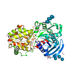
3CX4
DownloadVisualize
BU of 3cx4 by Molmil
Crystal Structure of E.coli GS mutant E377A in complex with ADP and oligosaccharides
Descriptor: (2R)-2-hydroxy-3-[4-(2-hydroxyethyl)piperazin-1-yl]propane-1-sulfonic acid, 2-{2-[2-2-(METHOXY-ETHOXY)-ETHOXY]-ETHOXY}-ETHANOL, ADENOSINE-5'-DIPHOSPHATE, ...
Authors:Sheng, F, Geiger, J.H.
Deposit date:2008-04-23
Release date:2009-04-28
Last modified:2023-08-30
Method:X-RAY DIFFRACTION (2.29 Å)
Cite:Oligosaccharide binding in Escherichia coli glycogen synthase.
Biochemistry, 48, 2009
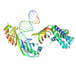
1RM1
DownloadVisualize
BU of 1rm1 by Molmil
Structure of a Yeast TFIIA/TBP/TATA-box DNA Complex
Descriptor: 5'-D(*AP*TP*CP*GP*AP*TP*CP*GP*AP*TP*AP*TP*AP*AP*AP*AP*CP*G)-3', 5'-D(P*CP*GP*TP*TP*TP*TP*AP*TP*AP*TP*CP*GP*AP*TP*CP*GP*AP*T)-3', TATA-box binding protein, ...
Authors:Jin, X, Gewirth, D.T, Geiger, J.H.
Deposit date:2003-11-26
Release date:2005-07-26
Last modified:2023-08-23
Method:X-RAY DIFFRACTION (2.5 Å)
Cite:High Resolution Structure of a Yeast TFIIA/TBP/TATA-box DNA Complex
TO BE PUBLISHED
1RM0
DownloadVisualize
BU of 1rm0 by Molmil
Crystal Structure of Myo-Inositol 1-Phosphate Synthase From Saccharomyces cerevisiae In Complex With NAD+ and 2-deoxy-D-glucitol 6-(E)-vinylhomophosphonate
Descriptor: (3,4,5,7-TETRAHYDROXY-HEPT-1-ENYL)-PHOSPHONIC ACID, 1,4-DIHYDRONICOTINAMIDE ADENINE DINUCLEOTIDE, MANGANESE (II) ION, ...
Authors:Jin, X, Foley, K.M, Geiger, J.H.
Deposit date:2003-11-26
Release date:2004-05-25
Last modified:2024-02-14
Method:X-RAY DIFFRACTION (2.05 Å)
Cite:The structure of the 1L-myo-inositol-1-phosphate synthase-NAD+-2-deoxy-D-glucitol 6-(E)-vinylhomophosphonate complex demands a revision of the enzyme mechanism.
J.Biol.Chem., 279, 2004
1YP2
DownloadVisualize
BU of 1yp2 by Molmil
Crystal structure of potato tuber ADP-glucose pyrophosphorylase
Descriptor: Glucose-1-phosphate adenylyltransferase small subunit, PARA-MERCURY-BENZENESULFONIC ACID, SULFATE ION
Authors:Jin, X, Ballicora, M.A, Preiss, J, Geiger, J.H.
Deposit date:2005-01-29
Release date:2005-03-15
Last modified:2024-10-30
Method:X-RAY DIFFRACTION (2.11 Å)
Cite:Crystal structure of potato tuber ADP-glucose pyrophosphorylase.
Embo J., 24, 2005
1YTB
DownloadVisualize
BU of 1ytb by Molmil
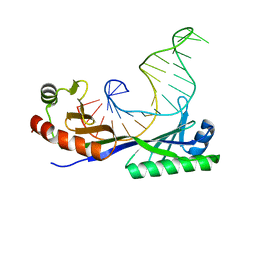
CRYSTAL STRUCTURE OF A YEAST TBP/TATA-BOX COMPLEX
Descriptor: DNA (29MER), PROTEIN (TATA BINDING PROTEIN (TBP))
Authors:Kim, Y, Geiger, J.H, Hahn, S, Sigler, P.B.
Deposit date:1994-09-28
Release date:1995-01-26
Last modified:2024-02-14
Method:X-RAY DIFFRACTION (1.8 Å)
Cite:Crystal structure of a yeast TBP/TATA-box complex.
Nature, 365, 1993

238582

PDB entries from 2025-07-09

PDB statisticsPDBj update infoContact PDBjnumon