Loading
PDBj
MenuPDBj@FacebookPDBj@TwitterPDBj@YouTubewwPDB FoundationwwPDB
RCSB PDBPDBeBMRBAdv. SearchSearch help
Search by PDB author
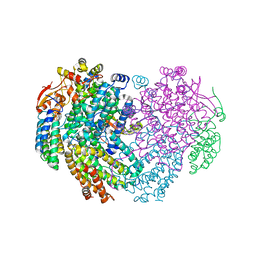
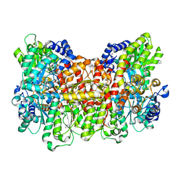
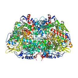
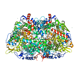
7SXM
DownloadVisualize
BU of 7sxm by Molmil
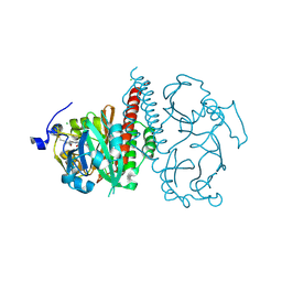
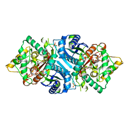
Structure of Xenon-derivatized Methyl-Coenzyme M Reductase from Methanothermobacter marburgensis
Descriptor: 1-THIOETHANESULFONIC ACID, ACETATE ION, Coenzyme B, ...
Authors:Chen, P.Y.-T, Drennan, C.L.
Deposit date:2021-11-23
Release date:2022-04-06
Last modified:2023-11-15
Method:X-RAY DIFFRACTION (2.503 Å)
Cite:XFEL serial crystallography reveals the room temperature structure of methyl-coenzyme M reductase.
J.Inorg.Biochem., 230, 2022
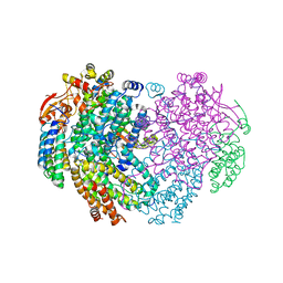
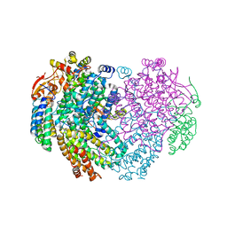
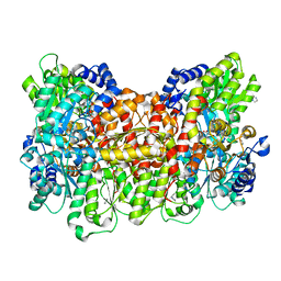
6OP1
DownloadVisualize
BU of 6op1 by Molmil
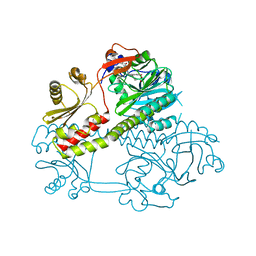
Selenium incorporated, carbon monoxide inhibited FeMo-cofactor of azotobacter vinelandii
Descriptor: 3-HYDROXY-3-CARBOXY-ADIPIC ACID, CALCIUM ION, CARBON MONOXIDE, ...
Authors:Arias, R.J, Rees, D.C.
Deposit date:2019-04-24
Release date:2019-08-14
Last modified:2023-10-11
Method:X-RAY DIFFRACTION (1.7 Å)
Cite:Localized Electronic Structure of Nitrogenase FeMoco Revealed by Selenium K-Edge High Resolution X-ray Absorption Spectroscopy.
J.Am.Chem.Soc., 141, 2019
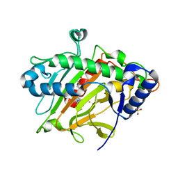
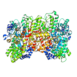
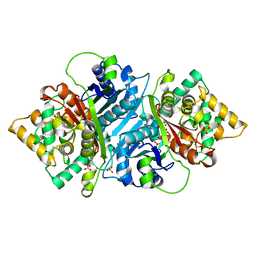
6ZW8
DownloadVisualize
BU of 6zw8 by Molmil
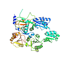
Isopenicillin N synthase in complex with Cd and ACV.
Descriptor: CADMIUM ION, GLYCEROL, Isopenicillin N synthase, ...
Authors:Rabe, P, Kamps, J.J.A.G, Sutherlin, K, Pharm, C, McDonough, M.A, Leissing, T.M, Aller, P, Butryn, A, Linyard, J, Lang, P, Brem, J, Fuller, F.D, Batyuk, A, Hunter, M.S, Pettinati, I, Clifton, I.J, Alonso-Mori, R, Gul, S, Young, I, Kim, I, Bhowmick, A, ORiordan, L, Brewster, A.S, Claridge, T.D.W, Sauter, N.K, Yachandra, V, Yano, J, Kern, J.F, Orville, A.M, Schofield, C.J.
Deposit date:2020-07-27
Release date:2021-06-09
Last modified:2024-01-31
Method:X-RAY DIFFRACTION (1.22 Å)
Cite:X-ray free-electron laser studies reveal correlated motion during isopenicillin N synthase catalysis.
Sci Adv, 7, 2021
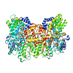
8CCN
DownloadVisualize
BU of 8ccn by Molmil
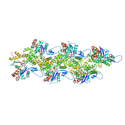
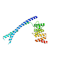
Filamentous actin II from Plasmodium falciparum
Descriptor: ADENOSINE-5'-DIPHOSPHATE, Actin-2, MAGNESIUM ION
Authors:Kursula, I, Lopez, A.J.
Deposit date:2023-01-27
Release date:2023-02-22
Last modified:2023-08-23
Method:ELECTRON MICROSCOPY (3.5 Å)
Cite:Structure and function of Plasmodium actin II in the parasite mosquito stages.
Plos Pathog., 19, 2023
8CCO
DownloadVisualize
BU of 8cco by Molmil
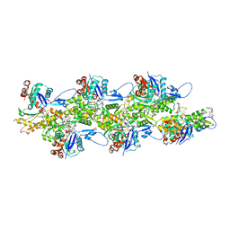
JAS-stabilized F-ActinII from Plasmodium falciparum
Descriptor: ADENOSINE-5'-DIPHOSPHATE, Actin-2, Jasplakinolide, ...
Authors:Kursula, I, Lopez, A.J.
Deposit date:2023-01-27
Release date:2023-03-01
Last modified:2023-08-23
Method:ELECTRON MICROSCOPY (3.28 Å)
Cite:Structure and function of Plasmodium actin II in the parasite mosquito stages.
Plos Pathog., 19, 2023
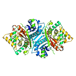
2YQ8
DownloadVisualize
BU of 2yq8 by Molmil
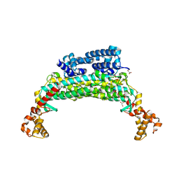
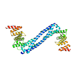
Crystal structure of the SeMet-labeled N-terminal domain and peptide substrate binding domain of alpha subunit of prolyl-4 hydroxylase type I from human.
Descriptor: PROLYL 4-HYDROXYLASE SUBUNIT ALPHA-1
Authors:Anantharajan, J, Koski, M.K, Wierenga, R.K.
Deposit date:2012-11-06
Release date:2013-10-09
Last modified:2024-10-16
Method:X-RAY DIFFRACTION (2.987 Å)
Cite:The Structural Motifs for Substrate Binding and Dimerization of the Alpha Subunit of Collagen Prolyl 4-Hydroxylase
Structure, 21, 2013
4BT9
DownloadVisualize
BU of 4bt9 by Molmil
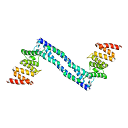
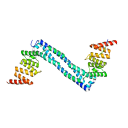
CRYSTAL STRUCTURE OF THE PEPTIDE(PRO-PRO-GLY)3 BOUND COMPLEX OF N- TERMINAL DOMAIN AND PEPTIDE SUBSTRATE BINDING DOMAIN OF PROLYL-4 HYDROXYLASE (RESIDUES 1-238) TYPE I FROM HUMAN
Descriptor: (PRO-PRO-GLY)3 PEPTIDE, PROLYL 4-HYDROXYLASE SUBUNIT ALPHA-1
Authors:Anantharajan, J, Koski, M.K, Wierenga, R.K.
Deposit date:2013-06-14
Release date:2013-10-09
Last modified:2023-12-20
Method:X-RAY DIFFRACTION (1.9 Å)
Cite:The Structural Motifs for Substrate Binding and Dimerization of the Alpha Subunit of Collagen Prolyl 4-Hydroxylase
Structure, 21, 2013
4BT8
DownloadVisualize
BU of 4bt8 by Molmil
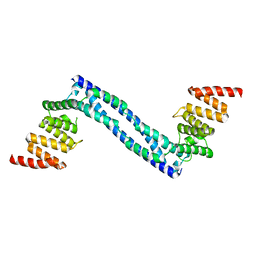
CRYSTAL STRUCTURE OF THE APO FORM OF N-TERMINAL DOMAIN AND PEPTIDE SUBSTRATE BINDING DOMAIN OF PROLYL-4 HYDROXYLASE TYPE I FROM HUMAN
Descriptor: PROLYL 4-HYDROXYLASE SUBUNIT ALPHA-1
Authors:Anantharajan, J, Koski, M.K, Wierenga, R.K.
Deposit date:2013-06-14
Release date:2013-10-09
Last modified:2023-12-20
Method:X-RAY DIFFRACTION (2.198 Å)
Cite:The Structural Motifs for Substrate Binding and Dimerization of the Alpha Subunit of Collagen Prolyl 4-Hydroxylase
Structure, 21, 2013
4BTA
DownloadVisualize
BU of 4bta by Molmil
CRYSTAL STRUCTURE OF THE PEPTIDE(PRO-PRO-GLY)3 BOUND COMPLEX OF N- TERMINAL DOMAIN AND PEPTIDE SUBSTRATE BINDING DOMAIN OF PROLYL-4 HYDROXYLASE (RESIDUES 1-244) TYPE I FROM HUMAN
Descriptor: PROLINE RICH PEPTIDE, PROLYL 4-HYDROXYLASE SUBUNIT ALPHA-1
Authors:Anantharajan, J, Koski, M.K, Pekkala, M, Wierenga, R.K.
Deposit date:2013-06-14
Release date:2013-10-09
Last modified:2023-12-20
Method:X-RAY DIFFRACTION (2.95 Å)
Cite:The Structural Motifs for Substrate Binding and Dimerization of the Alpha Subunit of Collagen Prolyl 4-Hydroxylase
Structure, 21, 2013
4BTB
DownloadVisualize
BU of 4btb by Molmil
CRYSTAL STRUCTURE OF THE PEPTIDE(PRO)9 BOUND COMPLEX OF N-TERMINAL DOMAIN AND PEPTIDE SUBSTRATE BINDING DOMAIN OF PROLYL-4 HYDROXYLASE (RESIDUES 1-238) TYPE I FROM HUMAN
Descriptor: POLY PROLINE PEPTIDE, PROLYL 4-HYDROXYLASE SUBUNIT ALPHA-1
Authors:Anantharajan, J, Koski, M.K, Wierenga, R.K.
Deposit date:2013-06-14
Release date:2013-10-09
Last modified:2023-12-20
Method:X-RAY DIFFRACTION (1.899 Å)
Cite:The Structural Motifs for Substrate Binding and Dimerization of the Alpha Subunit of Collagen Prolyl 4-Hydroxylase
Structure, 21, 2013
6YDI
DownloadVisualize
BU of 6ydi by Molmil
XFEL structure of the Soluble methane monooxygenase hydroxylase and regulatory subunit complex, from Methylosinus trichosporium OB3b, diferrous state
Descriptor: FE (II) ION, GLYCEROL, Methane monooxygenase, ...
Authors:Srinivas, V, Hogbom, M.
Deposit date:2020-03-20
Release date:2020-09-30
Last modified:2024-01-24
Method:X-RAY DIFFRACTION (1.95 Å)
Cite:High-Resolution XFEL Structure of the Soluble Methane Monooxygenase Hydroxylase Complex with its Regulatory Component at Ambient Temperature in Two Oxidation States.
J. Am. Chem. Soc., 142, 2020
6Y0P
DownloadVisualize
BU of 6y0p by Molmil
isopenicillin N synthase in complex with IPN and Fe using FT-SSX methods
Descriptor: FE (III) ION, ISOPENICILLIN N, Isopenicillin N synthase, ...
Authors:Rabe, P, Beale, J.H, Lang, P.A, Dirr, A.S, Leissing, T.M, Butryn, A, Aller, P, Kamps, J.J.A.G, Axford, D, McDonough, M.A, Orville, A.M, Owen, R, Schofield, C.J.
Deposit date:2020-02-10
Release date:2021-02-17
Last modified:2024-01-24
Method:X-RAY DIFFRACTION (1.98 Å)
Cite:X-ray free-electron laser studies reveal correlated motion during isopenicillin N synthase catalysis.
Sci Adv, 7, 2021
6OP3
DownloadVisualize
BU of 6op3 by Molmil
Selenium incorporated FeMo-cofactor of nitrogenase from Azotobacter vinelandii with low concentration of selenium
Descriptor: 3-HYDROXY-3-CARBOXY-ADIPIC ACID, CALCIUM ION, FE(8)-S(7) CLUSTER, ...
Authors:Arias, R.J, Rees, D.C.
Deposit date:2019-04-24
Release date:2019-08-14
Last modified:2023-10-11
Method:X-RAY DIFFRACTION (1.6 Å)
Cite:Localized Electronic Structure of Nitrogenase FeMoco Revealed by Selenium K-Edge High Resolution X-ray Absorption Spectroscopy.
J.Am.Chem.Soc., 141, 2019
6YY3
DownloadVisualize
BU of 6yy3 by Molmil
XFEL structure of the Soluble methane monooxygenase hydroxylase and regulatory subunit complex, from Methylosinus trichosporium OB3b, t=0 diferrous state prior to oxygen activation
Descriptor: FE (II) ION, GLYCEROL, Methane monooxygenase, ...
Authors:Srinivas, V, Hogbom, M.
Deposit date:2020-05-04
Release date:2020-09-30
Last modified:2024-01-24
Method:X-RAY DIFFRACTION (2 Å)
Cite:High-Resolution XFEL Structure of the Soluble Methane Monooxygenase Hydroxylase Complex with its Regulatory Component at Ambient Temperature in Two Oxidation States.
J. Am. Chem. Soc., 142, 2020
6OP2
DownloadVisualize
BU of 6op2 by Molmil
Selenium incorporated FeMo-cofactor of nitrogenase from azotobacter vinelandii at high concentration of selenium
Descriptor: 3-HYDROXY-3-CARBOXY-ADIPIC ACID, CALCIUM ION, FE(8)-S(7) CLUSTER, ...
Authors:Arias, R.J, Rees, D.C.
Deposit date:2019-04-24
Release date:2019-08-14
Last modified:2023-10-11
Method:X-RAY DIFFRACTION (1.9 Å)
Cite:Localized Electronic Structure of Nitrogenase FeMoco Revealed by Selenium K-Edge High Resolution X-ray Absorption Spectroscopy.
J.Am.Chem.Soc., 141, 2019
6YD0
DownloadVisualize
BU of 6yd0 by Molmil
XFEL structure of the Soluble methane monooxygenase hydroxylase and regulatory subunit complex, from Methylosinus trichosporium OB3b, diferric state
Descriptor: FE (III) ION, GLYCEROL, Methane monooxygenase, ...
Authors:Srinivas, V, Hogbom, M.
Deposit date:2020-03-19
Release date:2020-09-30
Last modified:2024-01-24
Method:X-RAY DIFFRACTION (1.95 Å)
Cite:High-Resolution XFEL Structure of the Soluble Methane Monooxygenase Hydroxylase Complex with its Regulatory Component at Ambient Temperature in Two Oxidation States.
J. Am. Chem. Soc., 142, 2020
6YDU
DownloadVisualize
BU of 6ydu by Molmil
XFEL structure of the Soluble methane monooxygenase hydroxylase and regulatory subunit complex, from Methylosinus trichosporium OB3b, reoxidized diferric state, 10s O2 exposure.
Descriptor: FE (III) ION, GLYCEROL, Methane monooxygenase, ...
Authors:Srinivas, V, Hogbom, M.
Deposit date:2020-03-21
Release date:2020-09-30
Last modified:2024-01-24
Method:X-RAY DIFFRACTION (1.95 Å)
Cite:High-Resolution XFEL Structure of the Soluble Methane Monooxygenase Hydroxylase Complex with its Regulatory Component at Ambient Temperature in Two Oxidation States.
J. Am. Chem. Soc., 142, 2020
6OP4
DownloadVisualize
BU of 6op4 by Molmil
Selenium-incorporated, carbon monoxide-inhibited, reactivated FeMo-cofactor of nitrogenase from Azotobacter vinelandii
Descriptor: 3-HYDROXY-3-CARBOXY-ADIPIC ACID, CALCIUM ION, FE(8)-S(7) CLUSTER, ...
Authors:Arias, R.J, Rees, D.C.
Deposit date:2019-04-24
Release date:2019-08-14
Last modified:2023-10-11
Method:X-RAY DIFFRACTION (2.3 Å)
Cite:Localized Electronic Structure of Nitrogenase FeMoco Revealed by Selenium K-Edge High Resolution X-ray Absorption Spectroscopy.
J.Am.Chem.Soc., 141, 2019
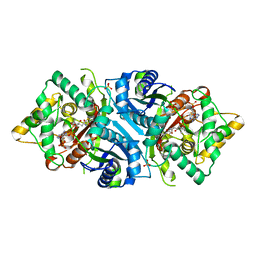
6Z5V
DownloadVisualize
BU of 6z5v by Molmil
CRYSTAL STRUCTURE OF RAT PEROXISOMAL MULTIFUNCTIONAL ENZYME TYPE-1 (RPMFE1) COMPLEXED WITH 3-KETODECANOYL-COA IN CROTONASE FOLD AND OXIDISED NICOTINAMIDE ADENINE DINUCLEOTIDE IN HAD FOLD
Descriptor: 3-KETO-DECANOYL-COA, GLYCEROL, NICOTINAMIDE-ADENINE-DINUCLEOTIDE, ...
Authors:Wierenga, R.K, Sridhar, S, Kiema, T.R.
Deposit date:2020-05-27
Release date:2020-12-09
Last modified:2024-01-24
Method:X-RAY DIFFRACTION (2.33 Å)
Cite:Crystallographic binding studies of rat peroxisomal multifunctional enzyme type 1 with 3-ketodecanoyl-CoA: capturing active and inactive states of its hydratase and dehydrogenase catalytic sites.
Acta Crystallogr D Struct Biol, 76, 2020
6Z5O
DownloadVisualize
BU of 6z5o by Molmil
CRYSTAL STRUCTURE OF RAT PEROXISOMAL MULTIFUNCTIONAL ENZYME TYPE-1 (RPMFE1) COMPLEXED WITH COENZYME-A AND OXIDISED NICOTINAMIDE ADENINE DINUCLEOTIDE
Descriptor: COENZYME A, GLYCEROL, NICOTINAMIDE, ...
Authors:Wierenga, R.K, Sridhar, S, Kiema, T.R.
Deposit date:2020-05-27
Release date:2020-12-09
Last modified:2024-01-24
Method:X-RAY DIFFRACTION (1.7 Å)
Cite:Crystallographic binding studies of rat peroxisomal multifunctional enzyme type 1 with 3-ketodecanoyl-CoA: capturing active and inactive states of its hydratase and dehydrogenase catalytic sites.
Acta Crystallogr D Struct Biol, 76, 2020
6Z5F
DownloadVisualize
BU of 6z5f by Molmil
CRYSTAL STRUCTURE OF RAT PEROXISOMAL MULTIFUNCTIONAL ENZYME TYPE-1 (RPMFE1) COMPLEXED WITH 3-KETODECANOYL-COA AND OXIDISED NICOTINAMIDE ADENINE DINUCLEOTIDE
Descriptor: 3-KETO-DECANOYL-COA, GLYCEROL, NICOTINAMIDE-ADENINE-DINUCLEOTIDE, ...
Authors:Wierenga, R.K, Sridhar, S, Kiema, T.R.
Deposit date:2020-05-26
Release date:2020-12-09
Last modified:2024-01-24
Method:X-RAY DIFFRACTION (2.25 Å)
Cite:Crystallographic binding studies of rat peroxisomal multifunctional enzyme type 1 with 3-ketodecanoyl-CoA: capturing active and inactive states of its hydratase and dehydrogenase catalytic sites.
Acta Crystallogr D Struct Biol, 76, 2020
6ZAI
DownloadVisualize
BU of 6zai by Molmil
Room temperature XFEL Isopenicillin N synthase structure in complex with Fe, O2 and ACV after exposure to dioxygen for 1600ms.
Descriptor: FE (II) ION, Isopenicillin N synthase, L-D-(A-AMINOADIPOYL)-L-CYSTEINYL-D-VALINE, ...
Authors:Rabe, P, Kamps, J.J.A.G, Sutherlin, K, Pharm, C, McDonough, M.A, Leissing, T.M, Aller, P, Butryn, A, Linyard, J, Lang, P, Brem, J, Fuller, F.D, Batyuk, A, Hunter, M.S, Pettinati, I, Clifton, I.J, Alonso-Mori, R, Gul, S, Young, I, Kim, I, Bhowmick, A, ORiordan, L, Brewster, A.S, Claridge, T.D.W, Sauter, N.K, Yachandra, V, Yano, J, Kern, J.F, Orville, A.M, Schofield, C.J.
Deposit date:2020-06-05
Release date:2021-06-09
Last modified:2024-01-24
Method:X-RAY DIFFRACTION (1.5501 Å)
Cite:X-ray free-electron laser studies reveal correlated motion during isopenicillin N synthase catalysis.
Sci Adv, 7, 2021
6ZAE
DownloadVisualize
BU of 6zae by Molmil
Room temperature XFEL Isopenicillin N synthase structure in complex with Fe and ACV under anaerobic conditions.
Descriptor: FE (II) ION, Isopenicillin N synthase, L-D-(A-AMINOADIPOYL)-L-CYSTEINYL-D-VALINE, ...
Authors:Rabe, P, Kamps, J.J.A.G, Sutherlin, K, Pharm, C, McDonough, M.A, Leissing, T.M, Aller, P, Butryn, A, Linyard, J, Lang, P, Brem, J, Fuller, F.D, Batyuk, A, Hunter, M.S, Pettinati, I, Clifton, I.J, Alonso-Mori, R, Gul, S, Young, I, Kim, I, Bhowmick, A, ORiordan, L, Brewster, A.S, Claridge, T.D.W, Sauter, N.K, Yachandra, V, Yano, J, Kern, J.F, Orville, A.M, Schofield, C.J.
Deposit date:2020-06-05
Release date:2021-06-09
Last modified:2024-01-24
Method:X-RAY DIFFRACTION (1.4 Å)
Cite:X-ray free-electron laser studies reveal correlated motion during isopenicillin N synthase catalysis.
Sci Adv, 7, 2021
6ZAQ
DownloadVisualize
BU of 6zaq by Molmil
Room temperature XFEL Isopenicillin N synthase structure in complex with Fe and IPN after dioxygen exposure
Descriptor: FE (II) ION, ISOPENICILLIN N, Isopenicillin N synthase, ...
Authors:Rabe, P, Kamps, J.J.A.G, Sutherlin, K, Pharm, C, McDonough, M.A, Leissing, T.M, Aller, P, Butryn, A, Linyard, J, Lang, P, Brem, J, Fuller, F.D, Batyuk, A, Hunter, M.S, Pettinati, I, Clifton, I.J, Alonso-Mori, R, Gul, S, Young, I, Kim, I, Bhowmick, A, ORiordan, L, Brewster, A.S, Claridge, T.D.W, Sauter, N.K, Yachandra, V, Yano, J, Kern, J.F, Orville, A.M, Schofield, C.J.
Deposit date:2020-06-05
Release date:2021-06-09
Last modified:2024-01-24
Method:X-RAY DIFFRACTION (1.6 Å)
Cite:X-ray free-electron laser studies reveal correlated motion during isopenicillin N synthase catalysis.
Sci Adv, 7, 2021
6ZAG
DownloadVisualize
BU of 6zag by Molmil
Room temperature XFEL Isopenicillin N synthase structure in complex with Fe and ACV after exposure to dioxygen for 500ms.
Descriptor: FE (II) ION, Isopenicillin N synthase, L-D-(A-AMINOADIPOYL)-L-CYSTEINYL-D-VALINE, ...
Authors:Rabe, P, Kamps, J.J.A.G, Sutherlin, K, Pharm, C, McDonough, M.A, Leissing, T.M, Aller, P, Butryn, A, Linyard, J, Lang, P, Brem, J, Fuller, F.D, Batyuk, A, Hunter, M.S, Pettinati, I, Clifton, I.J, Alonso-Mori, R, Gul, S, Young, I, Kim, I, Bhowmick, A, ORiordan, L, Brewster, A.S, Claridge, T.D.W, Sauter, N.K, Yachandra, V, Yano, J, Kern, J.F, Orville, A.M, Schofield, C.J.
Deposit date:2020-06-05
Release date:2021-06-09
Last modified:2024-01-24
Method:X-RAY DIFFRACTION (1.4001 Å)
Cite:X-ray free-electron laser studies reveal correlated motion during isopenicillin N synthase catalysis.
Sci Adv, 7, 2021

226707

PDB entries from 2024-10-30

PDB statisticsPDBj update infoContact PDBjnumon