Loading
PDBj
MenuPDBj@FacebookPDBj@TwitterPDBj@YouTubewwPDB FoundationwwPDB
RCSB PDBPDBeBMRBAdv. SearchSearch help
Search by PDB author
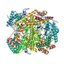
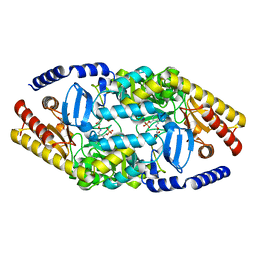
1PWX
DownloadVisualize
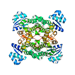
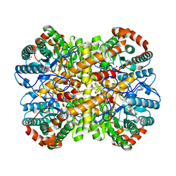
BU of 1pwx by Molmil
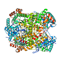
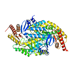
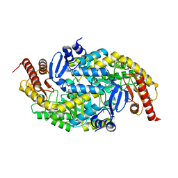
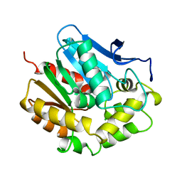
Crystal structure of the haloalcohol dehalogenase HheC complexed with bromide
Descriptor: BROMIDE ION, halohydrin dehalogenase
Authors:de Jong, R.M, Tiesinga, J.J.W, Rozeboom, H.J, Kalk, K.H, Tang, L, Janssen, D.B, Dijkstra, B.W.
Deposit date:2003-07-02
Release date:2003-10-07
Last modified:2024-04-03
Method:X-RAY DIFFRACTION (1.8 Å)
Cite:Structure and Mechanism of a Bacterial Haloalcohol Dehalogenase: a new variation of the short-chain dehydrogenase/reductase fold without an NAD(P)H binding site
EMBO J., 22, 2003
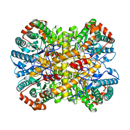
1PX0
DownloadVisualize
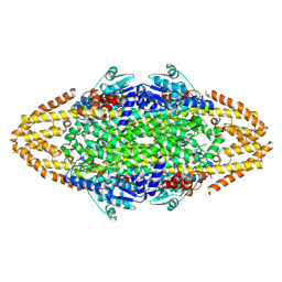
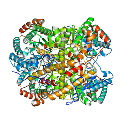
BU of 1px0 by Molmil
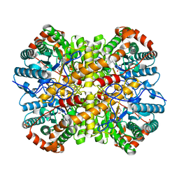
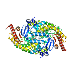
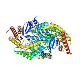
Crystal structure of the haloalcohol dehalogenase HheC complexed with the haloalcohol mimic (R)-1-para-nitro-phenyl-2-azido-ethanol
Descriptor: (R)-1-PARA-NITRO-PHENYL-2-AZIDO-ETHANOL, halohydrin dehalogenase
Authors:de Jong, R.M, Tiesinga, J.J.W, Rozeboom, H.J, Kalk, K.H, Tang, L, Janssen, D.B, Dijkstra, B.W.
Deposit date:2003-07-02
Release date:2003-10-07
Last modified:2023-08-16
Method:X-RAY DIFFRACTION (1.9 Å)
Cite:Structure and Mechanism of a Bacterial Haloalcohol Dehalogenase: a new variation of the short-chain dehydrogenase/reductase fold without an NAD(P)H binding site
EMBO J., 22, 2003
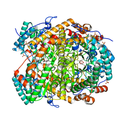
5NH6
DownloadVisualize
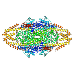
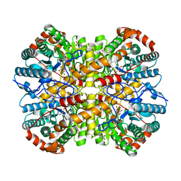
BU of 5nh6 by Molmil
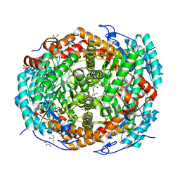
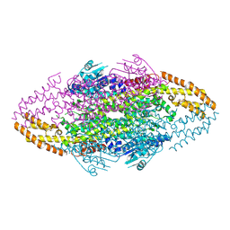
Crystal structure of xylose isomerase from Piromyces E2 Complexed with one Mg2+ ion and xylitol
Descriptor: MAGNESIUM ION, SULFATE ION, Xylitol, ...
Authors:Rozeboom, H.J, Janssen, D.B.
Deposit date:2017-03-21
Release date:2017-11-01
Last modified:2024-05-01
Method:X-RAY DIFFRACTION (1.75 Å)
Cite:Metal Dependence of the Xylose Isomerase from Piromyces sp. E2 Explored by Activity Profiling and Protein Crystallography.
Biochemistry, 56, 2017
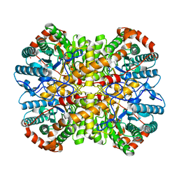
5NH4
DownloadVisualize
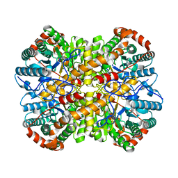
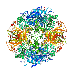
BU of 5nh4 by Molmil
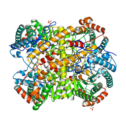
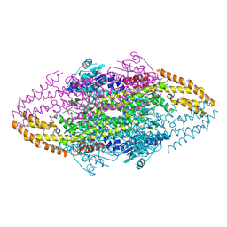
Crystal structure of xylose isomerase from Piromyces E2 in complex with one Mg2+ ions and glycerol
Descriptor: GLYCEROL, MAGNESIUM ION, SULFATE ION, ...
Authors:Rozeboom, H.J, Janssen, D.B.
Deposit date:2017-03-21
Release date:2017-11-01
Last modified:2024-01-17
Method:X-RAY DIFFRACTION (1.8 Å)
Cite:Metal Dependence of the Xylose Isomerase from Piromyces sp. E2 Explored by Activity Profiling and Protein Crystallography.
Biochemistry, 56, 2017
5NHD
DownloadVisualize
BU of 5nhd by Molmil
Crystal structure of xylose isomerase from Piromyces E2 in complex with 2 Ni2+ ions and xylose
Descriptor: D-xylose, NICKEL (II) ION, SULFATE ION, ...
Authors:Rozeboom, H.J, Janssen, D.B.
Deposit date:2017-03-21
Release date:2017-11-01
Last modified:2024-05-01
Method:X-RAY DIFFRACTION (1.8 Å)
Cite:Metal Dependence of the Xylose Isomerase from Piromyces sp. E2 Explored by Activity Profiling and Protein Crystallography.
Biochemistry, 56, 2017
5NHC
DownloadVisualize
BU of 5nhc by Molmil
Crystal structure of xylose isomerase from Piromyces E2 in complex with two Co2+ ions and xylulose
Descriptor: 4-HYDROXYPROLINE, COBALT (II) ION, D-XYLULOSE, ...
Authors:Rozeboom, H.J, Janssen, D.B.
Deposit date:2017-03-21
Release date:2017-11-01
Last modified:2024-05-01
Method:X-RAY DIFFRACTION (1.93 Å)
Cite:Metal Dependence of the Xylose Isomerase from Piromyces sp. E2 Explored by Activity Profiling and Protein Crystallography.
Biochemistry, 56, 2017
5NHE
DownloadVisualize
BU of 5nhe by Molmil
Crystal structure of xylose isomerase from Piromyces E2 in complex with two Cd2+ ions and xylose
Descriptor: CADMIUM ION, D-xylose, SULFATE ION, ...
Authors:Rozeboom, H.J, Janssen, D.B.
Deposit date:2017-03-21
Release date:2017-11-01
Last modified:2024-05-01
Method:X-RAY DIFFRACTION (1.86 Å)
Cite:Metal Dependence of the Xylose Isomerase from Piromyces sp. E2 Explored by Activity Profiling and Protein Crystallography.
Biochemistry, 56, 2017
5NHB
DownloadVisualize
BU of 5nhb by Molmil
Crystal structure of xylose isomerase from Piromyces E2 in complex with two Fe2+ ions
Descriptor: FE (II) ION, SULFATE ION, Xylose isomerase
Authors:Rozeboom, H.J, Janssen, D.B.
Deposit date:2017-03-21
Release date:2017-11-01
Last modified:2024-05-01
Method:X-RAY DIFFRACTION (2.4 Å)
Cite:Metal Dependence of the Xylose Isomerase from Piromyces sp. E2 Explored by Activity Profiling and Protein Crystallography.
Biochemistry, 56, 2017
5OX2
DownloadVisualize
BU of 5ox2 by Molmil
Crystal structure of thymoligase, a substrate-tailored peptiligase variant
Descriptor: Fragment of prodomain, SULFATE ION, Subtilisin BPN'
Authors:Rozeboom, H.J, Janssen, D.B.
Deposit date:2017-09-05
Release date:2018-01-10
Last modified:2018-04-18
Method:X-RAY DIFFRACTION (2.24 Å)
Cite:Design of a substrate-tailored peptiligase variant for the efficient synthesis of thymosin-alpha1.
Org. Biomol. Chem., 16, 2018
5NH9
DownloadVisualize
BU of 5nh9 by Molmil
Crystal structure of xylose isomerase from Piromyces E2 in complex with two Mn2+ ions and xylose
Descriptor: D-xylose, MANGANESE (II) ION, SULFATE ION, ...
Authors:Rozeboom, H.J, Janssen, D.B.
Deposit date:2017-03-21
Release date:2017-11-01
Last modified:2024-05-01
Method:X-RAY DIFFRACTION (2.08 Å)
Cite:Metal Dependence of the Xylose Isomerase from Piromyces sp. E2 Explored by Activity Profiling and Protein Crystallography.
Biochemistry, 56, 2017
5NH7
DownloadVisualize
BU of 5nh7 by Molmil
Crystal structure of xylose isomerase from Piromyces E2 in complex with two Mg2+ ions and xylose
Descriptor: D-xylose, MAGNESIUM ION, SULFATE ION, ...
Authors:Rozeboom, H.J, Janssen, D.B.
Deposit date:2017-03-21
Release date:2017-11-01
Last modified:2024-05-01
Method:X-RAY DIFFRACTION (1.9 Å)
Cite:Metal Dependence of the Xylose Isomerase from Piromyces sp. E2 Explored by Activity Profiling and Protein Crystallography.
Biochemistry, 56, 2017
5NHA
DownloadVisualize
BU of 5nha by Molmil
Crystal structure of xylose isomerase from Piromyces sp. E2 in complex with two Mn2+ ions and sorbitol
Descriptor: MANGANESE (II) ION, SULFATE ION, Xylose isomerase, ...
Authors:Rozeboom, H.J, Janssen, D.B.
Deposit date:2017-03-21
Release date:2017-11-01
Last modified:2024-05-01
Method:X-RAY DIFFRACTION (1.8 Å)
Cite:Metal Dependence of the Xylose Isomerase from Piromyces sp. E2 Explored by Activity Profiling and Protein Crystallography.
Biochemistry, 56, 2017
5NH8
DownloadVisualize
BU of 5nh8 by Molmil
Crystal structure of xylose isomerase from Piromyces E2 in complex with two Ca2+ ions and xylose
Descriptor: CALCIUM ION, D-xylose, SULFATE ION, ...
Authors:Rozeboom, H.J, Janssen, D.B.
Deposit date:2017-03-21
Release date:2017-11-01
Last modified:2024-05-01
Method:X-RAY DIFFRACTION (1.86 Å)
Cite:Metal Dependence of the Xylose Isomerase from Piromyces sp. E2 Explored by Activity Profiling and Protein Crystallography.
Biochemistry, 56, 2017
5NHM
DownloadVisualize
BU of 5nhm by Molmil
Crystal structure of apo xylose isomerase from Piromyces E2
Descriptor: ACETIC ACID, GLYCEROL, SULFATE ION, ...
Authors:Rozeboom, H.J, Janssen, D.B.
Deposit date:2017-03-21
Release date:2017-11-01
Last modified:2024-05-01
Method:X-RAY DIFFRACTION (1.67 Å)
Cite:Metal Dependence of the Xylose Isomerase from Piromyces sp. E2 Explored by Activity Profiling and Protein Crystallography.
Biochemistry, 56, 2017
5NH5
DownloadVisualize
BU of 5nh5 by Molmil
Crystal structure of native xylose isomerase from Piromyces E2
Descriptor: CALCIUM ION, FE (II) ION, GLYCEROL, ...
Authors:Rozeboom, H.J, Janssen, D.B.
Deposit date:2017-03-21
Release date:2017-11-01
Last modified:2024-01-17
Method:X-RAY DIFFRACTION (1.8 Å)
Cite:Metal Dependence of the Xylose Isomerase from Piromyces sp. E2 Explored by Activity Profiling and Protein Crystallography.
Biochemistry, 56, 2017
6G4C
DownloadVisualize
BU of 6g4c by Molmil
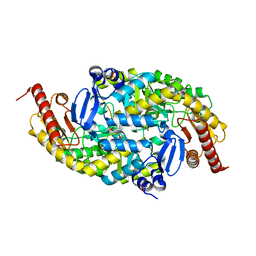
Crystal structure of the omega transaminase from Pseudomonas jessenii in the apo form, crystallized from ammonium phosphate
Descriptor: Aspartate aminotransferase family protein, PHOSPHATE ION
Authors:Rozeboom, H.J, Janssen, D.B.
Deposit date:2018-03-27
Release date:2019-04-10
Last modified:2024-05-01
Method:X-RAY DIFFRACTION (1.87 Å)
Cite:Biochemical properties of a Pseudomonas aminotransferase involved in caprolactam metabolism.
Febs J., 286, 2019
6G4B
DownloadVisualize
BU of 6g4b by Molmil
Crystal structure of the omega transaminase from Pseudomonas jessenii in the apo form, crystallized from succinate
Descriptor: Aspartate aminotransferase family protein, GLYCEROL, SUCCINIC ACID
Authors:Rozeboom, H.J, Janssen, D.B.
Deposit date:2018-03-27
Release date:2019-04-10
Last modified:2024-01-17
Method:X-RAY DIFFRACTION (1.8 Å)
Cite:Biochemical properties of a Pseudomonas aminotransferase involved in caprolactam metabolism.
Febs J., 286, 2019
6G4D
DownloadVisualize
BU of 6g4d by Molmil
Crystal structure of the omega TRANSAMINASE FROM PSEUDOMONAS Jessenii in complex with PLP
Descriptor: Aspartate aminotransferase family protein, GLYCEROL, PYRIDOXAL-5'-PHOSPHATE
Authors:Rozeboom, H.J, Janssen, D.B.
Deposit date:2018-03-27
Release date:2019-04-10
Last modified:2024-05-01
Method:X-RAY DIFFRACTION (2.15 Å)
Cite:Biochemical properties of a Pseudomonas aminotransferase involved in caprolactam metabolism.
Febs J., 286, 2019
6G4E
DownloadVisualize
BU of 6g4e by Molmil
Crystal structure of the omega TRANSAMINASE FROM PSEUDOMONAS Jessenii in complex with PLP and 6-aminohexanoate (6-ACA)
Descriptor: 6-AMINOHEXANOIC ACID, Aspartate aminotransferase family protein, GLYCEROL, ...
Authors:Rozeboom, H.J, Janssen, D.B.
Deposit date:2018-03-27
Release date:2019-04-10
Last modified:2024-05-01
Method:X-RAY DIFFRACTION (2.45 Å)
Cite:Biochemical properties of a Pseudomonas aminotransferase involved in caprolactam metabolism.
Febs J., 286, 2019
6G4F
DownloadVisualize
BU of 6g4f by Molmil
Crystal structure of the omega TRANSAMINASE FROM PSEUDOMONAS Jessenii in complex with PMP
Descriptor: 4'-DEOXY-4'-AMINOPYRIDOXAL-5'-PHOSPHATE, Aspartate aminotransferase family protein, GLYCEROL, ...
Authors:Rozeboom, H.J, Janssen, D.B.
Deposit date:2018-03-27
Release date:2019-04-10
Last modified:2024-05-01
Method:X-RAY DIFFRACTION (2.5 Å)
Cite:Biochemical properties of a Pseudomonas aminotransferase involved in caprolactam metabolism.
Febs J., 286, 2019
4V2Q
DownloadVisualize
BU of 4v2q by Molmil
Ironing out their differences: Dissecting the structural determinants of a phenylalanine aminomutase and ammonia lyase
Descriptor: PHENYLALANINE AMMONIA-LYASE
Authors:Heberling, M, Masman, M, Bartsch, S, Wybenga, G.G, Dijkstra, B.W, Marrink, S, Janssen, D.
Deposit date:2014-10-14
Release date:2014-12-10
Last modified:2024-01-10
Method:X-RAY DIFFRACTION (1.95 Å)
Cite:Ironing Out Their Differences: Dissecting the Structural Determinants of a Phenylalanine Aminomutase and Ammonia Lyase.
Acs Chem.Biol., 10, 2015
1CIJ
DownloadVisualize
BU of 1cij by Molmil
HALOALKANE DEHALOGENASE SOAKED WITH HIGH CONCENTRATION OF BROMIDE
Descriptor: BROMIDE ION, PROTEIN (HALOALKANE DEHALOGENASE)
Authors:Ridder, I.S, Rozeboom, H.J, Kalk, K.H, Dijkstra, B.W.
Deposit date:1999-03-31
Release date:1999-09-29
Last modified:2023-08-09
Method:X-RAY DIFFRACTION (2.3 Å)
Cite:Crystallographic and kinetic evidence of a collision complex formed during halide import in haloalkane dehalogenase.
Biochemistry, 38, 1999
1EDD
DownloadVisualize
BU of 1edd by Molmil
CRYSTALLOGRAPHIC AND FLUORESCENCE STUDIES OF THE INTERACTION OF HALOALKANE DEHALOGENASE WITH HALIDE IONS: STUDIES WITH HALIDE COMPOUNDS REVEAL A HALIDE BINDING SITE IN THE ACTIVE SITE
Descriptor: CHLORIDE ION, HALOALKANE DEHALOGENASE
Authors:Verschueren, K.H.G, Dijkstra, B.W.
Deposit date:1993-05-13
Release date:1993-10-31
Last modified:2024-02-07
Method:X-RAY DIFFRACTION (2.19 Å)
Cite:Crystallographic and fluorescence studies of the interaction of haloalkane dehalogenase with halide ions. Studies with halide compounds reveal a halide binding site in the active site.
Biochemistry, 32, 1993
4V2R
DownloadVisualize
BU of 4v2r by Molmil
Ironing out their differences: Dissecting the structural determinants of a phenylalanine aminomutase and ammonia lyase
Descriptor: PHENYLALANINE AMINOMUTASE (L-BETA-PHENYLALANINE FORMING)
Authors:Heberling, M, Masman, M, Bartsch, S, Wybenga, G.G, Dijkstra, B.W, Marrink, S, Janssen, D.
Deposit date:2014-10-14
Release date:2014-12-10
Last modified:2024-01-10
Method:X-RAY DIFFRACTION (2.2 Å)
Cite:Ironing out their differences: dissecting the structural determinants of a phenylalanine aminomutase and ammonia lyase.
ACS Chem. Biol., 10, 2015
2EDC
DownloadVisualize
BU of 2edc by Molmil
CRYSTALLOGRAPHIC AND FLUORESCENCE STUDIES OF THE INTERACTION OF HALOALKANE DEHALOGENASE WITH HALIDE IONS: STUDIES WITH HALIDE COMPOUNDS REVEAL A HALIDE BINDING SITE IN THE ACTIVE SITE
Descriptor: HALOALKANE DEHALOGENASE, IODIDE ION
Authors:Verschueren, K.H.G, Dijkstra, B.W.
Deposit date:1993-08-31
Release date:1994-01-31
Last modified:2024-02-14
Method:X-RAY DIFFRACTION (2.3 Å)
Cite:Crystallographic and fluorescence studies of the interaction of haloalkane dehalogenase with halide ions. Studies with halide compounds reveal a halide binding site in the active site.
Biochemistry, 32, 1993

221371

PDB entries from 2024-06-19

PDB statisticsPDBj update infoContact PDBjnumon