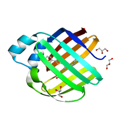
Loading
PDBj
MenuPDBj@FacebookPDBj@X(formerly Twitter)PDBj@BlueSkyPDBj@YouTubewwPDB FoundationwwPDBDonate
RCSB PDBPDBeBMRBAdv. SearchSearch help
Search by PDB author
4QYP
DownloadVisualize
BU of 4qyp by Molmil
The Crystal Structures of holo-wt human Cellular Retinol Binding protein II (hCRBPII) bound to Retinal
Descriptor: ACETATE ION, RETINAL, Retinol-binding protein 2
Authors:Nossoni, Z, Assar, Z, Yapici, I, Nosrati, M, Wang, W, Berbasova, T, Vasileiou, C, Borhan, B, Geiger, H.
Deposit date:2014-07-25
Release date:2014-12-10
Last modified:2023-09-20
Method:X-RAY DIFFRACTION (1.62 Å)
Cite:Structures of holo wild-type human cellular retinol-binding protein II (hCRBPII) bound to retinol and retinal.
Acta Crystallogr.,Sect.D, 70, 2014
6MOP
DownloadVisualize
BU of 6mop by Molmil
Crystal Structure of the All-trans Retinal-Bound R111K:Y134F:T54V:R132Q:P39Y:R59Y:L121E Mutant of Human Cellular Retinoic Acid Binding Protein II in the Dark at 1.9 Angstrom Resolution
Descriptor: Cellular retinoic acid-binding protein 2, RETINAL
Authors:Ghanbarpour, A, Geiger, J.
Deposit date:2018-10-04
Release date:2019-01-09
Last modified:2024-10-16
Method:X-RAY DIFFRACTION (1.9 Å)
Cite:Mimicking Microbial Rhodopsin Isomerization in a Single Crystal.
J. Am. Chem. Soc., 141, 2019
6MPK
DownloadVisualize
BU of 6mpk by Molmil
Crystal Structure of the 13-cis Retinal-Bound R111K:Y134F:T54V:R132Q:P39Y:R59Y:L121E:A32Y Mutant of Human Cellular Retinoic Acid Binding Protein II in the Dark at 1.58 Angstrom Resolution
Descriptor: Cellular retinoic acid-binding protein 2, GLYCEROL, RETINAL
Authors:Ghanbarpour, A, Geiger, J.
Deposit date:2018-10-07
Release date:2019-01-09
Last modified:2024-10-23
Method:X-RAY DIFFRACTION (1.58 Å)
Cite:Mimicking Microbial Rhodopsin Isomerization in a Single Crystal.
J. Am. Chem. Soc., 141, 2019
6MQY
DownloadVisualize
BU of 6mqy by Molmil
Crystal Structure of the 13-cis Product of All-trans Retinal-Bound R111K:Y134F:T54V:R132Q:P39Y:R59Y:L121E Mutant of Human Cellular Retinoic Acid Binding Protein II Irradiated with 400 nm Laser (30 seconds) at 1.9 Angstrom
Descriptor: Cellular retinoic acid-binding protein 2, GLYCEROL, RETINAL
Authors:Ghanbarpour, A, Geiger, J.
Deposit date:2018-10-11
Release date:2019-01-09
Last modified:2024-10-23
Method:X-RAY DIFFRACTION (1.9 Å)
Cite:Mimicking Microbial Rhodopsin Isomerization in a Single Crystal.
J. Am. Chem. Soc., 141, 2019
6MOX
DownloadVisualize
BU of 6mox by Molmil
Crystal Structure of the All-trans Retinal-Bound R111K:Y134F:T54V:R132Q:P39Q:R59Y:L121E Human Cellular Retinoic Acid Binding Protein II Mutant in the Dark at 2.18 Angstrom Resolution
Descriptor: Cellular retinoic acid-binding protein 2, RETINAL
Authors:Ghanbarpour, A, Geiger, J.
Deposit date:2018-10-04
Release date:2019-01-09
Last modified:2024-10-16
Method:X-RAY DIFFRACTION (2.18 Å)
Cite:Mimicking Microbial Rhodopsin Isomerization in a Single Crystal.
J. Am. Chem. Soc., 141, 2019
6MQX
DownloadVisualize
BU of 6mqx by Molmil
Crystal Structure of All-trans Retinal-Bound R111K:Y134F:T54V:R132Q:P39Y:R59Y:L121E Human Cellular Retinoic Acid Binding Protein II Irradiated with 400 nm Laser (30 seconds) and Subsequently Dark Adapted (25 minutes) at 2.0 Angstrom Resolution
Descriptor: Cellular retinoic acid-binding protein 2, GLYCEROL, RETINAL
Authors:Ghanbarpour, A, Geiger, J.
Deposit date:2018-10-11
Release date:2019-01-09
Last modified:2024-11-20
Method:X-RAY DIFFRACTION (2.01 Å)
Cite:Mimicking Microbial Rhodopsin Isomerization in a Single Crystal.
J. Am. Chem. Soc., 141, 2019
6MQI
DownloadVisualize
BU of 6mqi by Molmil
Crystal Structure of the All-trans Retinal bound R111K:Y134F:T54V:R132Q:P39Y:R59Y:L121Q mutant of Human Cellular Retinoic Acid Binding Protein II Irradiated with 400 nm Laser for 5 minutes at 1.87 Angstrom Resolution
Descriptor: Cellular retinoic acid-binding protein 2, RETINAL
Authors:Ghanbarpour, A, Geiger, J.
Deposit date:2018-10-09
Release date:2019-01-09
Last modified:2024-11-13
Method:X-RAY DIFFRACTION (1.87 Å)
Cite:Mimicking Microbial Rhodopsin Isomerization in a Single Crystal.
J. Am. Chem. Soc., 141, 2019
6MQZ
DownloadVisualize
BU of 6mqz by Molmil
Crystal Structure of the 13-cis Product of the All-trans Retinal-Bound R111K:Y134F:T54V:R132Q:P39Y:R59Y:L121E Mutant of Human Cellular Retinoic Acid Binding Protein II Irradiated with 400 nm Laser (5 minutes) at 2.07 Angstrom
Descriptor: Cellular retinoic acid-binding protein 2, GLYCEROL, RETINAL
Authors:Ghanbarpour, A, Geiger, J.
Deposit date:2018-10-11
Release date:2019-01-09
Last modified:2024-11-20
Method:X-RAY DIFFRACTION (2.07 Å)
Cite:Mimicking Microbial Rhodopsin Isomerization in a Single Crystal.
J. Am. Chem. Soc., 141, 2019
6MQW
DownloadVisualize
BU of 6mqw by Molmil
Crystal Structure of All-trans Retinal-Bound R111K:Y134F:T54V:R132Q:P39Y:R59Y:L121E Human Cellular Retinoic Acid Binding Protein II Irradiated with 400 nm laser (30 seconds) and subsequently dark adapted (10 minutes) at 2.1 Angstrom Resolution
Descriptor: Cellular retinoic acid-binding protein 2, RETINAL
Authors:Ghanbarpour, A, Geiger, J.
Deposit date:2018-10-11
Release date:2019-01-09
Last modified:2024-10-16
Method:X-RAY DIFFRACTION (2.09 Å)
Cite:Mimicking Microbial Rhodopsin Isomerization in a Single Crystal.
J. Am. Chem. Soc., 141, 2019
6MOV
DownloadVisualize
BU of 6mov by Molmil
Crystal Structure of the All-trans Retinal bound R111K:Y134F:T54V:R132Q:P39Y:R59Y:L121Q Human Cellular Retinoic Acid Binding Protein II in the Dark at 1.75 Angstrom Resolution
Descriptor: Cellular retinoic acid-binding protein 2, RETINAL
Authors:Ghanbarpour, A, Geiger, J.
Deposit date:2018-10-04
Release date:2019-01-09
Last modified:2024-11-13
Method:X-RAY DIFFRACTION (1.752 Å)
Cite:Mimicking Microbial Rhodopsin Isomerization in a Single Crystal.
J. Am. Chem. Soc., 141, 2019
6MR0
DownloadVisualize
BU of 6mr0 by Molmil
Crystal Structure of the All-trans Retinal Bound R111K:Y134F:T54V:R132Q:P39Q:R59Y:L121E Human Cellular Retinoic Acid Binding Protein II Mutant After 5 Minutes UV irradiation at 2.6 Angstrom Resolution
Descriptor: Cellular retinoic acid-binding protein 2, RETINAL
Authors:Ghanbarpour, A, Geiger, J.
Deposit date:2018-10-11
Release date:2019-01-09
Last modified:2024-10-16
Method:X-RAY DIFFRACTION (2.649 Å)
Cite:Mimicking Microbial Rhodopsin Isomerization in a Single Crystal.
J. Am. Chem. Soc., 141, 2019
6MOR
DownloadVisualize
BU of 6mor by Molmil
Crystal Structure of the All-Trans Retinal-Bound R111K:Y134F:T54V:R132Q:P39Y:R59Y:L121Y Mutant of Human Cellular Retinoic Acid Binding Protein II in the Dark at 1.79 Angstrom Resolution
Descriptor: Cellular retinoic acid-binding protein 2, RETINAL
Authors:Ghanbarpour, A, Geiger, J.
Deposit date:2018-10-04
Release date:2019-01-09
Last modified:2024-11-06
Method:X-RAY DIFFRACTION (1.79 Å)
Cite:Mimicking Microbial Rhodopsin Isomerization in a Single Crystal.
J. Am. Chem. Soc., 141, 2019
4QYN
DownloadVisualize
BU of 4qyn by Molmil
The Crystal Structures of holo-wt human Cellular Retinol Binding protein II (hCRBPII) bound to Retinol
Descriptor: ACETATE ION, RETINOL, Retinol-binding protein 2
Authors:Nossoni, Z, Assar, Z, Yapici, I, Nosrati, M, Wang, W, Berbasova, T, Vasileiou, C, Borhan, B, Geiger, H.
Deposit date:2014-07-24
Release date:2014-12-31
Last modified:2024-02-28
Method:X-RAY DIFFRACTION (1.19 Å)
Cite:Structures of holo wild-type human cellular retinol-binding protein II (hCRBPII) bound to retinol and retinal.
Acta Crystallogr.,Sect.D, 70, 2014
4QZT
DownloadVisualize
BU of 4qzt by Molmil
Crystal Structure of wild type Human Cellular Retinol Binding Protein II (hCRBPII) bound to retinol at 7 KeV beam energy
Descriptor: ACETATE ION, RETINOL, Retinol-binding protein 2
Authors:Assar, Z, Geiger, J.H.
Deposit date:2014-07-28
Release date:2014-12-10
Last modified:2023-09-20
Method:X-RAY DIFFRACTION (1.9 Å)
Cite:Structures of holo wild-type human cellular retinol-binding protein II (hCRBPII) bound to retinol and retinal.
Acta Crystallogr.,Sect.D, 70, 2014
4QZU
DownloadVisualize
BU of 4qzu by Molmil
Crystal Structure of wild type Human Cellular Retinol Binding Protein II (hCRBPII) bound to retinol at 11 KeV beam energy
Descriptor: ACETATE ION, GLYCEROL, RETINOL, ...
Authors:Assar, Z, Geiger, J.H.
Deposit date:2014-07-28
Release date:2014-12-10
Last modified:2023-09-20
Method:X-RAY DIFFRACTION (1.5 Å)
Cite:Structures of holo wild-type human cellular retinol-binding protein II (hCRBPII) bound to retinol and retinal.
Acta Crystallogr.,Sect.D, 70, 2014
4YKO
DownloadVisualize
BU of 4yko by Molmil
Crystal structure of the R111K:Y134F:T54V:R132Q:P39Q:R59Y:A32Y:F3Q mutant of human Cellular Retinoic Acid Binding Protein II with Retinal at 1.58 angstrom resolution
Descriptor: Cellular retinoic acid-binding protein 2, RETINAL
Authors:Nosrati, M, Geiger, J.
Deposit date:2015-03-04
Release date:2016-03-09
Last modified:2024-10-23
Method:X-RAY DIFFRACTION (1.57 Å)
Cite:A Photoisomerizing Rhodopsin Mimic Observed at Atomic Resolution.
J.Am.Chem.Soc., 138, 2016
7MFY
DownloadVisualize
BU of 7mfy by Molmil
The Crystal Structure of Q108K:K40L:T51V:T53S:R58W:Y19W:A33W:L117E Mutant of HCRBPII Bound with LizFluor
Descriptor: 4-{5-[(2E)-but-2-en-2-yl]thiophen-2-yl}-N,N-dimethylaniline, ACETATE ION, GLYCEROL, ...
Authors:Ghanbarpour, A, Geiger, J.
Deposit date:2021-04-12
Release date:2022-01-12
Last modified:2024-11-20
Method:X-RAY DIFFRACTION (1.26 Å)
Cite:Design of Large Stokes Shift Fluorescent Proteins Based on Excited State Proton Transfer of an Engineered Photobase.
J.Am.Chem.Soc., 143, 2021
7MFZ
DownloadVisualize
BU of 7mfz by Molmil
The Crystal Structure of Domain-Swapped Trimer Q108K:K40D:T53A:R58L:Q38F:Q4F Mutant of HCRBPII Bound with LizFluor3 Chromophore Showing Excited State Intermolecular Proton Transfer
Descriptor: 1-(4-{5-[(2E)-but-2-en-2-yl]thiophen-2-yl}phenyl)azetidine, ACETATE ION, GLYCEROL, ...
Authors:Ghanbarpour, A, Geiger, J.
Deposit date:2021-04-12
Release date:2022-01-12
Last modified:2024-10-09
Method:X-RAY DIFFRACTION (2.49 Å)
Cite:Design of Large Stokes Shift Fluorescent Proteins Based on Excited State Proton Transfer of an Engineered Photobase.
J.Am.Chem.Soc., 143, 2021
7LSQ
DownloadVisualize
BU of 7lsq by Molmil
The Crystal Structure of Q108K:K40E:T53A:R58W:Q38F:Q4F:Y19W Mutant of HCRBPII Bound with LizFluor Chromophore Showing Excited State Intermolecular Proton Transfer
Descriptor: 4-{5-[(2E)-but-2-en-2-yl]thiophen-2-yl}-N,N-dimethylaniline, ACETATE ION, Retinol-binding protein 2
Authors:Ghanbarpour, A, Geiger, J.
Deposit date:2021-02-18
Release date:2022-01-12
Last modified:2024-10-23
Method:X-RAY DIFFRACTION (1.67130542 Å)
Cite:Design of Large Stokes Shift Fluorescent Proteins Based on Excited State Proton Transfer of an Engineered Photobase.
J.Am.Chem.Soc., 143, 2021
6MCU
DownloadVisualize
BU of 6mcu by Molmil
Crystal structure of the holo retinal-bound domain-swapped dimer Q108K:T51D:A28H mutant of human Cellular Retinol Binding Protein II
Descriptor: GLYCEROL, RETINAL, Retinol-binding protein 2
Authors:Ghanbarpour, A, Geiger, J.
Deposit date:2018-09-02
Release date:2019-10-16
Last modified:2024-10-23
Method:X-RAY DIFFRACTION (2.572 Å)
Cite:Engineering the hCRBPII Domain-Swapped Dimer into a New Class of Protein Switches.
J.Am.Chem.Soc., 141, 2019
6MCV
DownloadVisualize
BU of 6mcv by Molmil
Crystal Structure of Holo Retinal-Bound Domain-Swapped Dimer of Wild Type Human Cellular Retinol Binding Protein II
Descriptor: RETINAL, Retinol-binding protein 2
Authors:Ghanbarpour, A, Geiger, J.
Deposit date:2018-09-02
Release date:2019-10-16
Last modified:2023-10-11
Method:X-RAY DIFFRACTION (3.3 Å)
Cite:Engineering the hCRBPII Domain-Swapped Dimer into a New Class of Protein Switches.
J.Am.Chem.Soc., 141, 2019
6MLB
DownloadVisualize
BU of 6mlb by Molmil
Crystal structure of the holo retinal-bound domain-swapped dimer Q108K:K40L:T51F mutant of human cellular retinol binding protein II
Descriptor: ACETATE ION, GLYCEROL, RETINAL, ...
Authors:Ghanbarpour, A, Geiger, J.
Deposit date:2018-09-27
Release date:2019-10-16
Last modified:2024-11-13
Method:X-RAY DIFFRACTION (2.15 Å)
Cite:Engineering the hCRBPII Domain-Swapped Dimer into a New Class of Protein Switches.
J.Am.Chem.Soc., 141, 2019
6MKV
DownloadVisualize
BU of 6mkv by Molmil
Crystal structure of Retinal-bound holo Q108K:K40L:T51W domain-swapped dimer of human cellular retinol binding protein 2
Descriptor: ACETATE ION, GLYCEROL, RETINAL, ...
Authors:Ghanbarpour, A, Geiger, J.
Deposit date:2018-09-26
Release date:2019-10-16
Last modified:2024-11-20
Method:X-RAY DIFFRACTION (2.107 Å)
Cite:Engineering the hCRBPII Domain-Swapped Dimer into a New Class of Protein Switches.
J.Am.Chem.Soc., 141, 2019
6ON5
DownloadVisualize
BU of 6on5 by Molmil
Crystal Structure of the Zn-bound Domain-Swapped Dimer Q108K:T51D:A28C:L36C:F57H Mutant of Human Cellular Retinol Binding Protein II
Descriptor: Retinol-binding protein 2, ZINC ION
Authors:Ghanbarpour, A, Geiger, J.
Deposit date:2019-04-20
Release date:2019-10-16
Last modified:2024-11-13
Method:X-RAY DIFFRACTION (1.638 Å)
Cite:Engineering the hCRBPII Domain-Swapped Dimer into a New Class of Protein Switches.
J.Am.Chem.Soc., 141, 2019
6ON8
DownloadVisualize
BU of 6on8 by Molmil
Crystal Structure of the Reduced Form of Apo Domain-Swapped Dimer Q108K:T51D:A28C:L36C:F57H Mutant of Human Cellular Retinol Binding Protein II
Descriptor: Retinol-binding protein 2
Authors:Ghanbarpour, A, Geiger, J.
Deposit date:2019-04-20
Release date:2019-10-16
Last modified:2023-10-11
Method:X-RAY DIFFRACTION (2.395 Å)
Cite:Engineering the hCRBPII Domain-Swapped Dimer into a New Class of Protein Switches.
J.Am.Chem.Soc., 141, 2019
<123


238582

PDB entries from 2025-07-09

PDB statisticsPDBj update infoContact PDBjnumon