Loading
PDBj
MenuPDBj@FacebookPDBj@TwitterPDBj@YouTubewwPDB FoundationwwPDB
RCSB PDBPDBeBMRBAdv. SearchSearch help
Search by PDB author
1V1A
DownloadVisualize
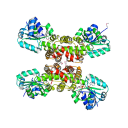
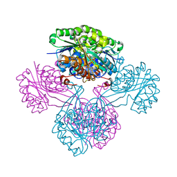
BU of 1v1a by Molmil
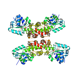
2-KETO-3-DEOXYGLUCONATE KINASE FROM THERMUS THERMOPHILUS WITH BOUND 2-KETO-3-DEOXYGLUCONATE AND ADP
Descriptor: 2-KETO-3-DEOXYGLUCONATE, 2-KETO-3-DEOXYGLUCONATE KINASE, ADENOSINE-5'-DIPHOSPHATE
Authors:Tahirov, T.H, Inagaki, E.
Deposit date:2004-04-12
Release date:2004-04-14
Last modified:2024-05-01
Method:X-RAY DIFFRACTION (2.1 Å)
Cite:Structure of Thermus thermophilus 2-Keto-3-deoxygluconate kinase: evidence for recognition of an open chain substrate.
J. Mol. Biol., 340, 2004
1V1S
DownloadVisualize
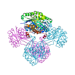
BU of 1v1s by Molmil
2-KETO-3-DEOXYGLUCONATE KINASE FROM THERMUS THERMOPHILUS (CRYSTAL FORM 2)
Descriptor: 2-KETO-3-DEOXYGLUCONATE KINASE
Authors:Tahirov, T.H, Inagaki, E.
Deposit date:2004-04-23
Release date:2004-04-26
Last modified:2023-12-13
Method:X-RAY DIFFRACTION (3.2 Å)
Cite:Structure of Thermus thermophilus 2-Keto-3-deoxygluconate kinase: evidence for recognition of an open chain substrate.
J. Mol. Biol., 340, 2004
2CVZ
DownloadVisualize
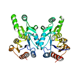
BU of 2cvz by Molmil
Structure of hydroxyisobutyrate dehydrogenase from thermus thermophilus HB8
Descriptor: 3-hydroxyisobutyrate dehydrogenase, NADPH DIHYDRO-NICOTINAMIDE-ADENINE-DINUCLEOTIDE PHOSPHATE
Authors:Lokanath, N.K, Kunishima, N, Miyano, M, RIKEN Structural Genomics/Proteomics Initiative (RSGI)
Deposit date:2005-06-14
Release date:2005-06-28
Last modified:2024-11-06
Method:X-RAY DIFFRACTION (1.8 Å)
Cite:Crystal Structure of Novel NADP-dependent 3-Hydroxyisobutyrate Dehydrogenase from Thermus thermophilus HB8
J.Mol.Biol., 352, 2005
1V7R
DownloadVisualize
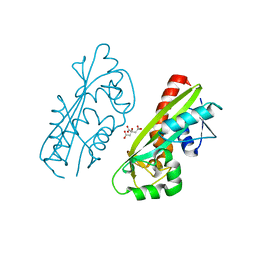
BU of 1v7r by Molmil
Structure of nucleotide triphosphate pyrophosphatase from pyrococcus horikoshii OT3
Descriptor: CITRIC ACID, hypothetical protein PH1917
Authors:Lokanath, N.K, Kunishima, N, RIKEN Structural Genomics/Proteomics Initiative (RSGI)
Deposit date:2003-12-22
Release date:2003-12-30
Last modified:2023-10-25
Method:X-RAY DIFFRACTION (1.4 Å)
Cite:Structures of dimeric nonstandard nucleotide triphosphate pyrophosphatase from Pyrococcus horikoshii OT3: functional significance of interprotomer conformational changes.
J.Mol.Biol., 375, 2008
1WR8
DownloadVisualize
BU of 1wr8 by Molmil
Crystal structure of hypothetical protein PH1421 from Pyrococcus horikoshii.
Descriptor: ACETATE ION, Phosphoglycolate phosphatase
Authors:Yamamoto, H, Kunishima, N, RIKEN Structural Genomics/Proteomics Initiative (RSGI)
Deposit date:2004-10-13
Release date:2004-10-26
Last modified:2024-03-13
Method:X-RAY DIFFRACTION (1.6 Å)
Cite:Structure of a haloacid dehalogenase superfamily phosphatase PH1421 from Pyrococcus horikoshii OT3: oligomeric state and thermoadaptation mechanism.
Acta Crystallogr.,Sect.D, 64, 2008
2DLF
DownloadVisualize
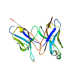
BU of 2dlf by Molmil
HIGH RESOLUTION CRYSTAL STRUCTURE OF THE FV FRAGMENT FROM AN ANTI-DANSYL SWITCH VARIANT ANTIBODY IGG2A(S) CRYSTALLIZED AT PH 6.75
Descriptor: PROTEIN (ANTI-DANSYL IMMUNOGLOBULIN IGG2A(S) (HEAVY CHAIN)), PROTEIN (ANTI-DANSYL IMMUNOGLOBULIN IGG2A(S)-KAPPA (LIGHT CHAIN)), SULFATE ION
Authors:Nakasako, M, Takahashi, H, Shimada, I, Arata, Y.
Deposit date:1998-12-17
Release date:1999-12-17
Last modified:2023-08-23
Method:X-RAY DIFFRACTION (1.55 Å)
Cite:The pH-dependent structural variation of complementarity-determining region H3 in the crystal structures of the Fv fragment from an anti-dansyl monoclonal antibody.
J.Mol.Biol., 291, 1999
1WP4
DownloadVisualize
BU of 1wp4 by Molmil
Structure of TT368 protein from Thermus Thermophilus HB8
Descriptor: 3-hydroxyisobutyrate dehydrogenase, NADPH DIHYDRO-NICOTINAMIDE-ADENINE-DINUCLEOTIDE PHOSPHATE, SULFATE ION
Authors:Lokanath, N.K, Kunishima, N, RIKEN Structural Genomics/Proteomics Initiative (RSGI)
Deposit date:2004-08-30
Release date:2005-08-30
Last modified:2023-10-25
Method:X-RAY DIFFRACTION (2 Å)
Cite:Crystal Structure of Novel NADP-dependent 3-Hydroxyisobutyrate Dehydrogenase from Thermus thermophilus HB8
J.Mol.Biol., 352, 2005
3GFL
DownloadVisualize
BU of 3gfl by Molmil
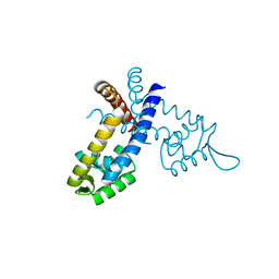
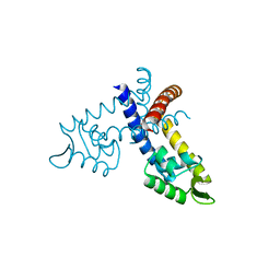
Crystal structure of the ST1710 mutant (R90A) protein
Descriptor: 146aa long hypothetical transcriptional regulator, CALCIUM ION
Authors:Kumarevel, T, Tanaka, T, Yokoyama, S, RIKEN Structural Genomics/Proteomics Initiative (RSGI)
Deposit date:2009-02-27
Release date:2009-08-25
Last modified:2023-11-01
Method:X-RAY DIFFRACTION (1.9 Å)
Cite:ST1710-DNA complex crystal structure reveals the DNA binding mechanism of the MarR family of regulators.
Nucleic Acids Res., 37, 2009
3GF2
DownloadVisualize
BU of 3gf2 by Molmil
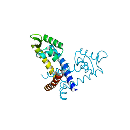
Crystal structure of the hypothetical regulator ST1710 complexed with sodium salicylate
Descriptor: 146aa long hypothetical transcriptional regulator, 2-HYDROXYBENZOIC ACID
Authors:Kumarevel, T, Tanaka, T, Yokoyama, S, RIKEN Structural Genomics/Proteomics Initiative (RSGI)
Deposit date:2009-02-26
Release date:2009-08-25
Last modified:2023-11-01
Method:X-RAY DIFFRACTION (1.8 Å)
Cite:ST1710-DNA complex crystal structure reveals the DNA binding mechanism of the MarR family of regulators.
Nucleic Acids Res., 37, 2009
3GEZ
DownloadVisualize
BU of 3gez by Molmil
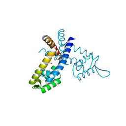
Crystal Structure of the hypothetical egulator from Sulfolobus tokodaii 7
Descriptor: 146aa long hypothetical transcriptional regulator, CALCIUM ION
Authors:Kumarevel, T, Tanaka, T, Yokoyama, S, RIKEN Structural Genomics/Proteomics Initiative (RSGI)
Deposit date:2009-02-26
Release date:2009-08-25
Last modified:2023-11-01
Method:X-RAY DIFFRACTION (2 Å)
Cite:ST1710-DNA complex crystal structure reveals the DNA binding mechanism of the MarR family of regulators.
Nucleic Acids Res., 37, 2009
3GFI
DownloadVisualize
BU of 3gfi by Molmil
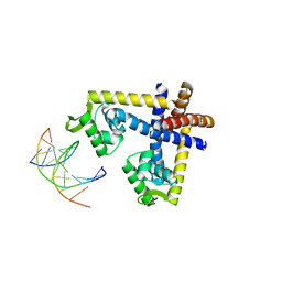
Crystal structure of ST1710 complexed with its promoter DNA
Descriptor: 146aa long hypothetical transcriptional regulator, 5'-D(*TP*AP*AP*CP*AP*AP*TP*AP*GP*CP*AP*AP*A)-3', 5'-D(*TP*TP*GP*CP*TP*AP*TP*TP*GP*T)-3'
Authors:Kumarevel, T, Tanaka, T, Yokoyama, S, RIKEN Structural Genomics/Proteomics Initiative (RSGI)
Deposit date:2009-02-26
Release date:2009-08-25
Last modified:2024-03-20
Method:X-RAY DIFFRACTION (2.1 Å)
Cite:ST1710-DNA complex crystal structure reveals the DNA binding mechanism of the MarR family of regulators.
Nucleic Acids Res., 37, 2009
3GFM
DownloadVisualize
BU of 3gfm by Molmil
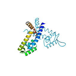
Crystal structure of the ST1710 mutant (K91A) protein
Descriptor: 146aa long hypothetical transcriptional regulator, CALCIUM ION
Authors:Kumarevel, T, Tanaka, T, Yokoyama, S, RIKEN Structural Genomics/Proteomics Initiative (RSGI)
Deposit date:2009-02-27
Release date:2009-08-25
Last modified:2023-11-01
Method:X-RAY DIFFRACTION (2.1 Å)
Cite:ST1710-DNA complex crystal structure reveals the DNA binding mechanism of the MarR family of regulators.
Nucleic Acids Res., 37, 2009
3GFJ
DownloadVisualize
BU of 3gfj by Molmil
Crystal structure of the ST1710 mutant (R89A) protein
Descriptor: 146aa long hypothetical transcriptional regulator, CALCIUM ION
Authors:Kumarevel, T, Tanaka, T, Yokoyama, S, RIKEN Structural Genomics/Proteomics Initiative (RSGI)
Deposit date:2009-02-27
Release date:2009-08-25
Last modified:2023-11-01
Method:X-RAY DIFFRACTION (2.2 Å)
Cite:ST1710-DNA complex crystal structure reveals the DNA binding mechanism of the MarR family of regulators.
Nucleic Acids Res., 37, 2009
2CYP
DownloadVisualize
BU of 2cyp by Molmil
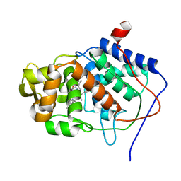
Crystal structure of yeast cytochrome C peroxidase refined at 1.7-angstroms resolution
Descriptor: CYTOCHROME C PEROXIDASE, OXYGEN ATOM, PROTOPORPHYRIN IX CONTAINING FE
Authors:Finzel, B.C, Poulos, T.L, Kraut, J.
Deposit date:1985-08-27
Release date:1986-01-21
Last modified:2024-02-14
Method:X-RAY DIFFRACTION (1.7 Å)
Cite:Crystal structure of yeast cytochrome c peroxidase refined at 1.7-A resolution
J.Biol.Chem., 259, 1984
<12


227111

PDB entries from 2024-11-06

PDB statisticsPDBj update infoContact PDBjnumon