Loading
PDBj
MenuPDBj@FacebookPDBj@X(formerly Twitter)PDBj@BlueSkyPDBj@YouTubewwPDB FoundationwwPDB
RCSB PDBPDBeBMRBAdv. SearchSearch help
Search by PDB author
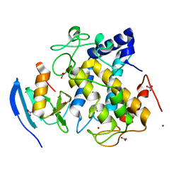
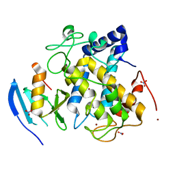
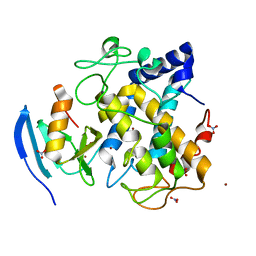
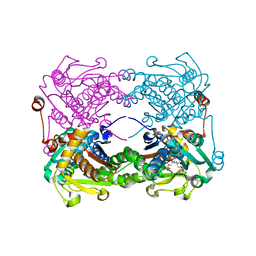
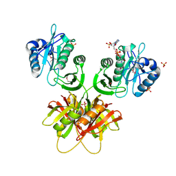
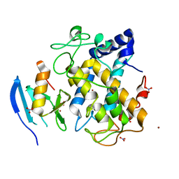
2ZMZ
DownloadVisualize
BU of 2zmz by Molmil
The 1.37-A crystal structure of the hydroxylamine-induced deoxy-form of the copper-bound tyrosinase in complex with a caddie protein from Streptomyces castaneoglobisporus
Descriptor: CADDIE, COPPER (I) ION, NITRATE ION, ...
Authors:Matoba, Y, Sugiyama, M.
Deposit date:2008-04-21
Release date:2009-05-05
Last modified:2023-11-01
Method:X-RAY DIFFRACTION (1.37 Å)
Cite:X-Ray Snapshots of a Hydroxylation Mechanism of Tyrosinase
To be Published
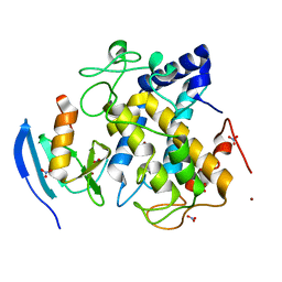
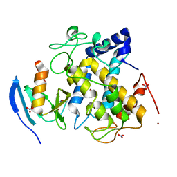
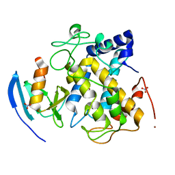
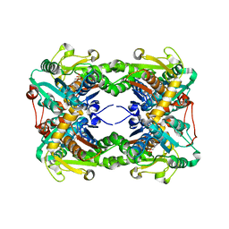
2ZWD
DownloadVisualize
BU of 2zwd by Molmil
Crystal structure of the copper-bound tyrosinase in complex with a caddie protein from streptomyces castaneoglobisporus obtained by soaking the deoxy-form crystal in dioxygen-saturated solution for 5 minutes
Descriptor: COPPER (I) ION, MelC, NITRATE ION, ...
Authors:Matoba, Y, Sugiyama, M.
Deposit date:2008-12-03
Release date:2009-12-15
Last modified:2023-11-01
Method:X-RAY DIFFRACTION (1.35 Å)
Cite:Crystallographic evidence of drastic movement of a copper ion toward the substrate tyrosine for starting hydroxylation reaction of tyrosinase
To be Published
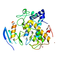
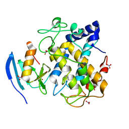
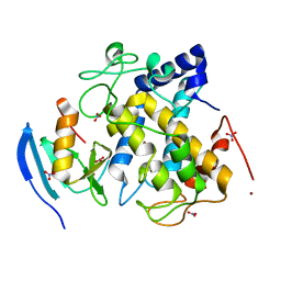
2ZMY
DownloadVisualize
BU of 2zmy by Molmil
Crystal structure of the met2-form of the copper-bound tyrosinase in complex with a caddie protein from Streptomyces castaneoglobisporus obtained by soaking in cupric sulfate solution for 80 hours
Descriptor: CADDIE, COPPER (II) ION, NITRATE ION, ...
Authors:Matoba, Y, Sugiyama, M.
Deposit date:2008-04-21
Release date:2009-05-05
Last modified:2023-11-01
Method:X-RAY DIFFRACTION (1.45 Å)
Cite:X-Ray Snapshots of a Hydroxylation Mechanism of Tyrosinase
To be Published
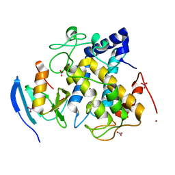
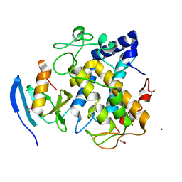
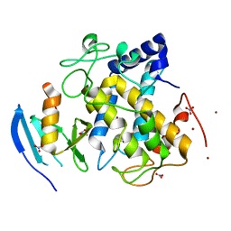
2ZMX
DownloadVisualize
BU of 2zmx by Molmil
Crystal structure of the met1-form of the copper-bound tyrosinase in complex with a caddie protein from Streptomyces castaneoglobisporus obtained by soaking in cupric sulfate solution for 36 hours
Descriptor: CADDIE, COPPER (II) ION, NITRATE ION, ...
Authors:Matoba, Y, Sugiyama, M.
Deposit date:2008-04-21
Release date:2009-04-07
Last modified:2023-11-01
Method:X-RAY DIFFRACTION (1.33 Å)
Cite:Crystallographic Evidence That the Dinuclear Copper Center of Tyrosinase Is Flexible during Catalysis
J.Biol.Chem., 281, 2006
2ZWG
DownloadVisualize
BU of 2zwg by Molmil
Crystal structure of the copper-bound tyrosinase in complex with a caddie protein from streptomyces castaneoglobisporus obtained by soaking the deoxy-form crystal in dioxygen-saturated solution for 12 hours
Descriptor: COPPER (II) ION, MelC, NITRATE ION, ...
Authors:Matoba, Y, Sugiyama, M.
Deposit date:2008-12-03
Release date:2009-12-15
Last modified:2024-10-30
Method:X-RAY DIFFRACTION (1.32 Å)
Cite:Crystallographic evidence of drastic movement of a copper ion toward the substrate tyrosine for starting hydroxylation reaction of tyrosinase
To be Published
3AWT
DownloadVisualize
BU of 3awt by Molmil
Crystal structure of Streptomyces tyrosinase in a complex with caddie soaked in a Cu(II)-containing solution for 20 hr: occupancy of Cu(II) is high
Descriptor: COPPER (II) ION, MelC, NITRATE ION, ...
Authors:Matoba, Y, Sugiyama, M.
Deposit date:2011-03-26
Release date:2011-06-29
Last modified:2023-11-01
Method:X-RAY DIFFRACTION (1.35 Å)
Cite:A molecular mechanism for copper transportation to tyrosinase that is assisted by a metallochaperone, caddie protein
J.Biol.Chem., 286, 2011
3AWY
DownloadVisualize
BU of 3awy by Molmil
Crystal structure of Streptomyces tyrosinase in a complex with caddie M84L mutant soaked in a Cu(II)-containing solution for 80 hr
Descriptor: COPPER (II) ION, MelC, NITRATE ION, ...
Authors:Matoba, Y, Sugiyama, M.
Deposit date:2011-03-26
Release date:2011-06-29
Last modified:2023-11-01
Method:X-RAY DIFFRACTION (1.58 Å)
Cite:A molecular mechanism for copper transportation to tyrosinase that is assisted by a metallochaperone, caddie protein
J.Biol.Chem., 286, 2011
3AWW
DownloadVisualize
BU of 3aww by Molmil
Crystal structure of Streptomyces tyrosinase in a complex with caddie soaked in a Cu(II)-containing solution for 80 hr: occupancy of CuA is high
Descriptor: COPPER (II) ION, MelC, NITRATE ION, ...
Authors:Matoba, Y, Sugiyama, M.
Deposit date:2011-03-26
Release date:2011-06-29
Last modified:2023-11-01
Method:X-RAY DIFFRACTION (1.35 Å)
Cite:A molecular mechanism for copper transportation to tyrosinase that is assisted by a metallochaperone, caddie protein
J.Biol.Chem., 286, 2011
3AWV
DownloadVisualize
BU of 3awv by Molmil
Crystal structure of Streptomyces tyrosinase in a complex with caddie soaked in a Cu(II)-containing solution for 80 hr: occupancy of CuA is low
Descriptor: COPPER (II) ION, MelC, NITRATE ION, ...
Authors:Matoba, Y, Sugiyama, M.
Deposit date:2011-03-26
Release date:2011-06-29
Last modified:2023-11-01
Method:X-RAY DIFFRACTION (1.4 Å)
Cite:A molecular mechanism for copper transportation to tyrosinase that is assisted by a metallochaperone, caddie protein
J.Biol.Chem., 286, 2011
3AWS
DownloadVisualize
BU of 3aws by Molmil
Crystal structure of Streptomyces tyrosinase in a complex with caddie soaked in a Cu(II)-containing solution for 20 hr: occupancy of Cu(II) is low
Descriptor: COPPER (II) ION, MelC, NITRATE ION, ...
Authors:Matoba, Y, Sugiyama, M.
Deposit date:2011-03-26
Release date:2011-06-29
Last modified:2023-11-01
Method:X-RAY DIFFRACTION (1.24 Å)
Cite:A molecular mechanism for copper transportation to tyrosinase that is assisted by a metallochaperone, caddie protein
J.Biol.Chem., 286, 2011
3AX0
DownloadVisualize
BU of 3ax0 by Molmil
Crystal structure of Streptomyces tyrosinase in a complex with caddie Y98F mutant soaked in a Cu(II)-containing solution for 80 hr
Descriptor: COPPER (II) ION, MelC, NITRATE ION, ...
Authors:Matoba, Y, Sugiyama, M.
Deposit date:2011-03-26
Release date:2011-06-29
Last modified:2023-11-01
Method:X-RAY DIFFRACTION (1.4 Å)
Cite:A molecular mechanism for copper transportation to tyrosinase that is assisted by a metallochaperone, caddie protein
J.Biol.Chem., 286, 2011
3AWZ
DownloadVisualize
BU of 3awz by Molmil
Crystal structure of Streptomyces tyrosinase in a complex with caddie H97Q mutant soaked in a Cu(II)-containing solution for 80 hr
Descriptor: COPPER (II) ION, MelC, NITRATE ION, ...
Authors:Matoba, Y, Sugiyama, M.
Deposit date:2011-03-26
Release date:2011-06-29
Last modified:2023-11-01
Method:X-RAY DIFFRACTION (1.43 Å)
Cite:A molecular mechanism for copper transportation to tyrosinase that is assisted by a metallochaperone, caddie protein
J.Biol.Chem., 286, 2011
3AWU
DownloadVisualize
BU of 3awu by Molmil
Crystal structure of Streptomyces tyrosinase in a complex with caddie soaked in a Cu(II)-containing solution for 40 h
Descriptor: COPPER (II) ION, MelC, NITRATE ION, ...
Authors:Matoba, Y, Sugiyama, M.
Deposit date:2011-03-26
Release date:2011-06-29
Last modified:2023-11-01
Method:X-RAY DIFFRACTION (1.16 Å)
Cite:A molecular mechanism for copper transportation to tyrosinase that is assisted by a metallochaperone, caddie protein
J.Biol.Chem., 286, 2011
3AWX
DownloadVisualize
BU of 3awx by Molmil
Crystal structure of Streptomyces tyrosinase in a complex with caddie H82Q mutant soaked in a Cu(II)-containing solution for 80 hr
Descriptor: COPPER (II) ION, MelC, NITRATE ION, ...
Authors:Matoba, Y, Sugiyama, M.
Deposit date:2011-03-26
Release date:2011-06-29
Last modified:2023-11-01
Method:X-RAY DIFFRACTION (1.25 Å)
Cite:A molecular mechanism for copper transportation to tyrosinase that is assisted by a metallochaperone, caddie protein
J.Biol.Chem., 286, 2011
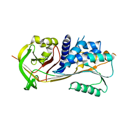
3WA6
DownloadVisualize
BU of 3wa6 by Molmil
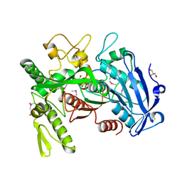
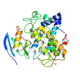
Crystal structure of tannase from Lactobacillus plantarum in the orthorhombic crystal
Descriptor: SULFATE ION, tannase
Authors:Matoba, Y, Tanaka, N, Sugiyama, M.
Deposit date:2013-04-27
Release date:2013-07-24
Last modified:2024-03-20
Method:X-RAY DIFFRACTION (1.8 Å)
Cite:Crystallographic and mutational analyses of tannase from Lactobacillus plantarum
Proteins, 81, 2013
3WSV
DownloadVisualize
BU of 3wsv by Molmil
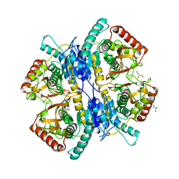
Crystal structure of minor L-lactate dehydrogenase from Enterococcus mundtii in the ligands-unbound form
Descriptor: GLYCEROL, L-lactate dehydrogenase
Authors:Matoba, Y, Sugiyama, M.
Deposit date:2014-03-27
Release date:2014-09-10
Last modified:2023-11-08
Method:X-RAY DIFFRACTION (2.38 Å)
Cite:An alternative allosteric regulation mechanism of an acidophilic l-lactate dehydrogenase from Enterococcus mundtii 15-1A.
Febs Open Bio, 4, 2014
3WSW
DownloadVisualize
BU of 3wsw by Molmil
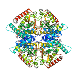
Crystal structure of minor L-lactate dehydrogenase from Enterococcus mundtii in the ligands-bound form
Descriptor: 1,6-di-O-phosphono-beta-D-fructofuranose, GLYCEROL, L-lactate dehydrogenase, ...
Authors:Matoba, Y, Sugiyama, M.
Deposit date:2014-03-27
Release date:2014-09-10
Last modified:2023-11-08
Method:X-RAY DIFFRACTION (2.3 Å)
Cite:An alternative allosteric regulation mechanism of an acidophilic l-lactate dehydrogenase from Enterococcus mundtii 15-1A.
Febs Open Bio, 4, 2014
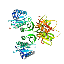
3WA7
DownloadVisualize
BU of 3wa7 by Molmil
Crystal structure of selenomethionine-labeled tannase from Lactobacillus plantarum in the orthorhombic crystal
Descriptor: ACETATE ION, GLYCEROL, SULFATE ION, ...
Authors:Matoba, Y, Tanaka, N, Sugiyama, M.
Deposit date:2013-04-27
Release date:2013-07-24
Last modified:2024-11-20
Method:X-RAY DIFFRACTION (1.7 Å)
Cite:Crystallographic and mutational analyses of tannase from Lactobacillus plantarum.
Proteins, 81, 2013
3X44
DownloadVisualize
BU of 3x44 by Molmil
Crystal structure of O-ureido-L-serine-bound K43A mutant of O-ureido-L-serine synthase
Descriptor: (E)-O-(carbamoylamino)-N-({3-hydroxy-2-methyl-5-[(phosphonooxy)methyl]pyridin-4-yl}methylidene)-L-serine, O-ureido-L-serine synthase
Authors:Matoba, Y, Uda, N, Oda, K, Sugiyama, M.
Deposit date:2015-03-13
Release date:2015-07-29
Last modified:2024-05-29
Method:X-RAY DIFFRACTION (1.9 Å)
Cite:The structural and mutational analyses of O-ureido-L-serine synthase necessary for D-cycloserine biosynthesis.
Febs J., 282, 2015
3X43
DownloadVisualize
BU of 3x43 by Molmil
Crystal structure of O-ureido-L-serine synthase
Descriptor: O-ureido-L-serine synthase, PYRIDOXAL-5'-PHOSPHATE
Authors:Matoba, Y, Uda, N, Oda, K, Sugiyama, M.
Deposit date:2015-03-13
Release date:2015-07-29
Last modified:2023-11-08
Method:X-RAY DIFFRACTION (2.25 Å)
Cite:The structural and mutational analyses of O-ureido-L-serine synthase necessary for D-cycloserine biosynthesis.
Febs J., 282, 2015
2ZV6
DownloadVisualize
BU of 2zv6 by Molmil
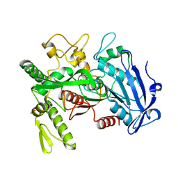
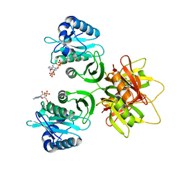
Crystal structure of human squamous cell carcinoma antigen 1
Descriptor: Serpin B3
Authors:Zheng, B, Matoba, Y, Katagiri, C, Hibino, T, Sugiyama, M.
Deposit date:2008-11-01
Release date:2009-02-24
Last modified:2023-11-01
Method:X-RAY DIFFRACTION (2.7 Å)
Cite:Crystal structure of SCCA1 and insight about the interaction with JNK1
Biochem.Biophys.Res.Commun., 380, 2009
2ZW6
DownloadVisualize
BU of 2zw6 by Molmil
Crystal structure of bleomycin N-acetyltransferase from bleomycin-producing Streptomyces verticillus ATCC15003
Descriptor: Bleomycin acetyltransferase, SULFATE ION
Authors:Oda, K, Matoba, Y, Sugiyama, M.
Deposit date:2008-12-01
Release date:2009-11-03
Last modified:2023-11-01
Method:X-RAY DIFFRACTION (2.5 Å)
Cite:Catalytic mechanism of bleomycin N-acetyltransferase proposed on the basis of its crystal structure.
J.Biol.Chem., 285, 2010
2ZW4
DownloadVisualize
BU of 2zw4 by Molmil
Crystal structure of bleomycin N-acetyltransferase complexed with coenzyme A in the orthorhombic crystal
Descriptor: Bleomycin acetyltransferase, COENZYME A, SULFATE ION
Authors:Oda, K, Matoba, Y, Sugiyama, M.
Deposit date:2008-12-01
Release date:2009-11-03
Last modified:2024-11-06
Method:X-RAY DIFFRACTION (2.7 Å)
Cite:Catalytic mechanism of bleomycin N-acetyltransferase proposed on the basis of its crystal structure.
J.Biol.Chem., 285, 2010
3A5J
DownloadVisualize
BU of 3a5j by Molmil
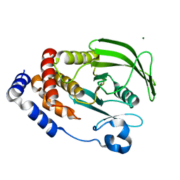
Crystal structure of protein-tyrosine phosphatase 1B
Descriptor: MAGNESIUM ION, Tyrosine-protein phosphatase non-receptor type 1
Authors:Ito, S.
Deposit date:2009-08-08
Release date:2010-08-11
Last modified:2023-11-01
Method:X-RAY DIFFRACTION (1.7 Å)
Cite:Post-translational modification of a non-catalytic Cys121 of PTP1B
To be Published
2ZW5
DownloadVisualize
BU of 2zw5 by Molmil
Crystal structure of bleomycin N-acetyltransferase complexed with coenzyme A in the trigonal crystal
Descriptor: Bleomycin acetyltransferase, COENZYME A
Authors:Oda, K, Matoba, Y, Sugiyama, M.
Deposit date:2008-12-01
Release date:2009-11-03
Last modified:2023-11-01
Method:X-RAY DIFFRACTION (2.4 Å)
Cite:Catalytic mechanism of bleomycin N-acetyltransferase proposed on the basis of its crystal structure.
J.Biol.Chem., 285, 2010

227561

PDB entries from 2024-11-20

PDB statisticsPDBj update infoContact PDBjnumon