Loading
PDBj
MenuPDBj@FacebookPDBj@TwitterPDBj@YouTubewwPDB FoundationwwPDB
RCSB PDBPDBeBMRBAdv. SearchSearch help
Search by PDB author
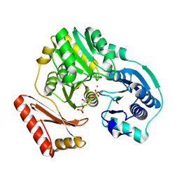
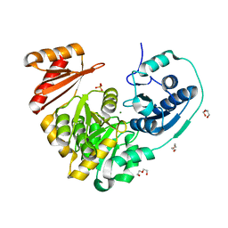
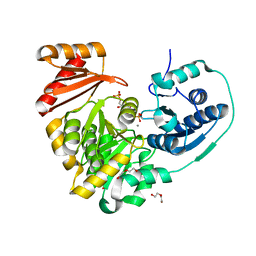
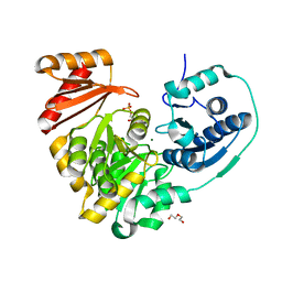
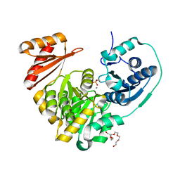
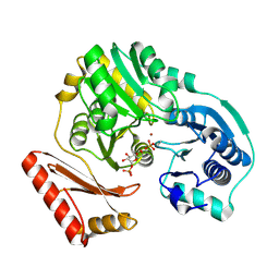
1PCM
DownloadVisualize
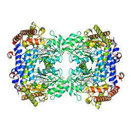
BU of 1pcm by Molmil
Enzyme-ligand complex of P. aeruginosa PMM/PGM
Descriptor: 6-O-phosphono-alpha-D-mannopyranose, Phosphomannomutase, ZINC ION
Authors:Regni, C, Tipton, P.A, Beamer, L.J.
Deposit date:2003-05-16
Release date:2004-01-20
Last modified:2023-08-16
Method:X-RAY DIFFRACTION (1.9 Å)
Cite:Structural basis of diverse substrate recognition by the enzyme PMM/PGM from P. aeruginosa.
Structure, 12, 2004
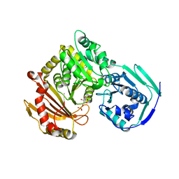
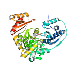
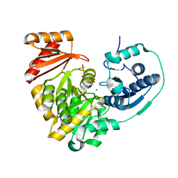
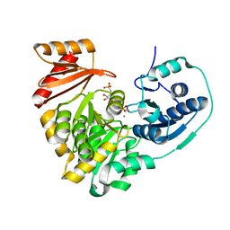
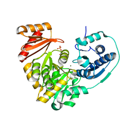
1P5D
DownloadVisualize
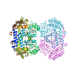
BU of 1p5d by Molmil
Enzyme-ligand complex of P. aeruginosa PMM/PGM
Descriptor: 1-O-phosphono-alpha-D-glucopyranose, Phosphomannomutase, ZINC ION
Authors:Regni, C, Tipton, P.A, Beamer, L.J.
Deposit date:2003-04-25
Release date:2004-01-20
Last modified:2024-04-03
Method:X-RAY DIFFRACTION (1.6 Å)
Cite:Structural basis of diverse substrate recognition by the enzyme PMM/PGM from P. aeruginosa.
Structure, 12, 2004
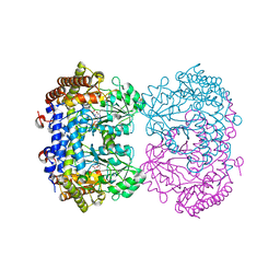
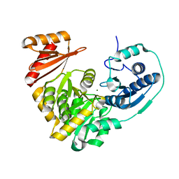
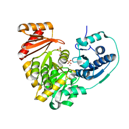
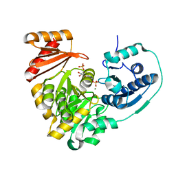
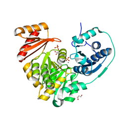
1P5G
DownloadVisualize
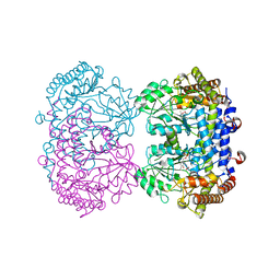
BU of 1p5g by Molmil
Enzyme-ligand complex of P. aeruginosa PMM/PGM
Descriptor: 6-O-phosphono-alpha-D-glucopyranose, Phosphomannomutase, ZINC ION
Authors:Regni, C, Tipton, P.A, Beamer, L.J.
Deposit date:2003-04-26
Release date:2004-01-20
Last modified:2023-08-16
Method:X-RAY DIFFRACTION (1.61 Å)
Cite:Structural basis of diverse substrate recognition by the enzyme PMM/PGM from P. aeruginosa.
Structure, 12, 2004
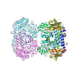
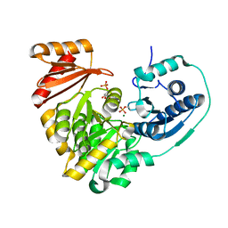
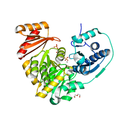
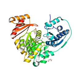
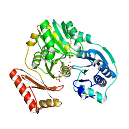
6UIQ
DownloadVisualize
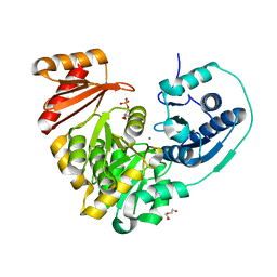
BU of 6uiq by Molmil
Crystal structure of wild-type human phosphoglucomutase 1 in complex with Glucose-6-Phosphate
Descriptor: 6-O-phosphono-alpha-D-glucopyranose, MAGNESIUM ION, phosphoglucomutase-1
Authors:Stiers, K.M, Beamer, L.J.
Deposit date:2019-10-01
Release date:2020-08-12
Last modified:2023-10-11
Method:X-RAY DIFFRACTION (2.3 Å)
Cite:A Hotspot for Disease-Associated Variants of Human PGM1 Is Associated with Impaired Ligand Binding and Loop Dynamics.
Structure, 26, 2018
6UXI
DownloadVisualize
BU of 6uxi by Molmil
Structure of serine hydroxymethyltransferase 8 from Glycine max cultivar Essex complexed with PLP-Glycine
Descriptor: 1,2-ETHANEDIOL, N-GLYCINE-[3-HYDROXY-2-METHYL-5-PHOSPHONOOXYMETHYL-PYRIDIN-4-YL-METHANE], Serine hydroxymethyltransferase
Authors:Korasick, D.A, Tanner, J.J, Beamer, L.J.
Deposit date:2019-11-07
Release date:2020-02-12
Last modified:2023-10-11
Method:X-RAY DIFFRACTION (2.1 Å)
Cite:Impaired folate binding of serine hydroxymethyltransferase 8 from soybean underlies resistance to the soybean cyst nematode.
J.Biol.Chem., 295, 2020
6UXK
DownloadVisualize
BU of 6uxk by Molmil
Structure of serine hydroxymethyltransferase 8 from Glycine max cultivar Forrest complexed with PLP
Descriptor: 1,2-ETHANEDIOL, Serine hydroxymethyltransferase
Authors:Korasick, D.A, Tanner, J.J, Beamer, L.J.
Deposit date:2019-11-07
Release date:2020-02-12
Last modified:2023-11-15
Method:X-RAY DIFFRACTION (2.1 Å)
Cite:Impaired folate binding of serine hydroxymethyltransferase 8 from soybean underlies resistance to the soybean cyst nematode.
J.Biol.Chem., 295, 2020
6UXJ
DownloadVisualize
BU of 6uxj by Molmil
Structure of serine hydroxymethyltransferase 8 from Glycine max cultivar Essex complexed with PLP-glycine and 5-formyltetrahydrofolate
Descriptor: 1,2-ETHANEDIOL, N-GLYCINE-[3-HYDROXY-2-METHYL-5-PHOSPHONOOXYMETHYL-PYRIDIN-4-YL-METHANE], N-[4-({[(6S)-2-amino-5-formyl-4-oxo-3,4,5,6,7,8-hexahydropteridin-6-yl]methyl}amino)benzoyl]-L-glutamic acid, ...
Authors:Korasick, D.A, Tanner, J.J, Beamer, L.J.
Deposit date:2019-11-07
Release date:2020-02-12
Last modified:2023-10-11
Method:X-RAY DIFFRACTION (1.4 Å)
Cite:Impaired folate binding of serine hydroxymethyltransferase 8 from soybean underlies resistance to the soybean cyst nematode.
J.Biol.Chem., 295, 2020
6UXH
DownloadVisualize
BU of 6uxh by Molmil
Structure of serine hydroxymethyltransferase 8 from Glycine max cultivar Essex complexed with PLP
Descriptor: 1,2-ETHANEDIOL, Serine hydroxymethyltransferase
Authors:Korasick, D.A, Tanner, J.J, Beamer, L.J.
Deposit date:2019-11-07
Release date:2020-02-12
Last modified:2023-11-15
Method:X-RAY DIFFRACTION (1.858 Å)
Cite:Impaired folate binding of serine hydroxymethyltransferase 8 from soybean underlies resistance to the soybean cyst nematode.
J.Biol.Chem., 295, 2020
6UXL
DownloadVisualize
BU of 6uxl by Molmil
Structure of serine hydroxymethyltransferase 8 from Glycine max cultivar Forrest complexed with PLP-Glycine
Descriptor: N-GLYCINE-[3-HYDROXY-2-METHYL-5-PHOSPHONOOXYMETHYL-PYRIDIN-4-YL-METHANE], Serine hydroxymethyltransferase
Authors:Korasick, D.A, Tanner, J.J, Beamer, L.J.
Deposit date:2019-11-07
Release date:2020-02-12
Last modified:2023-10-11
Method:X-RAY DIFFRACTION (2.35 Å)
Cite:Impaired folate binding of serine hydroxymethyltransferase 8 from soybean underlies resistance to the soybean cyst nematode.
J.Biol.Chem., 295, 2020
6NOL
DownloadVisualize
BU of 6nol by Molmil
Xanthomonas citri Dephospho-PGM in complex with mannose-1-phosphate
Descriptor: 1-O-phosphono-alpha-D-mannopyranose, DI(HYDROXYETHYL)ETHER, MAGNESIUM ION, ...
Authors:Stiers, K.M, Beamer, L.J.
Deposit date:2019-01-16
Release date:2019-04-10
Last modified:2023-10-11
Method:X-RAY DIFFRACTION (1.73 Å)
Cite:Structural and dynamical description of the enzymatic reaction of a phosphohexomutase.
Struct Dyn., 6, 2019
6NN1
DownloadVisualize
BU of 6nn1 by Molmil
Xanthomonas citri PGM Apo-Dephospho
Descriptor: DI(HYDROXYETHYL)ETHER, MAGNESIUM ION, PHOSPHATE ION, ...
Authors:Stiers, K.M, Beamer, L.J.
Deposit date:2019-01-14
Release date:2019-04-10
Last modified:2023-10-11
Method:X-RAY DIFFRACTION (1.5 Å)
Cite:Structural and dynamical description of the enzymatic reaction of a phosphohexomutase.
Struct Dyn., 6, 2019
6NQF
DownloadVisualize
BU of 6nqf by Molmil
Xanthomonas citri PGM Apo-Phospho at room temperature
Descriptor: CALCIUM ION, Phosphoglucomutase
Authors:Stiers, K.M, Beamer, L.J.
Deposit date:2019-01-21
Release date:2019-04-10
Last modified:2023-10-11
Method:X-RAY DIFFRACTION (1.9 Å)
Cite:Structural and dynamical description of the enzymatic reaction of a phosphohexomutase.
Struct Dyn., 6, 2019
6NQE
DownloadVisualize
BU of 6nqe by Molmil
Xanthomonas citri PGM Apo-Dephospho at room temperature
Descriptor: MAGNESIUM ION, Phosphoglucomutase
Authors:Stiers, K.M, Beamer, L.J.
Deposit date:2019-01-21
Release date:2019-04-10
Last modified:2023-10-11
Method:X-RAY DIFFRACTION (1.85 Å)
Cite:Structural and dynamical description of the enzymatic reaction of a phosphohexomutase.
Struct Dyn., 6, 2019
6NOQ
DownloadVisualize
BU of 6noq by Molmil
Xanthomonas citri Phospho-PGM in complex with mannose-1-phosphate
Descriptor: 1-O-phosphono-alpha-D-mannopyranose, MAGNESIUM ION, Phosphoglucomutase
Authors:Stiers, K.M, Beamer, L.J.
Deposit date:2019-01-16
Release date:2019-04-10
Last modified:2020-07-29
Method:X-RAY DIFFRACTION (1.61 Å)
Cite:Structural and dynamical description of the enzymatic reaction of a phosphohexomutase.
Struct Dyn., 6, 2019
6NP8
DownloadVisualize
BU of 6np8 by Molmil
Xanthomonas citri Phospho-PGM in complex with mannose-6-phosphate
Descriptor: 6-O-phosphono-alpha-D-mannopyranose, CALCIUM ION, DI(HYDROXYETHYL)ETHER, ...
Authors:Stiers, K.M, Beamer, L.J.
Deposit date:2019-01-17
Release date:2019-04-10
Last modified:2023-10-11
Method:X-RAY DIFFRACTION (1.41 Å)
Cite:Structural and dynamical description of the enzymatic reaction of a phosphohexomutase.
Struct Dyn., 6, 2019
6NNP
DownloadVisualize
BU of 6nnp by Molmil
Xanthomonas citri Dephospho-PGM in complex with glucose-6-phosphate
Descriptor: 6-O-phosphono-alpha-D-glucopyranose, MAGNESIUM ION, PHOSPHATE ION, ...
Authors:Stiers, K.M, Beamer, L.J.
Deposit date:2019-01-15
Release date:2019-04-10
Last modified:2023-10-11
Method:X-RAY DIFFRACTION (1.5 Å)
Cite:Structural and dynamical description of the enzymatic reaction of a phosphohexomutase.
Struct Dyn., 6, 2019
6NNS
DownloadVisualize
BU of 6nns by Molmil
Xanthomonas citri Phospho-PGM in complex with glucose-6-phosphate
Descriptor: 6-O-phosphono-alpha-D-glucopyranose, CALCIUM ION, Phosphoglucomutase
Authors:Stiers, K.M, Beamer, L.J.
Deposit date:2019-01-15
Release date:2019-04-10
Last modified:2023-10-11
Method:X-RAY DIFFRACTION (1.45 Å)
Cite:Structural and dynamical description of the enzymatic reaction of a phosphohexomutase.
Struct Dyn., 6, 2019
6NNU
DownloadVisualize
BU of 6nnu by Molmil
Xanthomonas citri Phospho-PGM in complex with glucose-1,6-phosphate
Descriptor: 1,6-di-O-phosphono-alpha-D-glucopyranose, CALCIUM ION, DI(HYDROXYETHYL)ETHER, ...
Authors:Stiers, K.M, Beamer, L.J.
Deposit date:2019-01-15
Release date:2019-04-10
Last modified:2023-10-11
Method:X-RAY DIFFRACTION (1.46 Å)
Cite:Structural and dynamical description of the enzymatic reaction of a phosphohexomutase.
Struct Dyn., 6, 2019
6NPX
DownloadVisualize
BU of 6npx by Molmil
Xanthomonas citri Dephospho-PGM in complex with mannose-6-phosphate
Descriptor: 6-O-phosphono-alpha-D-mannopyranose, DI(HYDROXYETHYL)ETHER, MAGNESIUM ION, ...
Authors:Stiers, K.M, Beamer, L.J.
Deposit date:2019-01-18
Release date:2019-04-10
Last modified:2023-10-11
Method:X-RAY DIFFRACTION (1.42 Å)
Cite:Structural and dynamical description of the enzymatic reaction of a phosphohexomutase.
Struct Dyn., 6, 2019
6NQG
DownloadVisualize
BU of 6nqg by Molmil
Xanthomonas citri Phospho-PGM in complex with glucopyranosyl-1-methyl-phosphonic acid at room temperature
Descriptor: (1S)-1,5-anhydro-1-(phosphonomethyl)-D-glucitol, CALCIUM ION, Phosphoglucomutase
Authors:Stiers, K.M, Beamer, L.J.
Deposit date:2019-01-21
Release date:2019-04-10
Last modified:2023-10-11
Method:X-RAY DIFFRACTION (2.05 Å)
Cite:Structural and dynamical description of the enzymatic reaction of a phosphohexomutase.
Struct Dyn., 6, 2019
6NQH
DownloadVisualize
BU of 6nqh by Molmil
Xanthomonas citri Dephospho-PGM in complex with xylose-1-phosphate
Descriptor: 1-O-phosphono-alpha-D-xylopyranose, MAGNESIUM ION, Phosphoglucomutase
Authors:Stiers, K.M, Beamer, L.J.
Deposit date:2019-01-21
Release date:2020-01-29
Last modified:2023-10-11
Method:X-RAY DIFFRACTION (1.45 Å)
Cite:Substate dynamics of the XcPGM reaction mechanism
To Be Published
6NNN
DownloadVisualize
BU of 6nnn by Molmil
Xanthomonas citri Dephospho-PGM in complex with glucose-1-phosphate
Descriptor: 1-O-phosphono-alpha-D-glucopyranose, DI(HYDROXYETHYL)ETHER, MAGNESIUM ION, ...
Authors:Stiers, K.M, Beamer, L.J.
Deposit date:2019-01-15
Release date:2019-04-10
Last modified:2023-10-11
Method:X-RAY DIFFRACTION (1.35 Å)
Cite:Structural and dynamical description of the enzymatic reaction of a phosphohexomutase.
Struct Dyn., 6, 2019
6NN2
DownloadVisualize
BU of 6nn2 by Molmil
Xanthomonas citri PGM Apo-Phospho
Descriptor: CALCIUM ION, HEXAETHYLENE GLYCOL, Phosphoglucomutase
Authors:Stiers, K.M, Beamer, L.J.
Deposit date:2019-01-14
Release date:2019-04-10
Last modified:2023-10-11
Method:X-RAY DIFFRACTION (1.44 Å)
Cite:Structural and dynamical description of the enzymatic reaction of a phosphohexomutase.
Struct Dyn., 6, 2019
6NNT
DownloadVisualize
BU of 6nnt by Molmil
Xanthomonas citri Dephospho-PGM in complex with glucose-1,6-bisphosphate
Descriptor: 1,6-di-O-phosphono-alpha-D-glucopyranose, MAGNESIUM ION, Phosphoglucomutase
Authors:Stiers, K.M, Beamer, L.J.
Deposit date:2019-01-15
Release date:2019-04-10
Last modified:2023-10-11
Method:X-RAY DIFFRACTION (1.45 Å)
Cite:Structural and dynamical description of the enzymatic reaction of a phosphohexomutase.
Struct Dyn., 6, 2019
6NNO
DownloadVisualize
BU of 6nno by Molmil
Xanthomonas citri Phospho-PGM in complex with glucose-1-phosphate
Descriptor: 1-O-phosphono-alpha-D-glucopyranose, CALCIUM ION, DI(HYDROXYETHYL)ETHER, ...
Authors:Stiers, K.M, Beamer, L.J.
Deposit date:2019-01-15
Release date:2019-04-10
Last modified:2023-10-11
Method:X-RAY DIFFRACTION (1.57 Å)
Cite:Structural and dynamical description of the enzymatic reaction of a phosphohexomutase.
Struct Dyn., 6, 2019

221371

PDB entries from 2024-06-19

PDB statisticsPDBj update infoContact PDBjnumon