Loading
PDBj
MenuPDBj@FacebookPDBj@X(formerly Twitter)PDBj@BlueSkyPDBj@YouTubewwPDB FoundationwwPDB
RCSB PDBPDBeBMRBAdv. SearchSearch help
Search by PDB author
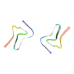
7XO2
DownloadVisualize
BU of 7xo2 by Molmil
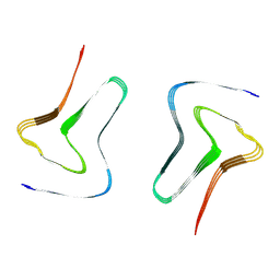
Minor polymorph in alpha-synuclein fibril seeded by cerebrospinal fluid from a mid-to-late stage (mid-PD-4) Parkinson's disease patient
Descriptor: Alpha-synuclein
Authors:Fan, Y, Sun, Y.P, Wang, J, Liu, C.
Deposit date:2022-04-30
Release date:2022-11-30
Last modified:2024-07-03
Method:ELECTRON MICROSCOPY (3 Å)
Cite:Conformational change of alpha-synuclein fibrils in cerebrospinal fluid from different clinical phases of Parkinson's disease.
Structure, 31, 2023
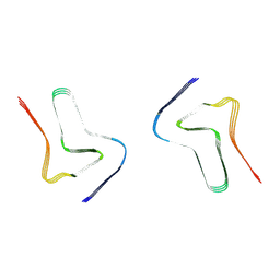
7XO0
DownloadVisualize
BU of 7xo0 by Molmil
Minor polymorph inalpha-synuclein fibril seeded by cerebrospinal fluid from a mid-to-late stage (mid-PD-1) Parkinson's disease patient
Descriptor: Alpha-synuclein
Authors:Fan, Y, Sun, Y.P, Wang, J, Liu, C.
Deposit date:2022-04-30
Release date:2022-11-30
Last modified:2024-07-03
Method:ELECTRON MICROSCOPY (3 Å)
Cite:Conformational change of alpha-synuclein fibrils in cerebrospinal fluid from different clinical phases of Parkinson's disease.
Structure, 31, 2023
7XO3
DownloadVisualize
BU of 7xo3 by Molmil
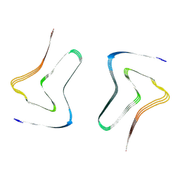
Type 1C alpha-synuclein fibril seeded by cerebrospinal fluid from a mid-to-late stage (mid-PD-4) Parkinson's disease patient
Descriptor: Alpha-synuclein
Authors:Fan, Y, Sun, Y.P, Wang, J, Liu, C.
Deposit date:2022-04-30
Release date:2022-11-30
Last modified:2024-07-03
Method:ELECTRON MICROSCOPY (2.6 Å)
Cite:Conformational change of alpha-synuclein fibrils in cerebrospinal fluid from different clinical phases of Parkinson's disease.
Structure, 31, 2023
7XO1
DownloadVisualize
BU of 7xo1 by Molmil
Major polymorph in alpha-synuclein fibril seeded by cerebrospinal fluid from a mid-to-late stage (mid-PD-1) Parkinson's disease patient
Descriptor: Alpha-synuclein
Authors:Fan, Y, Sun, Y.P, Wang, J, Liu, C.
Deposit date:2022-04-30
Release date:2022-11-30
Last modified:2024-07-03
Method:ELECTRON MICROSCOPY (3 Å)
Cite:Conformational change of alpha-synuclein fibrils in cerebrospinal fluid from different clinical phases of Parkinson's disease.
Structure, 31, 2023
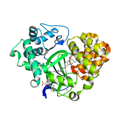
5YJO
DownloadVisualize
BU of 5yjo by Molmil
Crystal structure of SmyD3 in complex with covalent inhibitor 4
Descriptor: Histone-lysine N-methyltransferase SMYD3, S-ADENOSYLMETHIONINE, ZINC ION, ...
Authors:Baburajendran, N, Anna E, J.
Deposit date:2017-10-11
Release date:2018-10-17
Last modified:2024-11-06
Method:X-RAY DIFFRACTION (2.135 Å)
Cite:Discovery of Irreversible Inhibitors Targeting Histone Methyltransferase, SMYD3.
Acs Med.Chem.Lett., 10, 2019
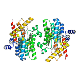
8WDN
DownloadVisualize
BU of 8wdn by Molmil
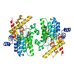
Crystal structure of PDE4D complexed with 7b-1
Descriptor: 1,2-ETHANEDIOL, 3-[(2~{S},3~{R})-7-ethoxy-2-(3-ethoxy-4-methoxy-phenyl)-3-(hydroxymethyl)-2,3-dihydro-1-benzofuran-5-yl]propan-1-ol, MAGNESIUM ION, ...
Authors:Liu, J.Y, Li, M.J, Xu, Y.C.
Deposit date:2023-09-15
Release date:2024-08-28
Method:X-RAY DIFFRACTION (1.55 Å)
Cite:Identification of Dihydrobenzofuran Neolignans as Novel PDE4 Inhibitors and Evaluation of Antiatopic Dermatitis Efficacy in DNCB-Induced Mice Model.
J.Med.Chem., 67, 2024
8WDO
DownloadVisualize
BU of 8wdo by Molmil
Crystal structure of PDE4D complexed with DCN
Descriptor: 1,2-ETHANEDIOL, 3-[(2~{S},3~{R})-2-(3,4-dimethoxyphenyl)-3-(hydroxymethyl)-7-methoxy-2,3-dihydro-1-benzofuran-5-yl]propan-1-ol, MAGNESIUM ION, ...
Authors:Liu, J.Y, Li, M.J, Xu, Y.C.
Deposit date:2023-09-15
Release date:2024-08-28
Method:X-RAY DIFFRACTION (1.67 Å)
Cite:Identification of Dihydrobenzofuran Neolignans as Novel PDE4 Inhibitors and Evaluation of Antiatopic Dermatitis Efficacy in DNCB-Induced Mice Model.
J.Med.Chem., 67, 2024
8HUE
DownloadVisualize
BU of 8hue by Molmil
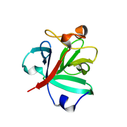
Crystal structure of FGF2-M2 mutant - D28E/C78I/C96I/S137P
Descriptor: 1,3,4,6-tetra-O-sulfo-beta-D-fructofuranose-(2-1)-2,3,4,6-tetra-O-sulfonato-alpha-D-glucopyranose, Fibroblast growth factor 2
Authors:Jung, Y.E, Cha, S.S, An, Y.J.
Deposit date:2022-12-23
Release date:2024-06-26
Last modified:2024-10-16
Method:X-RAY DIFFRACTION (1.48 Å)
Cite:Structural and biochemical investigation into stable FGF2 mutants with novel mutation sites and hydrophobic replacements for surface-exposed cysteines.
Plos One, 19, 2024
8HU7
DownloadVisualize
BU of 8hu7 by Molmil
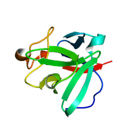
Crystal structure of FGF2-M1 mutant - D28E/C78L/C96I/S137P
Descriptor: Fibroblast growth factor 2
Authors:Jung, Y.E, Cha, S.S, An, Y.J.
Deposit date:2022-12-22
Release date:2024-06-26
Last modified:2024-10-16
Method:X-RAY DIFFRACTION (1.4 Å)
Cite:Structural and biochemical investigation into stable FGF2 mutants with novel mutation sites and hydrophobic replacements for surface-exposed cysteines.
Plos One, 19, 2024
7VWV
DownloadVisualize
BU of 7vwv by Molmil
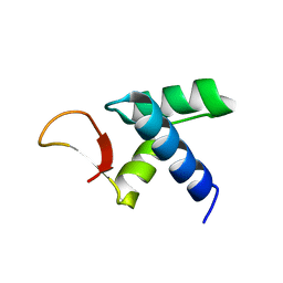
The crystal structure of African swine fever virus I73R
Descriptor: I73R
Authors:Shen, Z, Liang, R.
Deposit date:2021-11-11
Release date:2022-11-16
Last modified:2024-05-29
Method:X-RAY DIFFRACTION (2.59 Å)
Cite:African swine fever virus I73R is a critical virulence-related gene: A potential target for attenuation.
Proc.Natl.Acad.Sci.USA, 120, 2023
8CBL
DownloadVisualize
BU of 8cbl by Molmil
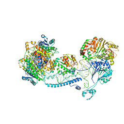
Structure of human mitochondrial RNase Z in complex with mitochondrial pre-tRNA-His(0,Ser)
Descriptor: 3-hydroxyacyl-CoA dehydrogenase type-2, Mitochondrial Precursor tRNA-His(0,Ser), NICOTINAMIDE-ADENINE-DINUCLEOTIDE, ...
Authors:MEYNIER, V, HARDWICK, S, CATALA, M, ROSKE, J, OERUM, S, CHIRGADZE, D, BARRAUD, P, YU, W, LUISI, B, TISNE, C.
Deposit date:2023-01-25
Release date:2024-06-12
Method:ELECTRON MICROSCOPY (2.79 Å)
Cite:Structural basis for human mitochondrial tRNA maturation.
Nat Commun, 15, 2024

227344

PDB entries from 2024-11-13

PDB statisticsPDBj update infoContact PDBjnumon