Loading
PDBj
MenuPDBj@FacebookPDBj@TwitterPDBj@YouTubewwPDB FoundationwwPDB
RCSB PDBPDBeBMRBAdv. SearchSearch help
Search by PDB author
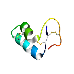
2EYB
DownloadVisualize
BU of 2eyb by Molmil
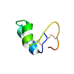
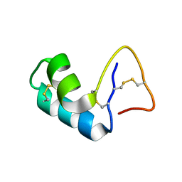
Water refined solution structure of crambin in ACETONE/WATER
Descriptor: CRAMBIN
Authors:Ahn, H.C, Markley, J.L.
Deposit date:2005-11-09
Release date:2006-05-23
Last modified:2022-03-09
Method:SOLUTION NMR
Cite:Three-dimensional structure of the water-insoluble protein crambin in dodecylphosphocholine micelles and its minimal solvent-exposed surface.
J.Am.Chem.Soc., 128, 2006
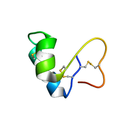
2EYA
DownloadVisualize
BU of 2eya by Molmil
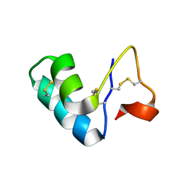
DMSO refined solution structure of crambin in acetone/water
Descriptor: Crambin
Authors:Ahn, H.C, Markley, J.L.
Deposit date:2005-11-09
Release date:2006-05-23
Last modified:2022-03-09
Method:SOLUTION NMR
Cite:Three-dimensional structure of the water-insoluble protein crambin in dodecylphosphocholine micelles and its minimal solvent-exposed surface.
J.Am.Chem.Soc., 128, 2006
2EYC
DownloadVisualize
BU of 2eyc by Molmil
DMSO refined solution structure of crambin in dpc micelles
Descriptor: CRAMBIN
Authors:Ahn, H.C, Markley, J.L.
Deposit date:2005-11-09
Release date:2006-05-23
Last modified:2022-03-09
Method:SOLUTION NMR
Cite:Three-dimensional structure of the water-insoluble protein crambin in dodecylphosphocholine micelles and its minimal solvent-exposed surface.
J.Am.Chem.Soc., 128, 2006
1YV8
DownloadVisualize
BU of 1yv8 by Molmil
Solution structure of crambin in acetone/water mixed solvent
Descriptor: Crambin
Authors:Ahn, H.-C, Markley, J.L.
Deposit date:2005-02-15
Release date:2006-03-07
Last modified:2011-07-13
Method:SOLUTION NMR
Cite:Three-Dimensional Structure of the Water-Insoluble Protein Crambin in Dodecylphosphocholine Micelles and Its Minimal Solvent-Exposed Surface
J.Am.Chem.Soc., 128, 2006
2L0F
DownloadVisualize
BU of 2l0f by Molmil
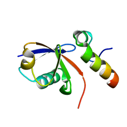
Solution NMR structure of human polymerase iota UBM2 (P692A mutant) in complex with ubiquitin
Descriptor: DNA polymerase iota, Ubiquitin
Authors:Cui, G, Benirschke, R, Mer, G.
Deposit date:2010-07-01
Release date:2010-11-03
Last modified:2024-05-01
Method:SOLUTION NMR
Cite:Structural Basis of Ubiquitin Recognition by Translesion Synthesis DNA Polymerase iota.
Biochemistry, 49, 2010
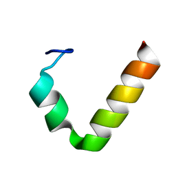
2L0G
DownloadVisualize
BU of 2l0g by Molmil
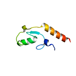
Solution NMR structure of ubiquitin-binding motif (UBM2) of human polymerase iota
Descriptor: DNA polymerase iota
Authors:Cui, G, Benirschke, R, Mer, G.
Deposit date:2010-07-01
Release date:2010-11-03
Last modified:2024-05-01
Method:SOLUTION NMR
Cite:Structural Basis of Ubiquitin Recognition by Translesion Synthesis DNA Polymerase iota.
Biochemistry, 49, 2010
1YVA
DownloadVisualize
BU of 1yva by Molmil
NMR solution structure of crambin in DPC micelles
Descriptor: Crambin
Authors:Ahn, H.-C, Markley, J.L.
Deposit date:2005-02-15
Release date:2006-03-07
Last modified:2022-03-02
Method:SOLUTION NMR
Cite:Three-Dimensional Structure of the Water-Insoluble Protein Crambin in Dodecylphosphocholine Micelles and Its Minimal Solvent-Exposed Surface
J.Am.Chem.Soc., 128, 2006
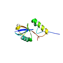
2KRE
DownloadVisualize
BU of 2kre by Molmil
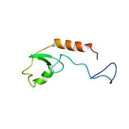
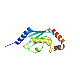
Solution structure of E4B/UFD2A U-Box domain
Descriptor: Ubiquitin conjugation factor E4 B
Authors:Nomine, Y, Wasielewski, E, Botuyan, M, Mer, G.
Deposit date:2009-12-16
Release date:2009-12-29
Last modified:2024-05-01
Method:SOLUTION NMR
Cite:Molecular Basis for the Association of Human E4B U Box Ubiquitin Ligase with E2-Conjugating Enzymes UbcH5c and Ubc4.
Structure, 18, 2010
2KTF
DownloadVisualize
BU of 2ktf by Molmil
Solution NMR structure of human polymerase iota UBM2 in complex with ubiquitin
Descriptor: DNA polymerase iota, Ubiquitin
Authors:Cui, G, Benirschke, R, Mer, G.
Deposit date:2010-02-01
Release date:2010-11-03
Last modified:2024-05-01
Method:SOLUTION NMR
Cite:Structural Basis of Ubiquitin Recognition by Translesion Synthesis DNA Polymerase iota.
Biochemistry, 49, 2010
3L1Z
DownloadVisualize
BU of 3l1z by Molmil
Crystal structure of the U-BOX domain of human E4B ubiquitin ligase in complex with UBCH5C E2 ubiquitin conjugating enzyme
Descriptor: Ubiquitin conjugation factor E4 B, Ubiquitin-conjugating enzyme E2 D3
Authors:Benirschke, R, Thompson, J.R, Mer, G.
Deposit date:2009-12-14
Release date:2010-05-05
Last modified:2023-09-06
Method:X-RAY DIFFRACTION (3.17 Å)
Cite:Molecular Basis for the Association of Human E4B U Box Ubiquitin Ligase with E2-Conjugating Enzymes UbcH5c and Ubc4.
Structure, 18, 2010
3L1X
DownloadVisualize
BU of 3l1x by Molmil
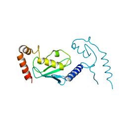
Crystal Structure of U-box Domain of Human E4B Ubiquitin Ligase
Descriptor: Ubiquitin conjugation factor E4 B
Authors:Benirschke, R, Thompson, J.R, Mer, G.
Deposit date:2009-12-14
Release date:2010-05-05
Last modified:2023-09-06
Method:X-RAY DIFFRACTION (2.6 Å)
Cite:Molecular Basis for the Association of Human E4B U Box Ubiquitin Ligase with E2-Conjugating Enzymes UbcH5c and Ubc4.
Structure, 18, 2010
3L1Y
DownloadVisualize
BU of 3l1y by Molmil
Crystal structure of human UBC4 E2 conjugating enzyme
Descriptor: Ubiquitin-conjugating enzyme E2 D2
Authors:Benirschke, R, Thompson, J.R, Mer, G.
Deposit date:2009-12-14
Release date:2010-05-05
Last modified:2023-09-06
Method:X-RAY DIFFRACTION (1.6 Å)
Cite:Molecular Basis for the Association of Human E4B U Box Ubiquitin Ligase with E2-Conjugating Enzymes UbcH5c and Ubc4.
Structure, 18, 2010

223166

PDB entries from 2024-07-31

PDB statisticsPDBj update infoContact PDBjnumon