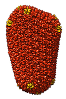
Landmark HIV Capsid Structures Released in PDB

Two complete HIV-capsid structures, both of unprecedented size, are described in this week's issue of Nature and released in the Protein Data Bank (PDB; wwpdb.org). This represents a significant advance in the field of structural biology and a milestone for the PDB.
PDB entries 3J3Q and 3J3Y are models based on cryo-electron microscopy data and use of a molecular dynamics flexible-fitting method. They contain 1356 and 1176 protein chains, respectively, and over two million atoms each. The HIV-1 capsid is the protein envelope that encloses and protects the RNA genome of the virus. An important subject of study, the full capsid has been a difficult target for structural characterization due to its extremely large size and morphological variability.
The wwPDB has anticipated structures of increasing size and complexity that exceed the limitations of the original PDB file format. These capsid structures have been curated following the recently announced wwPDB procedures for the deposition and release of large structures. Extremely large structures can now be deposited, annotated, and released as single files in PDBx/mmCIF and PDBML/XML formats.
The intact capsid structures and the cryo-electron tomography reconstruction from which they were generated can be downloaded from the PDB archive ftp sites in the USA, Europe and Japan as follows:
- USA
-
ftp://ftp.wwpdb.org/pub/pdb/data/large_structures/mmCIF/3j3q.cif.gz
ftp://ftp.wwpdb.org/pub/pdb/data/large_structures/XML/3j3q.xml.gz
ftp://ftp.wwpdb.org/pub/pdb/data/large_structures/mmCIF/3j3y.cif.gz
ftp://ftp.wwpdb.org/pub/pdb/data/large_structures/XML/3j3y.xml.gz
ftp://ftp.wwpdb.org/pub/emdb/structures/EMD-5639/map/emd_5639.map.gz
- Europe
-
ftp://ftp.ebi.ac.uk/pub/databases/pdb/data/large_structures/mmCIF/3j3q.cif.gz
ftp://ftp.ebi.ac.uk/pub/databases/pdb/data/large_structures/XML/3j3q.xml.gz
ftp://ftp.ebi.ac.uk/pub/databases/pdb/data/large_structures/mmCIF/3j3y.cif.gz
ftp://ftp.ebi.ac.uk/pub/databases/pdb/data/large_structures/XML/3j3y.xml.gz
ftp://ftp.ebi.ac.uk/pub/databases/emdb/structures/EMD-5639/map/emd_5639.map.gz
- Japan
-
ftp://ftp.pdbj.org/pub/pdb/data/large_structures/mmCIF/3j3q.cif.gz
ftp://ftp.pdbj.org/pub/pdb/data/large_structures/XML/3j3q.xml.gz
ftp://ftp.pdbj.org/pub/pdb/data/large_structures/mmCIF/3j3y.cif.gz
ftp://ftp.pdbj.org/pub/pdb/data/large_structures/XML/3j3y.xml.gz
ftp://ftp.pdbj.org/pub/emdb/structures/EMD-5639/map/emd_5639.map.gz
Reference
Mature HIV-1 capsid structure by cryo-electron microscopy and all-atom molecular dynamics.
Gongpu Zhao, Juan R. Perilla, Ernest L. Yufenyuy, Xin Meng, Bo Chen, Jiying Ning, Jinwoo Ahn, Angela M. Gronenborn, Klaus Schulten, Christopher Aiken, & Peijun Zhang,
Nature 497, 643-646 (2013) DOI: 10.1038/nature12162
Related Entries
EMDB entry EMD-5639 is the cryo-electron tomography reconstruction from which 3J3Q and 3J3Y were generated; related entry 3J34, derived from an 8.6 Ångström reconstruction of a capsid hexameric subunit in a helical assembly (EMD-5582), was used in the construction of both 3J3Q and 3J3Y. These entries can be accessed and analysed through the websites of the wwPDB partners in Europe (pdbe.org), the USA (rcsb.org) and Japan (pdbj.org).


