






 9YCT
9YCTSummary for 9YCT
| Entry DOI | 10.2210/pdb9yct/pdb |
| Related | 9P6L |
| EMDB information | 72786 |
| Descriptor | DNA (5'-D(*TP*TP*TP*TP*TP*T)-3'), UL52, UL5, ... (6 entities in total) |
| Functional Keywords | hsv, helicase-primase, pritelivir, viral protein |
| Biological source | Human alphaherpesvirus 1 strain R-15 More |
| Total number of polymer chains | 4 |
| Total formula weight | 295635.57 |
| Authors | Yu, Z.,Abraham, J. (deposition date: 2025-09-19, release date: 2025-12-31, Last modification date: 2026-07-15) |
| Primary citation | Yu, Z.,Sathyanarayana, P.,Liu, C.,Tan, J.M.J.,Yang, P.,Das, B.,Hu, S.,Fan, X.,Ji, C.,Weller, S.K.,Shekhar, M.,Coen, D.M.,Kranzusch, P.J.,Loparo, J.J.,Abraham, J. Mechanisms of HSV-1 helicase-primase inhibition and replication fork complex assembly. Cell, 189:478-494.e18, 2026 Cited by PubMed Abstract: Herpesviruses are widespread double-stranded DNA viruses that establish lifelong latency and cause various diseases. Although DNA-polymerase-targeting antivirals are effective, increasing drug resistance underscores the need for alternatives. Helicase-primase inhibitors (HPIs) are promising antivirals, but their mechanisms of action are poorly defined. Furthermore, how the helicase-primase (H/P) complex and DNA polymerase coordinate genome replication is not well understood for herpesviruses. Here, we report cryo-electron microscopy (cryo-EM) structures of the herpes simplex virus 1 H/P complex bound to HPIs, showing that these lock the H/P complex in an inactive state. Single-molecule assays reveal that HPIs cause H/P complexes to pause in unwinding activity on DNA. The structure of an HPI-bound replication fork complex, comprising the H/P complex (UL5, UL52, and UL8) and the polymerase holoenzyme (UL30 and UL42), reveals a previously uncharacterized interface bridging these complexes. These findings provide a structural framework for understanding herpesvirus replisome assembly and advancing inhibitor development. PubMed: 41468884DOI: 10.1016/j.cell.2025.11.041 PDB entries with the same primary citation |
| Experimental method | ELECTRON MICROSCOPY (3.7 Å) |
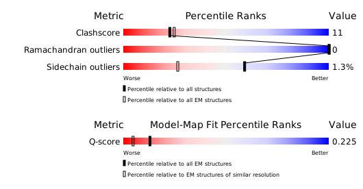
Structure validation
Download full validation report


