






 9WIW
Crystal structure of a Escherichia phage UPEC07 protein
Summary for 9WIW
| Entry DOI | 10.2210/pdb9wiw/pdb |
| Descriptor | Endoribonuclease, SULFATE ION (3 entities in total) |
| Functional Keywords | helix, dimer, sponge protein, anti typeiv thoeris, antimicrobial protein |
| Biological source | Escherichia phage T4 |
| Total number of polymer chains | 1 |
| Total formula weight | 13068.47 |
| Authors | |
| Primary citation | Chen, Y.,Wei, L.,Han, Z.,Zhao, R.,Zeng, Z.,Liu, Y.,Zhang, P.,Tan, K.,Fan, Y.,Sheng, L.,Wang, H.,Pinilla-Redondo, R.,Feng, Y.,Han, W. A phage-encoded sponge protein suppresses bacterial TIR-Caspase immune signaling. Nat Commun, 17:-, 2026 Cited by PubMed Abstract: Triggering immune response through generation of signal molecules is a common immune strategy across all domains of life. In the bacterial type IV Thoeris anti-phage system, a Toll/interleukin-1 receptor (TIR)-domain protein produces adenosine 5'-diphosphate-cyclo[N7:1"]-ribose (N7-cADPR) as the immune signal to activate a Caspase-like effector. Here, we identified an inhibitor of type IV Thoeris through a phage mating assay that allows a sensitive phage to acquire anti-defense genes from related resistant phages. The inhibitor (hereafter TadIV-1) functions as a sponge that sequesters the N7-cADPR signal to inhibit Caspase activation. Structural analyses of TadIV-1 indicate a distinctive signal binding mechanism, wherein the binding pocket comprises its N-terminal flexible loop. In addition, phages lacking TadIV-1 can escape type IV Thoeris sensing through mutation in the capsid vertex protein. Collectively, this work expands the phage anti-defense arsenal with a unique immune signal sequestration mechanism and provides insights into phage invasion recognition mechanism of type IV Thoeris. PubMed: 41968157DOI: 10.1038/s41467-026-71527-4 PDB entries with the same primary citation |
| Experimental method | X-RAY DIFFRACTION (2.56 Å) |
Structure validation
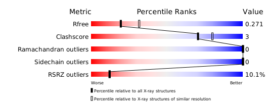
Download full validation report


