






 9WIK
9WIKSummary for 9WIK
| Entry DOI | 10.2210/pdb9wik/pdb |
| EMDB information | 65999 |
| Descriptor | Guanine nucleotide-binding protein G(i) subunit alpha-1, Guanine nucleotide-binding protein G(I)/G(S)/G(T) subunit beta-1, Guanine nucleotide-binding protein G(I)/G(S)/G(O) subunit gamma-2, ... (6 entities in total) |
| Functional Keywords | gpcr, signaling protein, membrane protein |
| Biological source | Homo sapiens (human) More |
| Total number of polymer chains | 5 |
| Total formula weight | 193418.04 |
| Authors | Suzuki, S.,Nishikawa, K.,Tran, D.P.,Akio, K.,Fujiyoshi, Y. (deposition date: 2025-08-28, release date: 2026-07-01, Last modification date: 2026-07-29) |
| Primary citation | Suzuki, S.,Tran, D.P.,Nishikawa, K.,Kitao, A.,Fujiyoshi, Y. Mechanistic insight into signal bias by the agonist-dependent conformational dynamics of GPR84. Nat Commun, 2026 Cited by PubMed Abstract: GPR84 is an orphan class A GPCR primarily expressed in immune cells, where it plays key roles in inflammation and metabolism. Here, we present the cryo-electron microscopy structures of the GPR84-Gi complex bound to the G protein-biased agonist DL-175, and the inactive state of GPR84 bound to the antagonist GLPG1205. Combined with signaling assays and molecular dynamics simulations, these structures elucidate the conformational landscape spanning the inactive and G protein-biased active states of GPR84, providing a mechanistic basis for biased agonism. Notably, steric interactions between DL-175 and L336 selectively preclude the conformational changes required for efficient β-arrestin recruitment without compromising G protein activation. These structural insights provide a structural context for the rational design of GPR84-targeted therapeutics with precisely tuned signaling profiles. PubMed: 42463693DOI: 10.1038/s41467-026-75728-9 PDB entries with the same primary citation |
| Experimental method | ELECTRON MICROSCOPY (3.3 Å) |
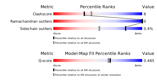
Structure validation
Download full validation report


