






 9WID
9WID
AMP-PNP bound E.coli CnoX-GroEL complex, state II
Summary for 9WID
| Entry DOI | 10.2210/pdb9wid/pdb |
| Related | 9WCW |
| EMDB information | 65990 |
| Descriptor | Chaperonin GroEL, Chaperedoxin, MAGNESIUM ION, ... (5 entities in total) |
| Functional Keywords | complex, chaperone |
| Biological source | Escherichia coli K-12 More |
| Total number of polymer chains | 28 |
| Total formula weight | 1256579.13 |
| Authors | |
| Primary citation | Kim, J.,Jung, M.,Roh, S.H. Structural interplay of the redox co-chaperone CnoX to GroEL/ES chaperonin. Life Sci Alliance, 9:-, 2026 Cited by PubMed Abstract: Protein folding by the bacterial chaperonin GroEL/ES relies on ATP-driven conformational cycles that promote substrate encapsulation and folding. Under oxidative stress, the redox-active co-chaperone CnoX protects oxidized proteins and associates with GroEL, yet the structural basis of its interaction with the GroEL/ES remains incompletely understood. Using single-particle cryo-electron microscopy, we resolved four distinct nucleotide-bound conformational states of CnoX-associated GroEL/ES complexes. CnoX remains tethered to GroEL through its C-terminal TPR domain despite substantial rearrangements of the GroEL apical domains. We further captured a GroEL/ES-CnoX ternary assembly in which CnoX and GroES simultaneously occupy the same GroEL ring, demonstrating that their binding sites are structurally distinct and non-overlapping. Comparison of two GroES-bound states reveals how apical-domain compaction occludes the CnoX-binding surface and coincides with loss of CnoX from the cis-ring. Together, these structures define how CnoX is accommodated and excluded across distinct GroEL/ES conformations and provide a structural framework for understanding the interplay between redox co-chaperones and chaperonin assemblies. PubMed: 42567683DOI: 10.26508/lsa.202603764 PDB entries with the same primary citation |
| Experimental method | ELECTRON MICROSCOPY (3.46 Å) |
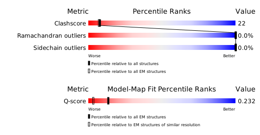
Structure validation
Download full validation report


