






 9W3A
9W3A
Crystal structure of PfiAT toxin-antitoxin complex
Summary for 9W3A
| Entry DOI | 10.2210/pdb9w3a/pdb |
| Descriptor | PfiT protein 1, PfiA protein 1 (3 entities in total) |
| Functional Keywords | pfiat, toxin-antitoxin, pare family, toxin |
| Biological source | Pseudomonas aeruginosa More |
| Total number of polymer chains | 10 |
| Total formula weight | 108735.56 |
| Authors | Wang, X.X.,Chen, R. (deposition date: 2025-07-29, release date: 2026-02-11, Last modification date: 2026-04-15) |
| Primary citation | Chen, R.,Zhang, Y.,Guo, Y.,Gu, J.,Lin, S.,Wang, X. Phosphorylation of PfiA modulates Pf4 phage production through PfiA/PfiT stoichiometric reconfiguration in Pseudomonas aeruginosa. Sci Adv, 12:eaeb5480-eaeb5480, 2026 Cited by PubMed Abstract: Filamentous Pf bacteriophages are widely distributed in and profoundly influence biofilm formation and host virulence. The Pf4 prophage encodes a type II toxin-antitoxin (TA) system, PfiAT, modulating Pf4 propagation; however, its mechanistic role remains unclear. Here, through structural and biochemical analysis, we demonstrate that the PfiT toxin (ParE/RelE superfamily) has a unique C-terminal extension essential for TA complex formation. The antitoxin PfiA harbors a previously uncharacterized DNA binding domain, and its phosphorylation during biofilm formation shifts the PfiAT complex stoichiometry from a noncanonical PfiAPfiT to a canonical PfiAPfiT assembly. This phosphorylation is mediated by the prophage Pf6-encoded kinase toxin PfkA/PfkB at T5 in PfiA's DNA binding domain. This posttranslational modification eliminates the pool of free toxins through complex reorganization, thereby neutralizing PfiT toxicity and enabling rapid Pf4 propagation during biofilm development. This study uncovers the cross-talk of TA systems from two coresident prophages and the role of posttranslational modification of TA system in mediating phage-phage and phage-host dynamics. PubMed: 41931616DOI: 10.1126/sciadv.aeb5480 PDB entries with the same primary citation |
| Experimental method | X-RAY DIFFRACTION (2.3 Å) |
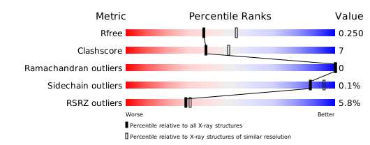
Structure validation
Download full validation report


