






 9VM3
9VM3
Structure of DOCK6-Cdc42 complex
Summary for 9VM3
| Entry DOI | 10.2210/pdb9vm3/pdb |
| Related | 9VM2 |
| EMDB information | 65175 |
| Descriptor | Dedicator of cytokinesis protein 6, Cell division control protein 42 homolog (2 entities in total) |
| Functional Keywords | dock, gef, rho, small gtpase, rac, cdc42, signaling protein |
| Biological source | Homo sapiens (human) More |
| Total number of polymer chains | 4 |
| Total formula weight | 503481.75 |
| Authors | Kukimoto-Niino, M.,Katsura, K.,Ishizuka-Katsura, Y.,Yonemochi, M.,Hanada, K.,Shirouzu, M. (deposition date: 2025-06-27, release date: 2026-06-03, Last modification date: 2026-09-09) |
| Primary citation | Kukimoto-Niino, M.,Katsura, K.,Yoshimura, K.,Ishizuka-Katsura, Y.,Miyamoto, Y.,Yonemochi, M.,Hanada, K.,Yamauchi, J.,Wong, R.W.,Shirouzu, M. Structural basis for auto-inhibition of the Rac1/Cdc42 guanine nucleotide exchange factor DOCK6 by oligomer formation. Commun Biol, 9:-, 2026 Cited by PubMed Abstract: The guanine nucleotide exchange factor DOCK6 is important for neurite outgrowth, as well as cell migration and invasion, through the activation of Rac1 and Cdc42-members of the Rho family of GTPases that regulate the actin cytoskeleton. However, the precise molecular mechanisms by which DOCK6 regulates the intracellular GTPase signaling remain unclear. Here, we present cryo-electron microscopy structures of DOCK6 alone and in complex with Rac1 and Cdc42. The DOCK6-Rac1 and DOCK6-Cdc42 complexes exhibit similar homodimeric structures, with local differences in the catalytic domain of DOCK6 owing to distinct interactions with Rac1 and Cdc42. In contrast, apo-DOCK6 exhibits a closed auto-inhibited conformation in tetrameric and octameric assemblies, with the catalytic and membrane-binding domains contacting each other between two DOCK6 dimers. High-speed atomic force microscopy reveals transitions among multiple oligomers in solution. Biochemical and cellular functional analyses demonstrate that the N-terminal region of DOCK6 plays an auto-inhibitory role, supporting the structural findings. Overall, we propose a mechanism by which DOCK6 activity is spatiotemporally regulated within cells through oligomerization. These findings provide a framework for future studies of DOCK-family GEFs and their broader roles in cell regulation and human disease. PubMed: 42215701DOI: 10.1038/s42003-026-10383-w PDB entries with the same primary citation |
| Experimental method | ELECTRON MICROSCOPY (4.52 Å) |
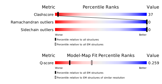
Structure validation
Download full validation report


