






 9TW2
9TW2
Cryo-EM structure of human VPS34-CI in complex with GABARAP
Summary for 9TW2
| Entry DOI | 10.2210/pdb9tw2/pdb |
| EMDB information | 56357 |
| Descriptor | Phosphatidylinositol 3-kinase catalytic subunit type 3, ZINC ION, Phosphoinositide 3-kinase regulatory subunit 4, ... (10 entities in total) |
| Functional Keywords | lipid kinase, complex, autophagy, lipid binding protein |
| Biological source | Homo sapiens (human) More |
| Total number of polymer chains | 7 |
| Total formula weight | 440492.32 |
| Authors | Dessus, A.N.,Williams, R.L. (deposition date: 2026-01-13, release date: 2026-08-05, Last modification date: 2026-09-16) |
| Primary citation | Dessus, A.N.,Ohashi, Y.,Bourguet, M.,Morgan, T.E.,Nunez, A.,Manifava, M.,Ktistakis, N.T.,Williams, R.L. A GABARAP-PtdIns3K-C1 positive feedback loop at the heart of the phagophore nucleation. Nat Commun, 17:-, 2026 Cited by PubMed Abstract: In mammalian cells, autophagosomes can reach diameters of over 1000 nm within 30 min after triggering starvation, but how such substantial amounts of membranes can be synthesized remains elusive. The phagophore initiation needs the lipid kinase PIK3C3-Complex 1 (PtdIns3K-C1), which produces phosphatidylinositol-3-phosphate (PtdIns3P). PtdIns3P recruits WIPI2 that facilitates lipidation of mammalian ATG8 (mATG8) family proteins on phagophores. Here we show that recombinant membrane-coupled GABARAP binds to and potently activates PtdIns3K-C1. By a combination of cryo-electron microscopy, structural mass spectrometry, activity assays and mutagenesis, we show that GABARAP activates PtdIns3K-C1 through two binding sites. We propose that once GABARAP is indirectly recruited by PtdIns3P generated by basal activity of PtdIns3K-C1, a positive feedback loop is formed where PtdIns3K-C1 interacts with GABARAP and becomes activated to produce more PtdIns3P, thereby further stimulating GABARAP lipidation. This mechanism would be central for autophagosome biogenesis, where enlarged membranes need to be rapidly synthesized. PubMed: 42693081DOI: 10.1038/s41467-026-76135-w PDB entries with the same primary citation |
| Experimental method | ELECTRON MICROSCOPY (3.58 Å) |
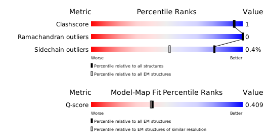
Structure validation
Download full validation report


