






 9RC2
9RC2
DvhD, periplasmic domain
Summary for 9RC2
| Entry DOI | 10.2210/pdb9rc2/pdb |
| Related | 9RBZ 9RC0 |
| Descriptor | HD domain/sensory box protein (1 entity in total) |
| Functional Keywords | biofilm regulator, second messenger signaling, transmembrane, signaling protein |
| Biological source | Nitratidesulfovibrio vulgaris str. Hildenborough |
| Total number of polymer chains | 1 |
| Total formula weight | 29595.42 |
| Authors | Font, M.E.,Sondermann, H. (deposition date: 2025-05-27, release date: 2026-03-04, Last modification date: 2026-04-29) |
| Primary citation | Font, M.E.,Karbelkar, A.A.,Lormand, J.D.,Mortensen, S.,Garcia-Garcia, M.J.,O'Toole, G.A.,Sondermann, H. Structural analyses uncover protease-adhesin interactions and c-di-GMP receptor regulation in sulfate-reducing bacteria. Nat Commun, 17:-, 2026 Cited by PubMed Abstract: Desulfovibrio vulgaris is a sulfate-reducing organism with biofim-forming capacity relevant for bioremediation and microbe-induced corrosion. Biofilm formation of D. vulgaris depends on two large adhesins that are regulated by proteins encoded in the Dvh operon, which resembles the gammaproteobacterial Lap system in composition but differs in the sequence and domain organization of its regulatory proteins, DvhG and DvhD. We show that DvhG is a calcium-dependent protease that targets the periplasmic domains of both adhesins via extensive interactions. Additionally, structures of DvhD establish this HD-GYP domain-containing protein as a c-di-GMP-dependent switch with a periplasmic dCache domain. Our data support a model in which DvhD controls DvhG activity through a c-di-GMP-dependent mechanism that is molecularly distinct, but functionally analogous to LapD. Together, our results reveal how conserved regulatory logic can be implemented through distinct molecular architectures, highlighting the evolutionary flexibility of c-di-GMP signaling networks in controlling surface attachment across diverse bacterial lineages. PubMed: 41998003DOI: 10.1038/s41467-026-71936-5 PDB entries with the same primary citation |
| Experimental method | X-RAY DIFFRACTION (3 Å) |
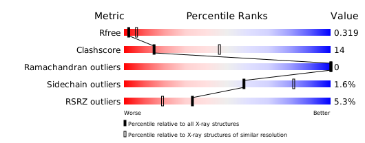
Structure validation
Download full validation report


