






 9PYS
9PYS
NMR RDC refinement of the helical domain of the SARS-CoV-2 monomeric Main Protease (MPROH41Q,10-306)
Summary for 9PYS
| Entry DOI | 10.2210/pdb9pys/pdb |
| NMR Information | BMRB: 31263 |
| Descriptor | 3C-like proteinase nsp5 (1 entity in total) |
| Functional Keywords | protease, helical, nsp5, sars-cov-2, hydrolase |
| Biological source | Severe acute respiratory syndrome coronavirus 2 (2019-nCoV, SARS-CoV-2) |
| Total number of polymer chains | 1 |
| Total formula weight | 33815.53 |
| Authors | Smith, M.J.,Ying, J.,Shen, Y.,Louis, J.M.,Bax, A. (deposition date: 2025-08-08, release date: 2025-09-17, Last modification date: 2026-02-18) |
| Primary citation | Smith, M.J.,Ying, J.,Shen, Y.,Louis, J.M.,Bax, A. Solution Domain Dynamics of Monomeric SARS-CoV‐2 Main Protease Revealed by Optimized NMR Residual Dipolar Coupling Measurements. ACS Phys Chem Au, 6:81-94, 2026 Cited by PubMed Abstract: The main protease (MPro) of SARS-CoV-2, released from a monomeric precursor, is essential for viral replication, and multiple antiviral compounds target the active site of the mature dimer. However, the solution structure and dynamics of the monomer remain poorly understood. Here, we utilize residual dipolar couplings (RDCs) to characterize conformational and dynamical differences between monomeric and dimeric MPro. While most protein NMR studies rely primarily on H-N RDCs, we demonstrate that at least four backbone RDCs can be measured at high accuracy from a single aligned sample. We compare frequency-based and intensity-based methods and introduce a mixed-time evolution scheme that improves resolution in 2D N-C' RDC measurements. These methods are applied to a 9-residue N-terminal deletion mutant of the SARS-CoV-2 MPro, revealing that the monomeric active site loop conformation closely resembles that of the SARS-CoV monomer X-ray structure, which differs substantially from the dimer. The C-terminal helical domain also undergoes large amplitude motions relative to the catalytic domain. In contrast, AlphaFold2 models of the deletion mutant predict structures adopting only the dimer-like conformation. Refinement of the monomeric X-ray structure (PDB: 2QCY) against our experimental RDCs resulted in backbone rearrangements up to ca. 1 Å, improved MolProbity statistics, and better agreement with independent RDCs not included in the refinement. These findings highlight the power of RDCs for probing conformational states and dynamics and may aid future identification and characterization of compounds targeting the precursor monomer active site. PubMed: 41624715DOI: 10.1021/acsphyschemau.5c00081 PDB entries with the same primary citation |
| Experimental method | SOLUTION NMR |
Structure validation
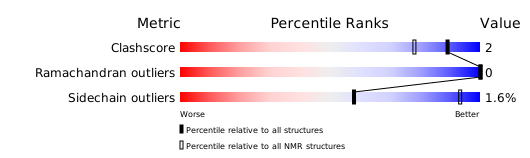
Download full validation report


